
- EMET Education
- 3D classifications
- Kellgren-Lawrence classification
Classification · Knee osteoarthritis
Kellgren-Lawrence Classification: 3D Visualization
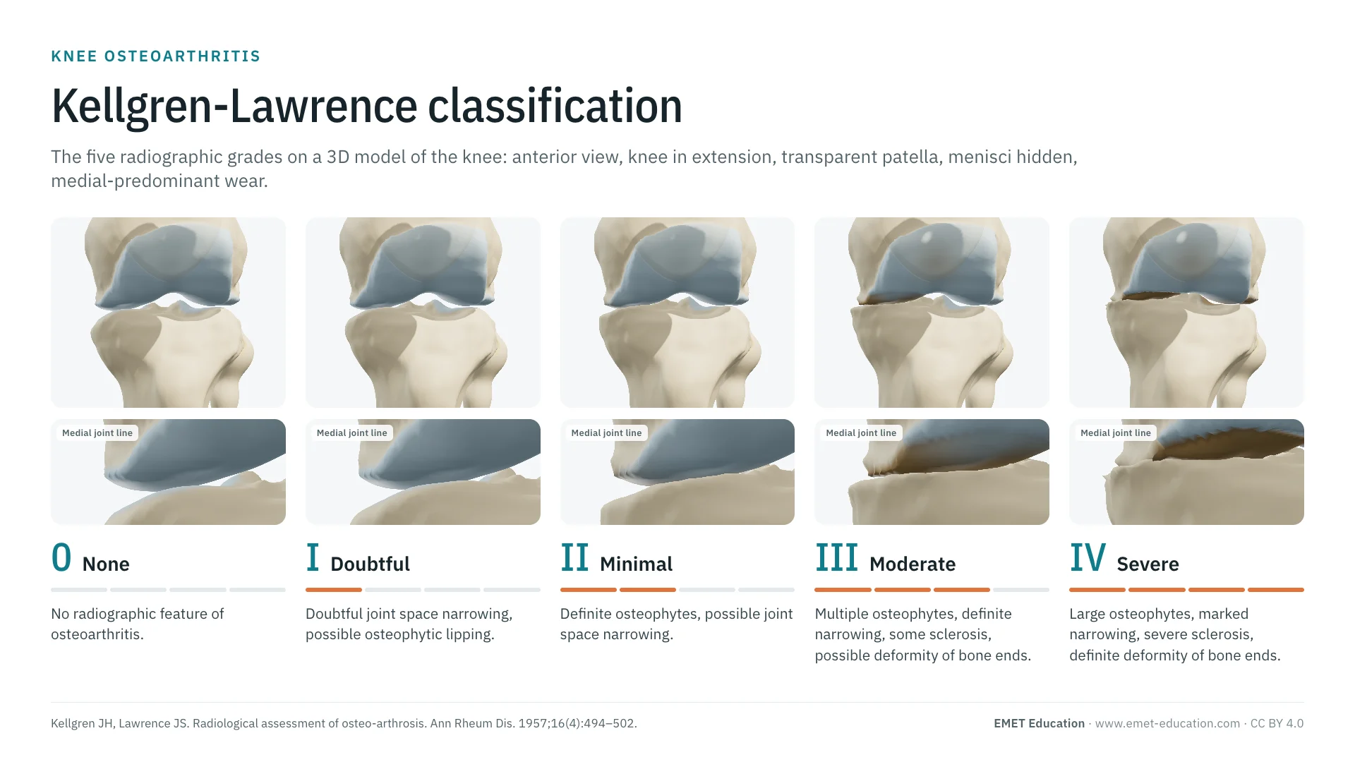
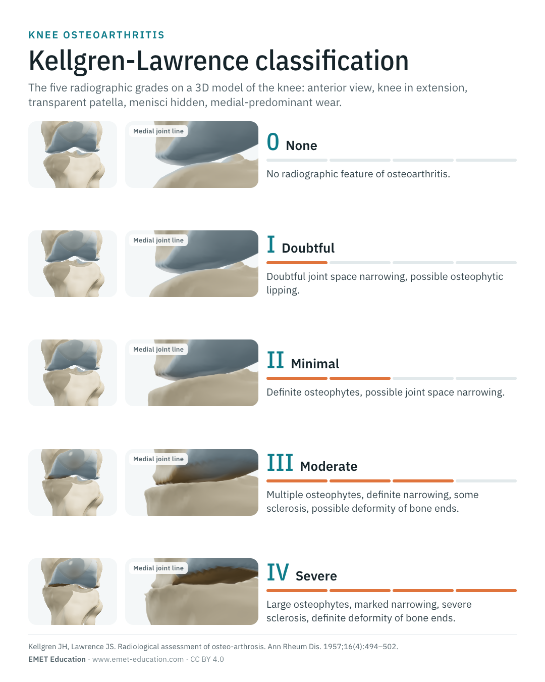
The five radiographic grades of osteoarthritis, from 0 to IV, shown on a 3D model of the knee. With the original article, reliability data and free infographics for teaching and publication.
Designed by an orthopedic surgeon
The 3D model opens in your browser, at the grade shown, with no sign-up. Infographics are free to reuse under CC BY 4.0.
- 1957Published by Kellgren and Lawrence in the Annals of the Rheumatic Diseases
- 0–IVFive grades, from no osteoarthritis to severe osteoarthritis
- 12,640Citations of the original article (OpenAlex, October 4, 2026)
- KL ≥ 2The usual definition of radiographic osteoarthritis in epidemiological studies
The five grades
From grade 0 to grade IV, in 3D
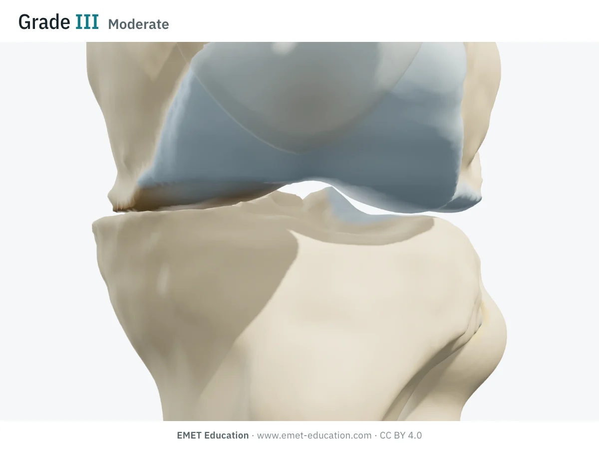
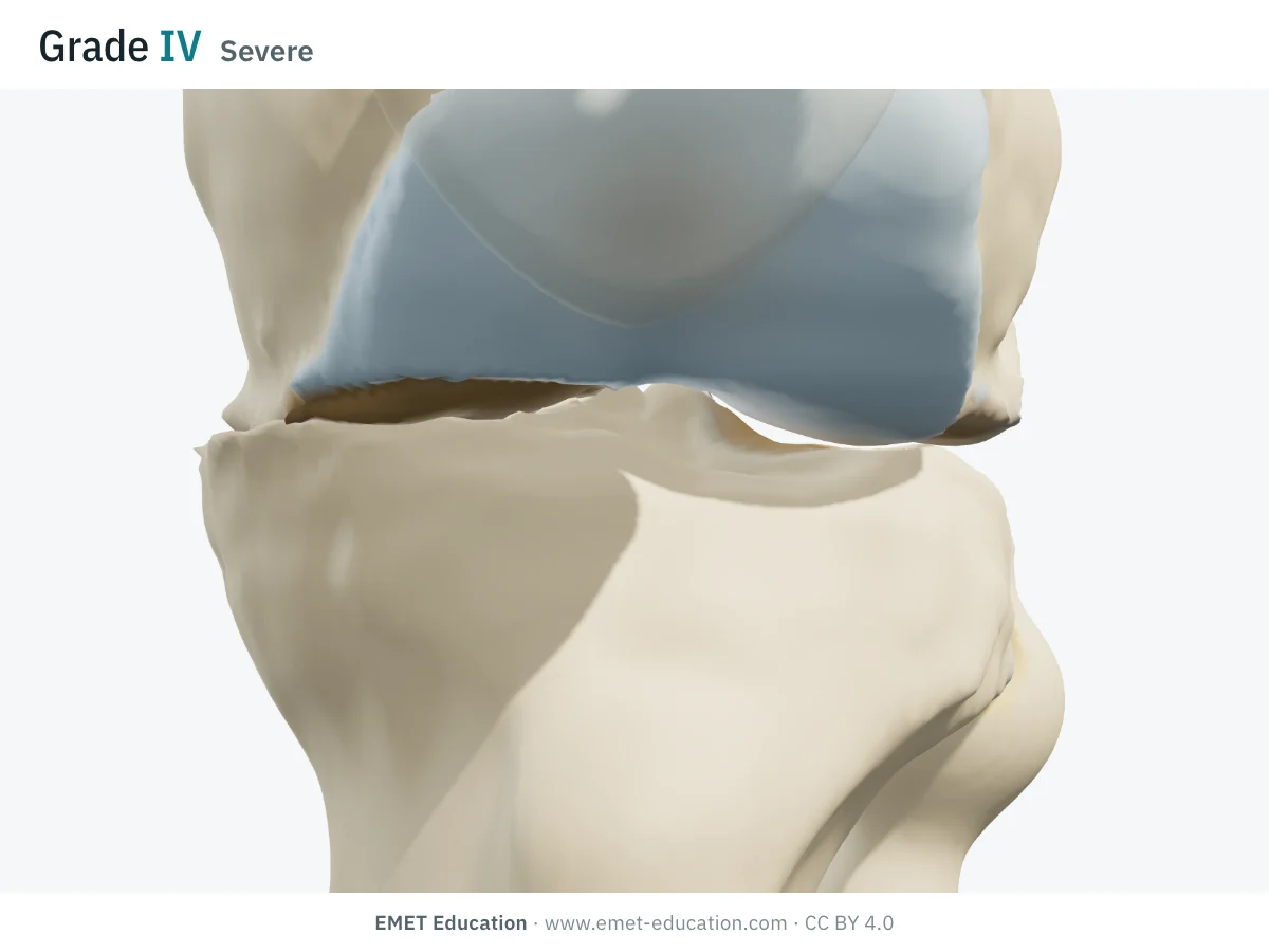
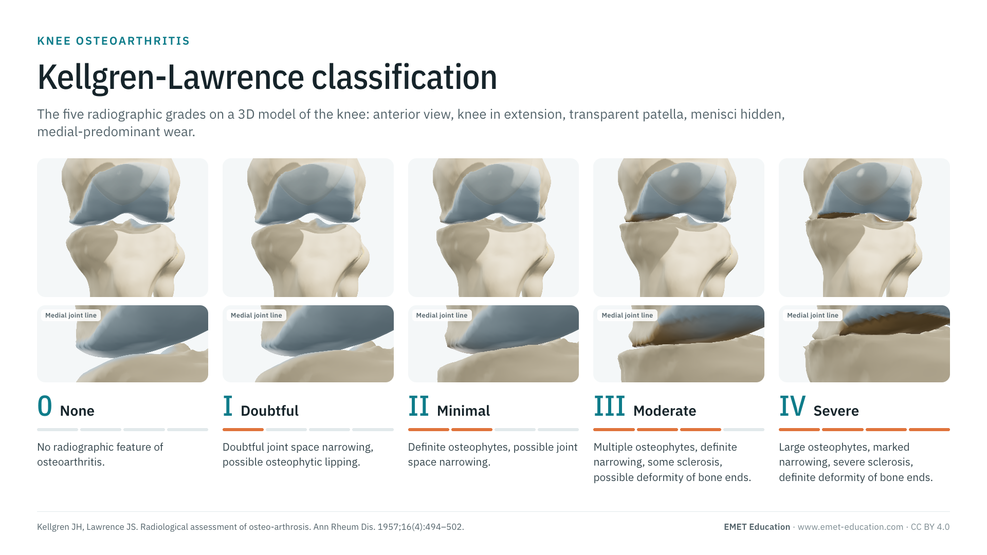
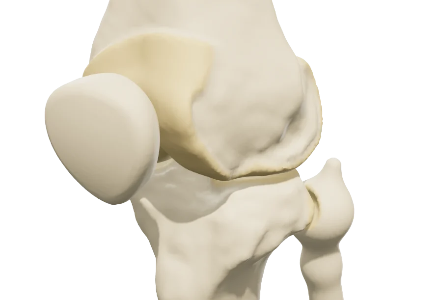
Each image is a rendering of the EMET Education knee model at that grade: anterior view, knee in extension, patella shown transparent and menisci hidden so that the joint line can be seen. Wear predominates in the medial femorotibial compartment, the most often involved, with lesser lateral and patellofemoral involvement at the higher grades. The 3D shows what the radiograph infers: cartilage loss behind the narrowing, osteophytes, then bone exposure and collapse.
-
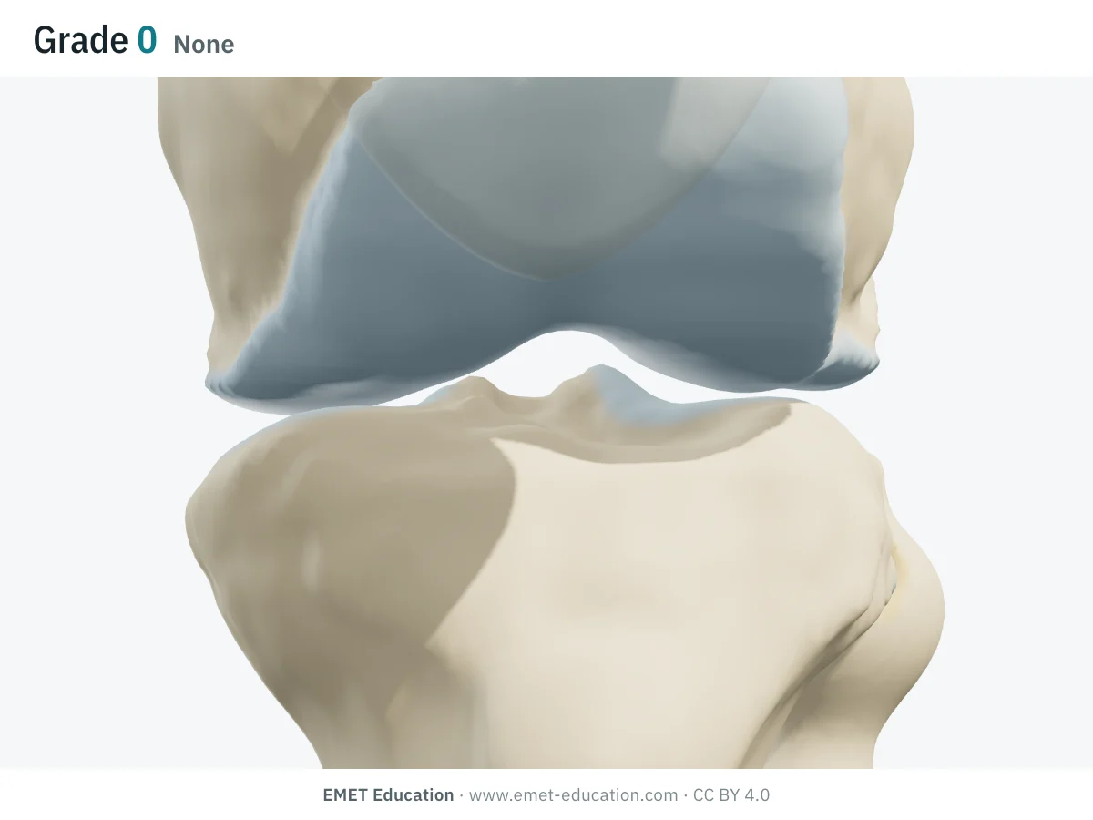
0None
- Radiographic criteria
- No radiographic feature of osteoarthritis.
- In the 3D model
- Intact articular cartilage, no osteophyte.
-
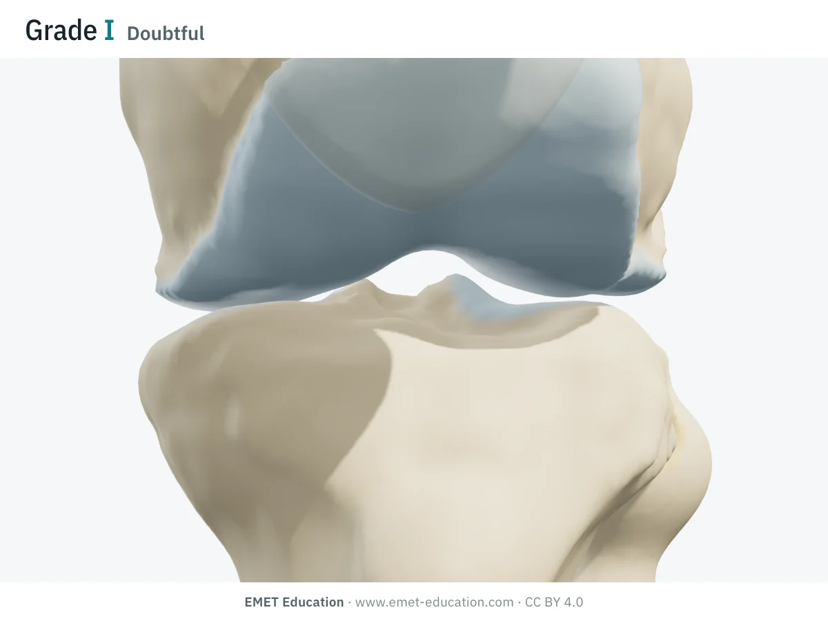
IDoubtful
- Radiographic criteria
- Doubtful joint space narrowing, possible osteophytic lipping.
- In the 3D model
- Medial compartment: cartilage thinned by about a fifth in the weight-bearing zone, dull surface, early osteophytes about 1 mm high. Lateral and patellofemoral compartments intact.
-
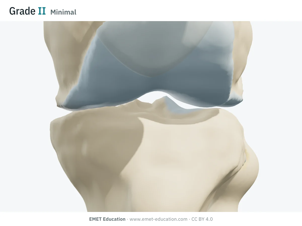
IIMinimal
- Radiographic criteria
- Definite osteophytes, possible joint space narrowing.
- In the 3D model
- Medial compartment: cartilage thinned by half in the weight-bearing zone, osteophytes about 2.5 mm at its margins. Early changes in the lateral compartment.
-
IIIModerate
- Radiographic criteria
- Multiple moderate osteophytes, definite joint space narrowing, some sclerosis, possible deformity of the bone ends.
- In the 3D model
- Medial compartment: cartilage almost gone in the center of the weight-bearing zone, subchondral bone exposed, osteophytes about 4 mm, narrowed joint space. Lateral and patellofemoral compartments at an early stage.
-
IVSevere
- Radiographic criteria
- Large osteophytes, marked joint space narrowing, severe sclerosis, definite deformity of the bone ends.
- In the 3D model
- Medial compartment: bone exposed over a wide area and collapsed (about 2 mm on the tibia, 1 mm on the femur), osteophytes about 6 mm, joint space closed. Lateral compartment worn by half, early patellofemoral wear.
Schematic rendering, for teaching: the model illustrates each grade on a single knee, it does not reproduce a radiograph. Criteria reworded from the original description; the 1957 figures and the 1963 atlas are not reproduced.
The original article
Kellgren and Lawrence, 1957
J. H. Kellgren and J. S. Lawrence published Radiological assessment of osteo-arthrosis in the Annals of the Rheumatic Diseases in 1957 (16:494–502, doi:10.1136/ard.16.4.494). Their aim was epidemiological: to grade, reproducibly, the radiographs of population samples.
The grading rests on the radiographic features they took as evidence of osteoarthritis:
- osteophytes at the joint margins or, in the knee, on the tibial spines;
- periarticular ossicles, mainly around the interphalangeal joints;
- narrowing of the joint cartilage with sclerosis of the subchondral bone;
- small pseudocystic areas with sclerotic walls in the subchondral bone;
- altered shape of the bone ends.
Each joint received a global grade from 0 to 4: none, doubtful, minimal, moderate, severe. The scale was adopted by the World Health Organization in 1961 as the radiological definition of osteoarthritis for epidemiological studies, and illustrated in the Atlas of Standard Radiographs of Arthritis (1963). As of October 4, 2026, OpenAlex counts 12,640 citations of the original article.
Reliability
How reproducible is the grade?
Interobserver agreement varies widely between studies, from fair to almost perfect, depending on the description used, the radiographic view and the readers' experience.
| Study | Coefficient | Interobserver agreement |
|---|---|---|
| Wing et al., 2021 | Intraclass correlation coefficient | 0.85 |
| Günther & Sun, 1999 | Intraclass correlation coefficient | 0.81 |
| Köse et al., 2018 | Cohen's kappa | 0.47 |
| Schiphof et al., 2011 | Cohen's kappa | 0.41 |
| Wright et al. (MARS Group), 2014 | Intraclass correlation coefficient | 0.38 |
| Gonçalves et al., 2016 | Krippendorff's alpha | 0.33 |
Two practical findings: experienced readers agree more often with themselves than trainees do, and the radiographic view matters. In the MARS cohort, the six radiographic scales studied, Kellgren-Lawrence included, were more reproducible on a 45° posteroanterior flexion weight-bearing radiograph (Rosenberg view; pooled ICC 0.63) than on an anteroposterior radiograph (0.55), and correlated better with the cartilage damage seen at arthroscopy.
Variants and related scales
Five descriptions, several scales
The wording of the grades has drifted since 1957. Schiphof et al. found five different descriptions of the Kellgren-Lawrence grades in the major cohort studies; for grade 2, one replaced "definite osteophytes and possible narrowing" with "definite osteophyte, unimpaired joint space", another with "minimal osteophytes, possible narrowing, cysts and sclerosis". On 3,071 people of the Rotterdam Study, these variants changed mostly the lowest cut-off (grade ≥ 1), and little the usual cut-offs (≥ 2 and ≥ 3).
When reporting a grade, say which description you used. Other scales grade different things:
| Scale | What it grades | Typical use |
|---|---|---|
| Kellgren-Lawrence (1957) | A global grade per joint (0 to IV), combining osteophytes, narrowing, sclerosis and deformity. | Epidemiology, trial eligibility, everyday reporting. |
| Ahlbäck (1968) | Tibiofemoral narrowing up to obliteration of the joint space, then bone attrition. | Advanced disease, before surgery. |
| OARSI atlas (2007) | Each feature separately (joint space narrowing, osteophytes) in each compartment, on a reference atlas. | Research: follow-up of individual features. |
| IKDC | Remaining joint space, from A (normal) to D (severely abnormal), on a weight-bearing radiograph. | Sports and knee surgery; best balance of reliability and arthroscopic correlation in the MARS study. |
| Compartment-specific Kellgren-Lawrence (2024) | A Kellgren-Lawrence grade for each compartment, patellofemoral included. | Describing which compartments are involved (interobserver α 0.51 to 0.56). |
Use
In research and in clinical practice
In research
Kellgren-Lawrence grade 2 or higher is the usual definition of radiographic knee osteoarthritis in epidemiological studies, and the grade is a common eligibility criterion or outcome in cohorts and trials, and a reference label for automated grading of radiographs. Its main limit there is the one Schiphof described: unless the description is stated, two studies "using Kellgren-Lawrence" may not grade the same way.
In clinical practice
The grade summarizes the structural severity of the joint; it does not measure pain or disability. Radiographic and clinical osteoarthritis are discordant: in a systematic review, 15% to 76% of people with knee pain had radiographic osteoarthritis, and 15% to 81% of people with radiographic osteoarthritis had pain. The radiograph should not be read in isolation.
In practice, a total knee replacement is usually discussed for advanced structural disease, grade III or IV, when pain and functional limitation persist despite nonoperative treatment; the grade is one element of that decision, not the decision.
Grading pitfalls
Five pitfalls when grading a knee
- Grade on a weight-bearing radiograph. Narrowing is underestimated without load. The 45° posteroanterior flexion view (Rosenberg) shows posterior wear of the condyles better and gave more reproducible grades than the anteroposterior view in the MARS study.
- Do not dismiss "doubtful" osteophytes. In a 10-year follow-up of women, 62% of knees graded 1 for a doubtful osteophyte developed definite osteophytic osteoarthritis, against 22% of knees without any sign (Hart and Spector, 2003).
- State the description you use, especially for grade 2, whose wording differs most between studies.
- Say what you graded. The original grade is global, for the whole joint. Grading the worst compartment, or each compartment, is a different measure; the patellofemoral joint needs a lateral or axial view and is not covered by the frontal grade.
- Keep the grade on the radiograph. Kellgren-Lawrence is a radiographic scale: it does not apply to MRI, which sees cartilage, menisci and bone marrow directly.
Self-test
Which grade?
Five views of the same knee from the medial side, in random order. Find the grade of each one; the answer comes with the criteria.
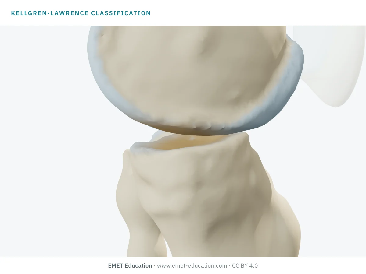
Which Kellgren-Lawrence grade is this?
Download and cite
Free infographics, for teaching and publication
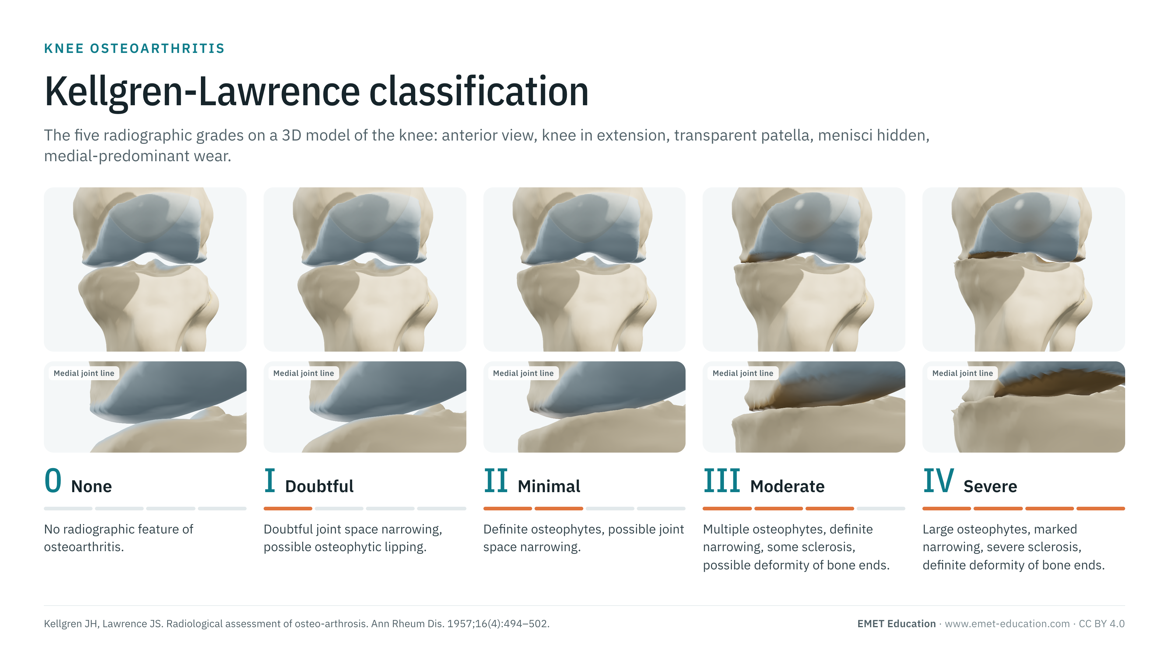
Use them in a course, a presentation, a social media post or a scientific article, under the Creative Commons Attribution 4.0 license: free reuse, including commercial, provided EMET Education is credited with a link to this page.
Cite this figure
Ready to paste. For a journal figure, add the original reference (Kellgren and Lawrence, 1957).
EMET Education. Kellgren-Lawrence classification of knee osteoarthritis: 3D visualization [Internet]. 2026 [cited]. Available from: https://www.emet-education.com/en/classifications/kellgren-lawrence/. CC BY 4.0.EMET Education. (2026). Kellgren-Lawrence classification of knee osteoarthritis: 3D visualization [Infographic]. https://www.emet-education.com/en/classifications/kellgren-lawrence/ (CC BY 4.0)<a href="https://www.emet-education.com/en/classifications/kellgren-lawrence/"><img src="https://www.emet-education.com/en/classifications/kellgren-lawrence/img/infographic.webp" alt="Kellgren-Lawrence classification in 3D" width="960"></a><br>Figure: <a href="https://www.emet-education.com/en/classifications/kellgren-lawrence/">EMET Education</a>, CC BY 4.0Questions
Frequently asked questions
What is the difference between Kellgren-Lawrence grade 2 and grade 3?
Grade 2 has definite osteophytes with at most possible narrowing of the joint space. Grade 3 adds definite narrowing, with multiple moderate osteophytes, some subchondral sclerosis and possible deformity of the bone ends. In practice, narrowing that becomes definite marks the change from 2 to 3.
Which grade defines radiographic knee osteoarthritis?
Grade 2 or higher (KL ≥ 2) in most epidemiological studies. Grade 1 remains debated: its "doubtful" osteophytes are associated with later osteoarthritis, but its definition varies most between descriptions.
Which radiograph should be graded?
A weight-bearing radiograph. The 45° posteroanterior flexion view (Rosenberg) shows the posterior part of the condyles, which an extension view misses, and gave more reproducible grades than the anteroposterior view in a multicenter study.
Does the Kellgren-Lawrence grade correlate with pain?
Only loosely. Depending on the study, 15% to 81% of people with radiographic knee osteoarthritis report pain, and 15% to 76% of people with knee pain have radiographic osteoarthritis. The grade describes structure; it must be read with the clinical picture.
Is the classification only for the knee?
No. Kellgren and Lawrence graded several joints, and the scale is still used for the hip and the hand. This page shows the knee, the joint where it is most used.
Can I reuse these images in a course or an article?
Yes. The images and infographics on this page are under the Creative Commons Attribution 4.0 license: free reuse, including commercial, with credit to EMET Education and a link to this page. The "Cite this figure" box gives ready-to-paste citations.
How were the 3D images made?
They are renderings of the EMET Education knee osteoarthritis model, set to each grade: the model thins the cartilage, grows the osteophytes and collapses the bone according to the grade, compartment by compartment (here medial-predominant), and the femur rests on the tibia, so the wear sets the varus tilt. The patella is shown transparent and the menisci hidden so that the joint line can be seen. It is a schematic model for teaching and patient explanation; it does not reproduce a radiograph.
Sources
Author and references
- Kellgren JH, Lawrence JS. Radiological assessment of osteo-arthrosis. Ann Rheum Dis. 1957;16(4):494–502. doi:10.1136/ard.16.4.494
- Kellgren JH, Jeffrey MR, Ball J, editors. The epidemiology of chronic rheumatism. Vol 2: Atlas of standard radiographs of arthritis. Oxford: Blackwell Scientific Publications; 1963.
- Kohn MD, Sassoon AA, Fernando ND. Classifications in brief: Kellgren-Lawrence classification of osteoarthritis. Clin Orthop Relat Res. 2016;474(8):1886–93. doi:10.1007/s11999-016-4732-4
- Schiphof D, Boers M, Bierma-Zeinstra SM. Differences in descriptions of Kellgren and Lawrence grades of knee osteoarthritis. Ann Rheum Dis. 2008;67(7):1034–6. doi:10.1136/ard.2007.079020
- Schiphof D, de Klerk BM, Kerkhof HJ, et al. Impact of different descriptions of the Kellgren and Lawrence classification criteria on the diagnosis of knee osteoarthritis. Ann Rheum Dis. 2011;70(8):1422–7. doi:10.1136/ard.2010.147520
- Alarcón Perico D, Camacho Uribe A, Jaimes Niño S, et al. A proposed modification to the Kellgren and Lawrence classification for knee osteoarthritis using a compartment-specific approach. J Exp Orthop. 2024;11(1):e12008. doi:10.1002/jeo2.12008
- Wright RW; MARS Group. Osteoarthritis classification scales: interobserver reliability and arthroscopic correlation. J Bone Joint Surg Am. 2014;96(14):1145–51. doi:10.2106/JBJS.M.00929
- Rosenberg TD, Paulos LE, Parker RD, et al. The forty-five-degree posteroanterior flexion weight-bearing radiograph of the knee. J Bone Joint Surg Am. 1988;70(10):1479–83. doi:10.2106/00004623-198870100-00006
- Ahlbäck S. Osteoarthrosis of the knee. A radiographic investigation. Acta Radiol Diagn (Stockh). 1968;Suppl 277:7–72.
- Altman RD, Gold GE. Atlas of individual radiographic features in osteoarthritis, revised. Osteoarthritis Cartilage. 2007;15 Suppl A:A1–56. doi:10.1016/j.joca.2006.11.009
- Felson DT, McAlindon TE, Anderson JJ, et al. Defining radiographic osteoarthritis for the whole knee. Osteoarthritis Cartilage. 1997;5(4):241–50. doi:10.1016/S1063-4584(97)80020-9
- Hart DJ, Spector TD. Kellgren & Lawrence grade 1 osteophytes in the knee: doubtful or definite? Osteoarthritis Cartilage. 2003;11(2):149–50. doi:10.1053/joca.2002.0853
- Bedson J, Croft PR. The discordance between clinical and radiographic knee osteoarthritis: a systematic search and summary of the literature. BMC Musculoskelet Disord. 2008;9:116. doi:10.1186/1471-2474-9-116
3D models
Show the grade to your patient
The images on this page come from an interactive model. In clinic, set the grade and the compartments from your patient's radiograph, then show the treatment discussed.
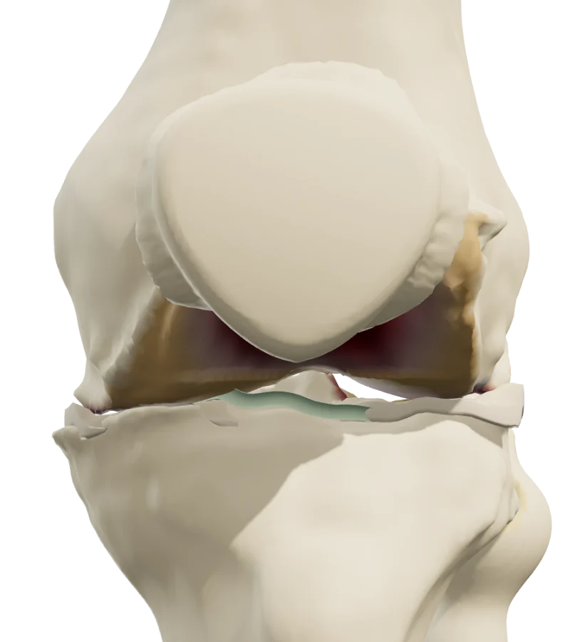 KneeKnee osteoarthritisKellgren-Lawrence grade per compartment, alignment, effusion, injections, total knee replacement.
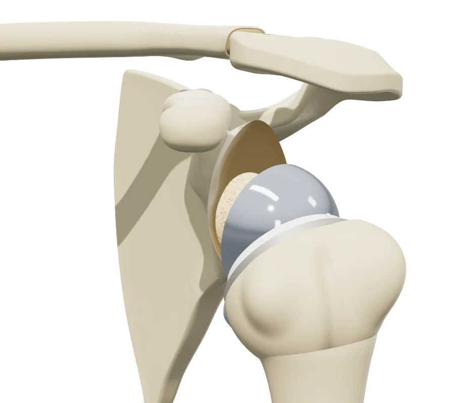 ShoulderShoulder osteoarthritisSamilson-Prieto and Hamada grades, humeral head osteonecrosis, shoulder replacement.
 KneeAll knee modelsOsteoarthritis and cruciate ligament injuries.
Explain the grade in 3D
The knee osteoarthritis model opens in your browser, at the grade you choose, with no sign-up.
Share this page with a colleague or a resident: