
- EMET Education
- Musculoskeletal
3D models
Musculoskeletal 3D models
Shoulder, elbow and knee 3D models to show your patient their lesion and the treatment you recommend. They leave with their model on their phone.
Designed by an orthopedic surgeon
Open in your browser, no sign-up. For orthopedic surgeons, emergency physicians, sports medicine physicians, physiatrists, rheumatologists and primary care physicians.
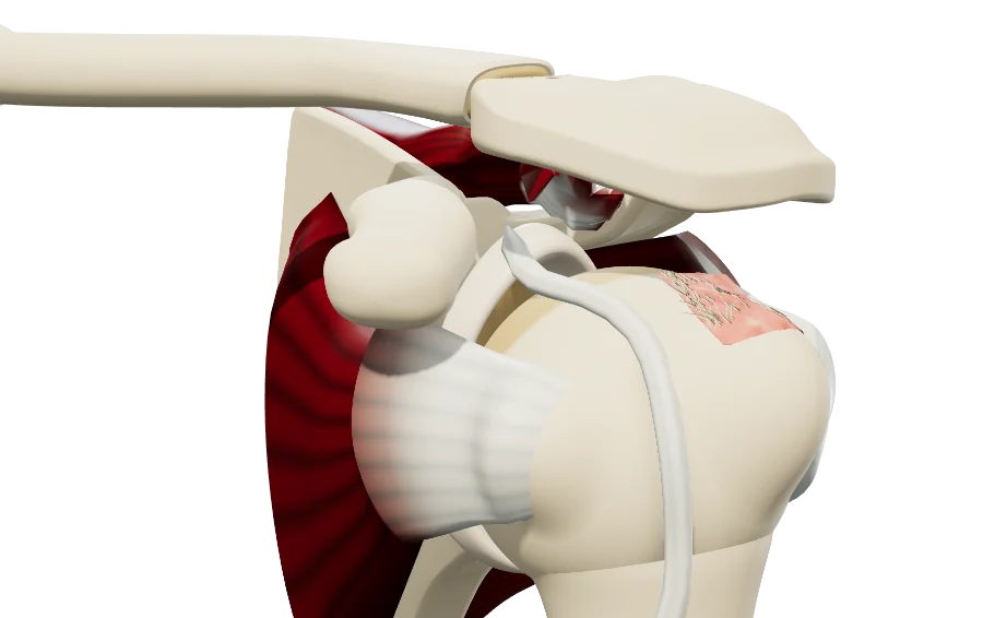
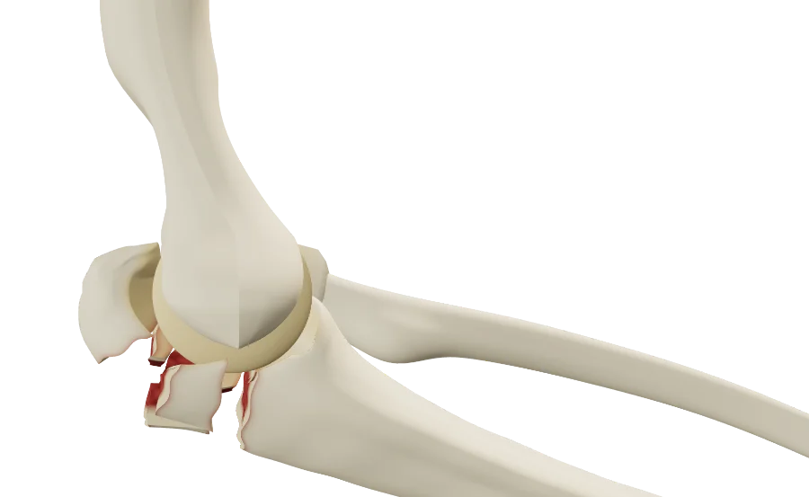
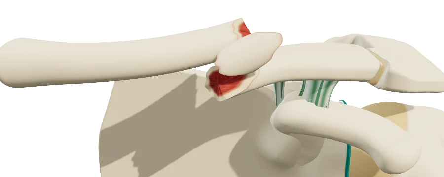
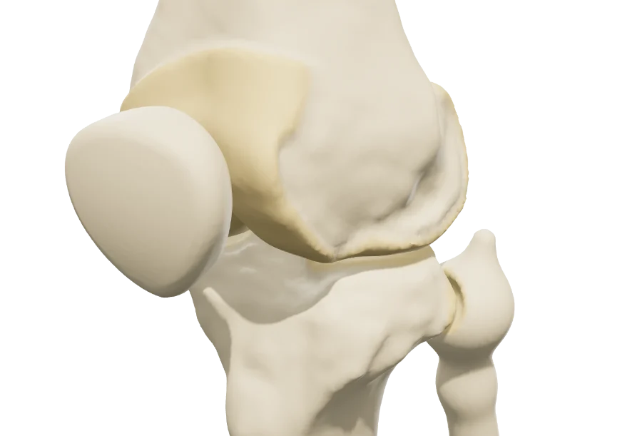
- The patient understandsTheir lesion, reproduced from their workup, on a model they handle themselves.
- Reassured, ready to decideThey review the proposed treatment at home before giving consent.
- Documented informationVisit saved and dated: a record of the information you provided.
- In their language🇫🇷 🇬🇧 🇪🇸 🇷🇺
The catalog
The models, region by region
Each model reproduces a specific condition, set from your patient's workup with the reference classifications, then plays the treatments offered: nonoperative, rehabilitation, injection, arthroscopy, fracture fixation, arthroplasty.
Shoulder
The models share the same shoulder anatomy: scapula, humerus, clavicle, and depending on the model the rotator cuff, the capsule, the labrum or the coracoclavicular ligaments. You set the lesion from the exam and imaging, then walk through the proposed treatment: arthroscopic, arthroplasty, fracture fixation or nonoperative.
ShoulderRotator cuffFull- and partial-thickness tears, calcific tendinitis, long head of the biceps, repair.
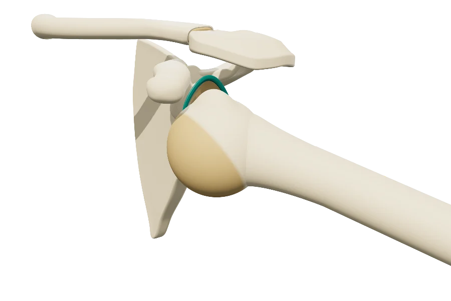 ShoulderShoulder instabilityDislocation, Bankart, Hill-Sachs; Latarjet, arthroscopic Bankart repair, remplissage.
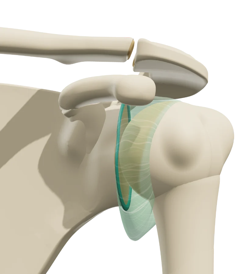 ShoulderPainful unstable shoulderStretched capsule, labral lesions, no true dislocation.
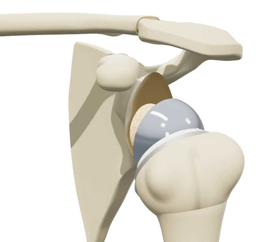 ShoulderShoulder osteoarthritisCentered or eccentric glenohumeral osteoarthritis, osteonecrosis; three arthroplasties.
ShoulderClavicle fractureShaft and lateral quarter, AO/OTA, Robinson, Neer; nonoperative treatment, plate, coracoclavicular button.
ShoulderShoulder osteoarthritisCentered or eccentric glenohumeral osteoarthritis, osteonecrosis; three arthroplasties.
ShoulderClavicle fractureShaft and lateral quarter, AO/OTA, Robinson, Neer; nonoperative treatment, plate, coracoclavicular button.
Elbow
The models share a common elbow anatomy: the three bones, flexion and pronation-supination, the tendons and the nerves. Fracture, tendon or nerve: the lesion is set from the workup, and the treatment plays step by step.
ElbowOlecranon fractureMayo classification, tension-band wiring and locking plate.
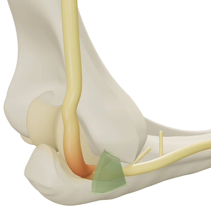 ElbowCubital tunnel syndromeUlnar nerve compression at the elbow, nerve instability, neurolysis and transposition.
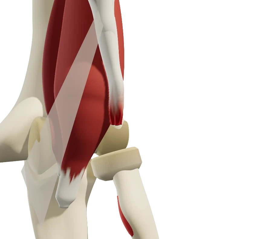 ElbowDistal biceps ruptureRuptured, retracted tendon; nonoperative treatment or reattachment to the radial tuberosity.
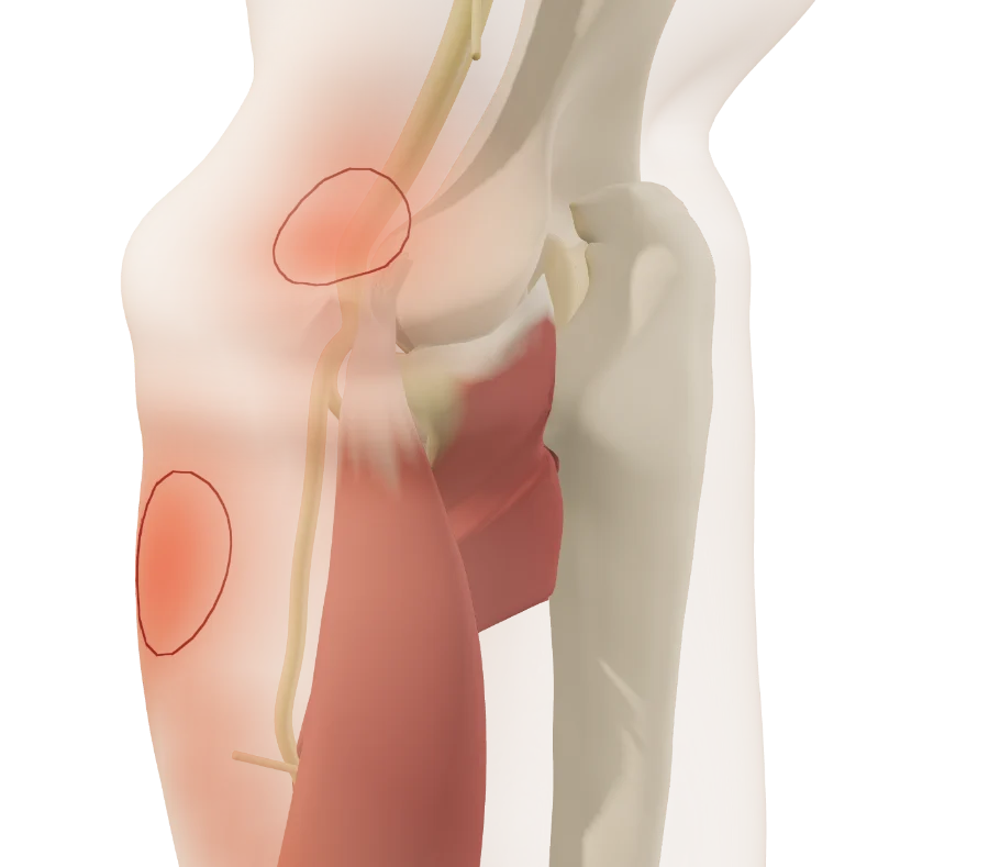 ElbowLateral epicondylalgia and posterior interosseous nerveExtensor carpi radialis brevis tendinopathy and posterior interosseous nerve compression.
Knee
The knee is under construction: femur, tibia, patella and fibula modeled from real bones, flexion kinematics, cruciate ligaments and menisci. It already opens in clinic.
How it works
In the visit, in three steps
Structured information in a few minutes, which the patient can review at home.
-
1
Show
Open the model for the region and walk through the anatomy: bones, joint, tendons, ligaments, nerves.
-
2
Reproduce
Set the lesion from the exam and imaging: location, type, stage, classification. Then walk through the proposed treatment, step by step.
-
3
Share
Give the patient the QR code or an image: they find their own case, in their language.
Who it's for
For every physician who explains
A patient who sees their injury understands the proposed treatment better, follows it better, and gives informed consent. The same models serve every stage of care.
- Orthopedic surgeonThe surgical indication, the proposed procedure step by step, informed consent.
- Sports medicine and PM&RThe injury, its mechanism, nonoperative treatment and what rehabilitation will restore.
- RheumatologistOsteoarthritis and tendinopathies, injections, referral to surgery.
- Emergency and primary careThe fracture or dislocation seen acutely, initial treatment, next steps in care.
Try it with your next case: the models open in your browser, nothing to install.
See the shoulder modelsPatient information
Quality information, documented
Each visit created with an account keeps a record of the information provided: what was shown, when, and whether the patient reviewed it. The link given to the patient carries no identifying data.
“Document the informed consent conversation and the patient’s (or surrogate’s) decision in the medical record in some manner.”
The models support the consent conversation and the written consent form; they do not replace them. Data and privacy: legal information (in French).
Questions
Questions from colleagues
For which physicians?
For any physician who explains a musculoskeletal injury to a patient: orthopedic surgeon, sports medicine physician, physiatrist, rheumatologist, emergency physician, primary care physician. The settings use specialist vocabulary; the explanations given to the patient are written for them.
Which regions and which conditions?
The shoulder, the elbow and the knee to begin with: tendons, ligaments, nerves, fractures, instability and osteoarthritis. Each region has its own page detailing what each model shows; new models join the catalog.
Are the models free?
Yes: the free plan opens every model, to show patients their diagnosis: most conditions and lesions. Treatments and other features are paid options.
Do I need an account?
Not to open a model, set the lesion and walk through the treatment. After about fifteen adjustments, you will be asked to create a free account: it saves your visits, picks them up from one time to the next, and records when the patient opens the link.
What equipment do I need?
Just a web browser, nothing to install: computer, tablet or TV screen in the exam room; the patient opens it on their phone. One click saves an image, without the interface, for the chart.
What happens to patient data?
No patient names are stored. The link given to the patient carries only the state of the model: lesion, treatment, language and visit date. A name written on the image you hand out stays on that image.
Which languages?
English, Spanish, French and Russian. The patient receives the model in the language chosen during the visit, which helps with the roughly 28 million people in the U.S. with limited English proficiency, alongside a qualified interpreter, not instead of one.
Design
Who designed these models
Try them with your next patient
The models open in your browser, nothing to install, no sign-up.
Know a colleague who could use them? Email this page ·