
- EMET Education
- 3D classifications
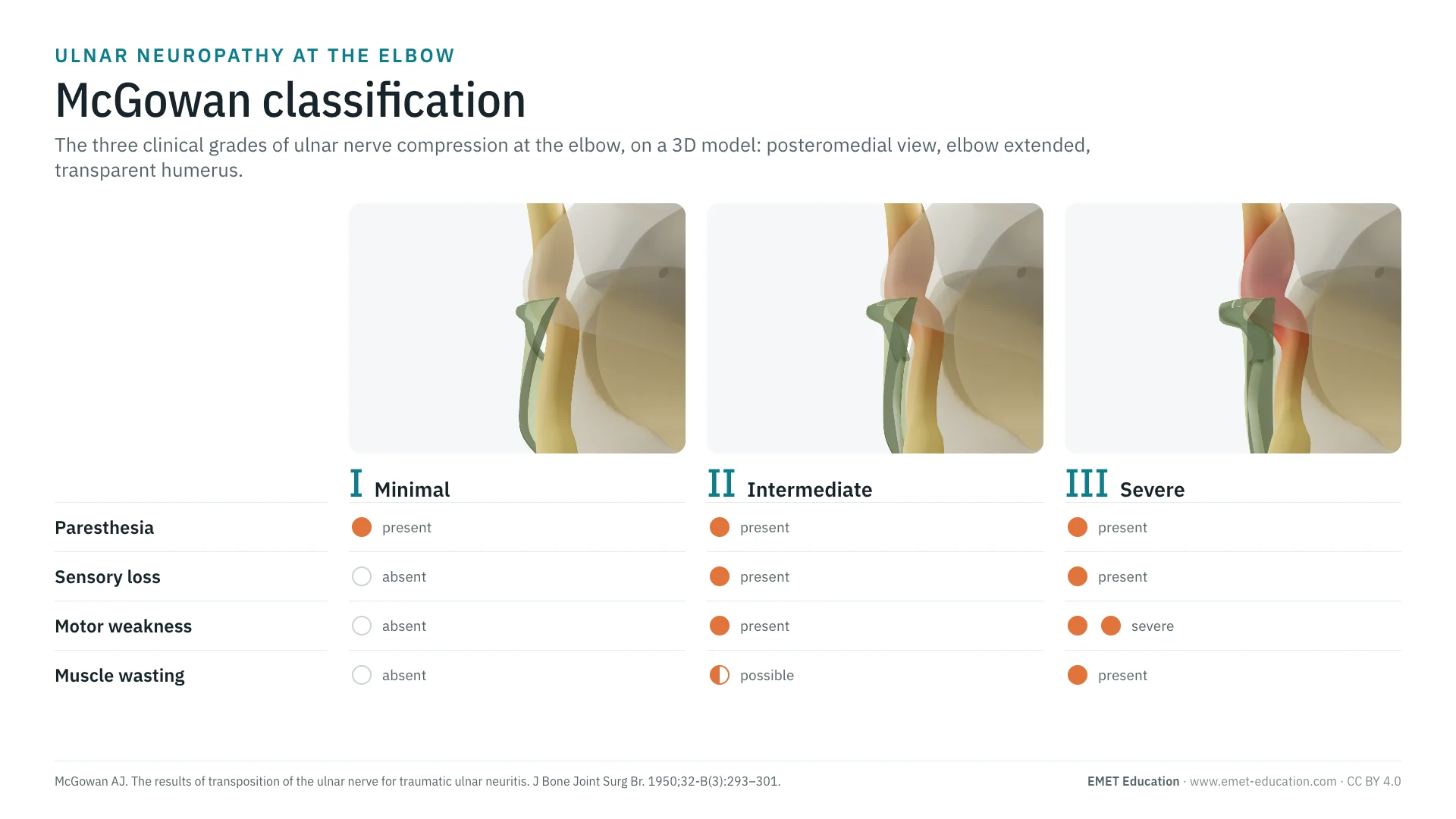
- McGowan classification
Classification · Ulnar neuropathy at the elbow
McGowan Classification: 3D Visualization
The three clinical grades of ulnar nerve compression at the elbow, from tingling alone to muscle wasting, illustrated on a 3D model of the cubital tunnel. With the original article, reliability, prognosis and free infographics.
Designed by an orthopedic surgeon
The 3D model opens in your browser, at the grade shown, with no sign-up. Infographics are free to reuse under CC BY 4.0.
- 1950Published by McGowan in the Journal of Bone and Joint Surgery (British volume)
- I–IIIThree clinical grades, from sensory symptoms alone to muscle wasting
- 453Citations of the original article (OpenAlex, October 4, 2026)
- κ 0.54Interobserver agreement of the modified grade: moderate (Choi 2022)
The three grades
From the healthy nerve to grade III, in 3D
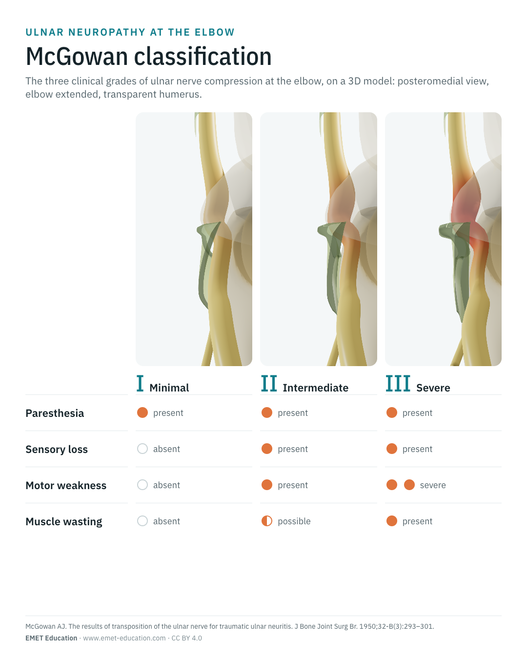
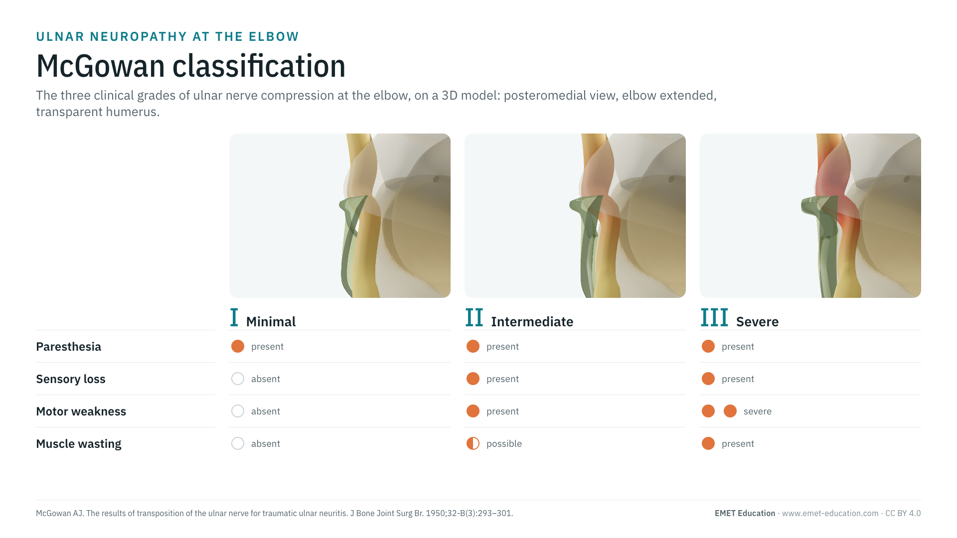
Each image is a rendering of the EMET Education ulnar neuropathy model: posteromedial view of the extended elbow, transparent humerus so that the nerve can be seen in its groove. On the left the whole elbow, on the right the nerve under Osborne's ligament. The McGowan classification is clinical: the 3D illustrates the compression that goes with each grade, from a barely constricted nerve to a congested pseudoneuroma.
-
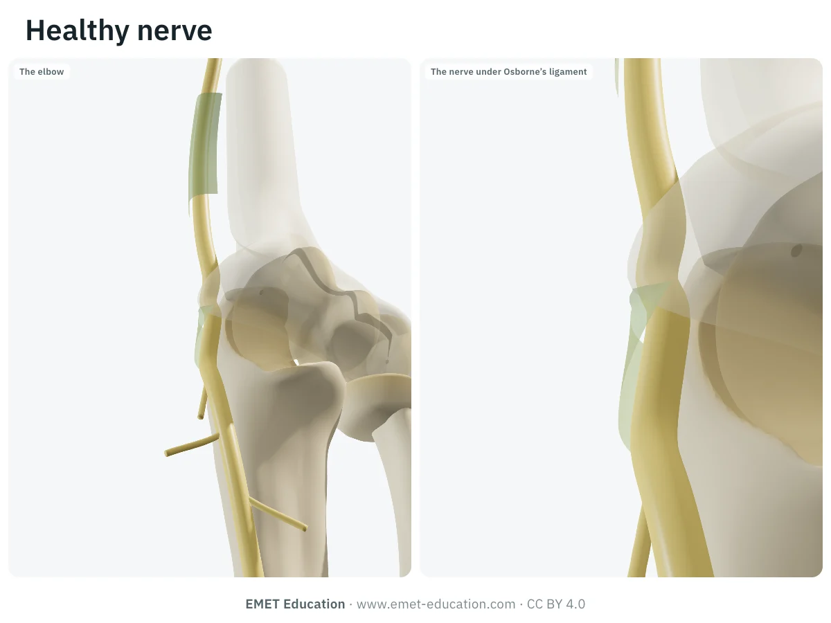
Healthy nerve (reference)
- In the 3D model
- Osborne's ligament slack with the elbow extended, nerve free and of even caliber in its groove.
-
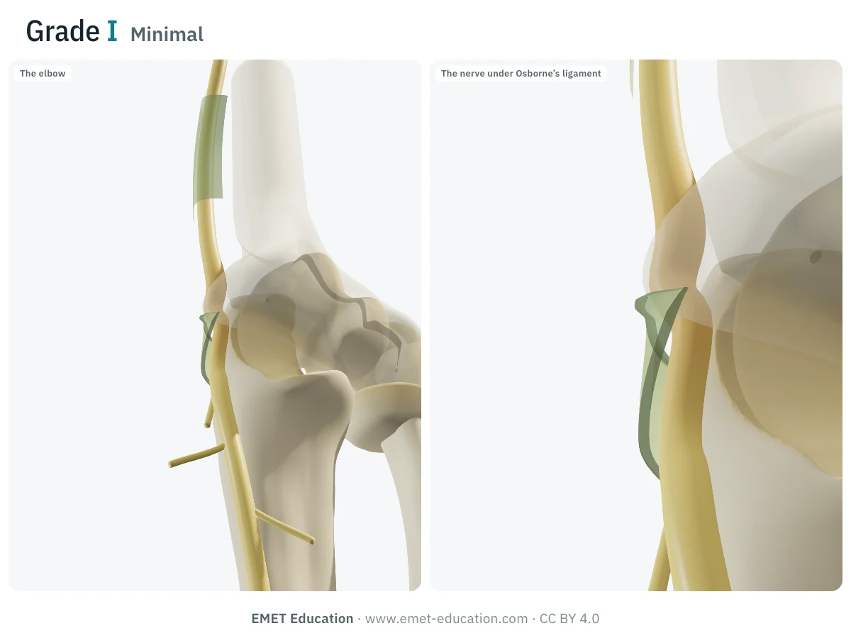
IMinimal
- Criteria
- Paresthesia and subjective clumsiness, no motor deficit.
- On examination
- Tinel sign at the groove and elbow flexion test often positive; sensation and strength objectively normal.
- In the 3D model
- The band tightens, the nerve starts to swell just above it.
-
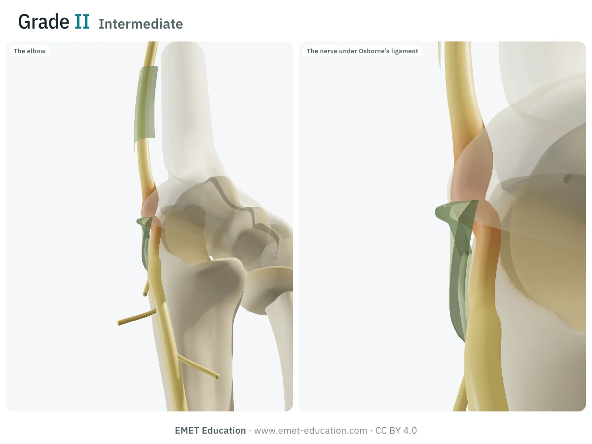
IIIntermediate
- Criteria
- Paresthesia and sensory loss, motor weakness, with or without early wasting of the interossei.
- On examination
- Sensory loss in the little finger and the ulnar side of the ring finger; weak finger abduction and thumb adduction (Froment sign possible); early wasting of the first dorsal interosseous.
- In the 3D model
- Nerve flattened under the band, swollen and congested above it.
-
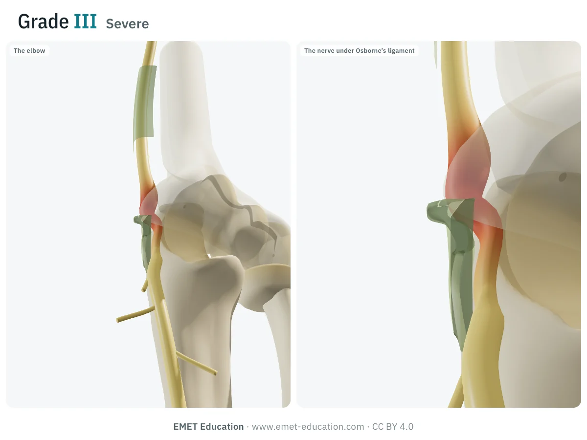
IIISevere
- Criteria
- Paresthesia and sensory loss, severe motor deficit and muscle wasting, up to paralysis of one or more interossei.
- On examination
- Wasting of the first web space, clawing of the ring and little fingers, Froment and Wartenberg signs, impaired two-point discrimination.
- In the 3D model
- Thickened band, nerve markedly flattened under it, red pseudoneuroma above.
Schematic rendering, for teaching: the appearance of the nerve is not a McGowan criterion, which is graded from history and examination. Criteria reworded from the original description and the 2021 EFORT review.
The original article
McGowan, 1950
A. J. McGowan published The results of transposition of the ulnar nerve for traumatic ulnar neuritis in the Journal of Bone and Joint Surgery, British volume, in 1950 (32-B:293–301, doi:10.1302/0301-620X.32B3.293). To judge the results of anterior transposition, the severity of the lesion had first to be graded before surgery.
He distinguished three degrees of lesion:
- minimal: sensory symptoms, without detectable weakness of the intrinsic muscles;
- intermediate: weakness and some wasting of the interossei;
- severe: paralysis of one or more interossei, with wasting.
Simple and purely clinical, the scale remains the most cited way to describe the severity of ulnar neuropathy at the elbow; Goldberg et al. refined it in 1989 by splitting grade II according to the extent of motor deficit. As of October 4, 2026, OpenAlex counts 453 citations of the original article.
Reliability
How reproducible is the grade?
Few studies have measured agreement between observers. It is moderate, like that of the other clinical classifications of ulnar neuropathy at the elbow.
| Study | Measure | Value |
|---|---|---|
| Choi et al., 2022 | Interobserver kappa, modified McGowan (39 patients, 2 surgeons) | 0.54 |
| Choi et al., 2022 | Intraobserver kappa, modified McGowan | 0.59 |
| Power et al., 2022 | Weighted kappa between classifications (Akahori, McGowan-Goldberg, Dellon, Gu; 44 patients) | 0.46 to 0.71 |
In the study by Choi et al., the grade correlated moderately with grip strength, monofilament testing, two-point discrimination and motor conduction velocity, but not with patient-reported disability (DASH, Boston questionnaire), and it separated the grades poorly on several measures.
Variants and related classifications
McGowan and the others
All these scales are hierarchical: sensory symptoms alone, then with weakness, then with wasting or paralysis. They differ in how much they rely on detailed sensory testing and on electrophysiology, and they grade patients in a similar way (correlations of 0.60 to 0.85 in the study by Power et al.).
When reporting a grade, say which scale you used:
| Classification | What it grades | Typical use |
|---|---|---|
| McGowan (1950) | Three clinical grades: sensory only, weakness, wasting or paralysis. | The most cited; reports, surgical studies. |
| McGowan modified by Goldberg (1989) | Grade II split into IIA and IIB by the extent of motor deficit. | Better predicting the outcome of the intermediate grade. |
| Dellon (1989) | Mild, moderate or severe involvement, from sensation (vibration, two-point discrimination) and motor function (measured weakness, wasting). | Comparing the results of surgical techniques. |
| Akahori (1986), Gu (2011) | Clinical criteria completed by nerve conduction velocity. | Japan and China; surgical decision. |
| Power et al. (2022) | Clinical signs and electrophysiology, including compound muscle action potential amplitude. | The only one to predict DASH improvement after surgery in the original study. |
Use
In clinical practice and in research
In research
The grade is mainly used to describe and compare operated series. In the literature analysis by Bartels et al. (3,024 operated patients, 1970–1997), simple decompression was most often used for grade 1, transposition and epicondylectomy for grade 3; for grade 2, techniques did not differ. Randomized trials found no difference between simple decompression and anterior transposition (meta-analysis by Zlowodzki et al.). No classification alone reliably directs treatment or predicts outcome (2021 EFORT review).
In clinical practice
The grade gives an idea of prognosis. After medial epicondylectomy, Goldberg et al. found normal function in 92% of grade I, 45% of grade IIA and 11% of grade IIB; in grade III, one patient in six improved to grade II and five remained grade III. Sensory recovery is usual in mild and moderate forms; useful motor recovery is unlikely in severe forms.
This argues against waiting for muscle wasting: the onset of motor deficit usually prompts discussion of surgery, whereas purely sensory forms are first managed with elbow protection measures and follow-up.
Grading pitfalls
Five pitfalls when grading an ulnar neuropathy
- Confirm the site before grading. C8 radiculopathy, thoracic outlet syndrome or compression in Guyon's canal give similar signs. Sensory loss on the dorsum of the hand, supplied by the dorsal branch that arises above the wrist, points to the elbow; electrophysiology confirms the level.
- Grade on examination, not on the complaint. Perceived clumsiness remains grade I; grade II requires objective sensory loss and weakness. Test finger abduction, the Froment sign and look at the first web space.
- Refine grade II. It is broad: Goldberg's IIA / IIB split, by the extent of motor deficit, separates very different prognoses (45% versus 11% normal function).
- Look for nerve instability. Subluxation or dislocation of the nerve in flexion is not graded by McGowan, but it changes the procedure discussed.
- Do not confuse grade and disability. The grade does not reflect perceived disability (no correlation with the DASH in the study by Choi et al.): a patient questionnaire complements it.
Self-test
Which grade?
McGowan is graded from history and examination: six clinical cases, in random order. The answer comes with the deciding argument.
Which McGowan grade is this?
Download and cite
Free infographics, for teaching and publication
Use them in a course, a presentation, a social media post or a scientific article, under the Creative Commons Attribution 4.0 license: free reuse, including commercial, provided EMET Education is credited with a link to this page.
Cite this figure
Ready to paste. For a journal figure, add the original reference (McGowan, 1950).
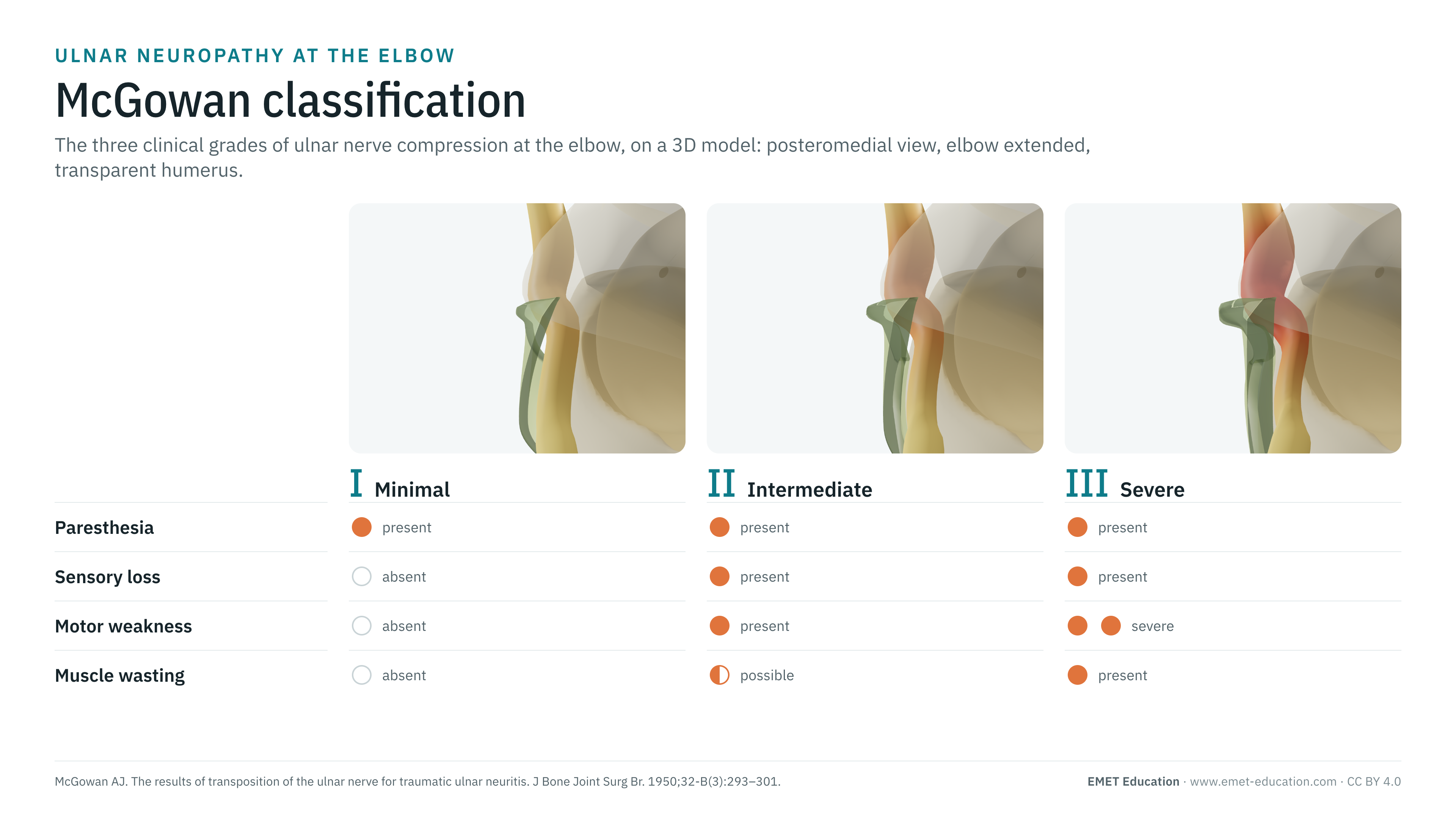
EMET Education. McGowan classification of ulnar neuropathy at the elbow: 3D visualization [Internet]. 2026 [cited]. Available from: https://www.emet-education.com/en/classifications/mcgowan/. CC BY 4.0.EMET Education. (2026). McGowan classification of ulnar neuropathy at the elbow: 3D visualization [Infographic]. https://www.emet-education.com/en/classifications/mcgowan/ (CC BY 4.0)<a href="https://www.emet-education.com/en/classifications/mcgowan/"><img src="https://www.emet-education.com/en/classifications/mcgowan/img/infographic.webp" alt="McGowan classification in 3D" width="960"></a><br>Figure: <a href="https://www.emet-education.com/en/classifications/mcgowan/">EMET Education</a>, CC BY 4.0Questions
Frequently asked questions
What is the difference between McGowan grade I and grade II?
Grade I is purely sensory and subjective: paresthesia, perceived clumsiness, normal examination. Grade II requires an objective deficit: sensory loss and motor weakness, with or without early wasting of the interossei.
Does the McGowan grade decide the treatment?
No, not on its own. It helps judge urgency and prognosis: motor deficit usually prompts discussion of surgery. The choice between simple decompression and transposition depends mainly on nerve stability; randomized trials have found no difference between the two.
Is electrodiagnostic testing needed to grade?
No: McGowan is a clinical classification. Electrodiagnostic testing confirms the diagnosis and the level of compression, and other classifications include it (Akahori, Gu, Power et al.).
What is the prognosis by grade?
In the series by Goldberg et al., function returned to normal in 92% of grade I, 45% of grade IIA and 11% of grade IIB; grade III recovered little. Useful motor recovery is unlikely in severe forms.
Is the grade reproducible?
Moderately: kappa of 0.54 between two surgeons and 0.59 for the same surgeon two weeks apart, for the modified classification (Choi et al., 2022).
Can I reuse these images in a course or an article?
Yes. The images and infographics on this page are under the Creative Commons Attribution 4.0 license: free reuse, including commercial, with credit to EMET Education and a link to this page. The "Cite this figure" box gives ready-to-paste citations.
How were the 3D images made?
They are renderings of the EMET Education ulnar neuropathy model, set to each grade: Osborne's ligament tightens then thickens, the nerve flattens under it and swells above it (pseudoneuroma), more and more congested. The humerus is transparent so that the nerve can be seen. It is a schematic illustration for teaching and patient explanation: the appearance of the nerve is not a McGowan criterion.
Sources
Author and references
- McGowan AJ. The results of transposition of the ulnar nerve for traumatic ulnar neuritis. J Bone Joint Surg Br. 1950;32-B(3):293–301. doi:10.1302/0301-620X.32B3.293
- Goldberg BJ, Light TR, Blair SJ. Ulnar neuropathy at the elbow: results of medial epicondylectomy. J Hand Surg Am. 1989;14(2 Pt 1):182–8. doi:10.1016/0363-5023(89)90003-8
- Dellon AL. Review of treatment results for ulnar nerve entrapment at the elbow. J Hand Surg Am. 1989;14(4):688–700. doi:10.1016/0363-5023(89)90192-5
- Burahee AS, Sanders AD, Shirley C, Power DM. Cubital tunnel syndrome. EFORT Open Rev. 2021;6(9):743–50. doi:10.1302/2058-5241.6.200129
- Choi SW, Bae JY, Shin YH, Moon SH, Kim JK. Reliability and validity of the modified McGowan grade in patients with cubital tunnel syndrome. Arch Orthop Trauma Surg. 2022;142(7):1697–703. doi:10.1007/s00402-022-04367-8
- Power HA, Peters BR, Patterson JMM, Padovano WM, Mackinnon SE. Classifying the severity of cubital tunnel syndrome: a preoperative grading system incorporating electrodiagnostic parameters. Plast Reconstr Surg. 2022;150(1):115e–126e. doi:10.1097/PRS.0000000000009255
- Bartels RH, Menovsky T, Van Overbeeke JJ, Verhagen WI. Surgical management of ulnar nerve compression at the elbow: an analysis of the literature. J Neurosurg. 1998;89(5):722–7. doi:10.3171/jns.1998.89.5.0722
- Zlowodzki M, Chan S, Bhandari M, Kalliainen L, Schubert W. Anterior transposition compared with simple decompression for treatment of cubital tunnel syndrome: a meta-analysis of randomized, controlled trials. J Bone Joint Surg Am. 2007;89(12):2591–8. doi:10.2106/JBJS.G.00183
- Bacle G, Marteau E, Freslon M, et al. Cubital tunnel syndrome: comparative results of a multicenter study of 4 surgical techniques with a mean follow-up of 92 months. Orthop Traumatol Surg Res. 2014;100(4 Suppl):S205–8. doi:10.1016/j.otsr.2014.03.009
- Akahori O. Cubital tunnel syndrome: grade of palsy and prognosis, and selection of operation [in Japanese]. Orthop Surg Traumatol. 1986;29:1745–51.
- Gu Y. Current status and suggestion of clinical classification of carpal and cubital tunnel syndromes [in Chinese]. Zhongguo Gu Yu Guan Jie Sun Shang Za Zhi. 2011;31:818–9.
- Childress HM. Recurrent ulnar-nerve dislocation at the elbow. Clin Orthop Relat Res. 1975;(108):168–73.
- Cambon-Binder A. Ulnar neuropathy at the elbow. Orthop Traumatol Surg Res. 2021;107(1S):102754. doi:10.1016/j.otsr.2020.102754
3D models
Show the compression to your patient
The images on this page come from an interactive model. In clinic, set the compression, nerve instability or a space-occupying lesion, flex the elbow, then show the release or the transposition.
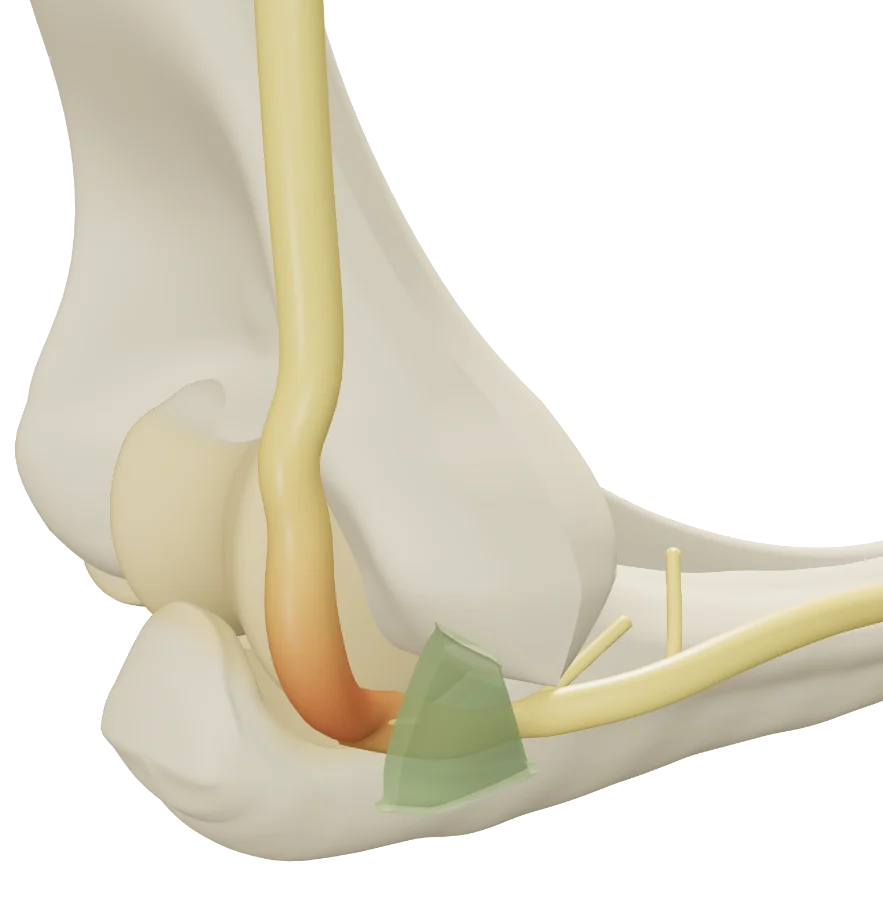
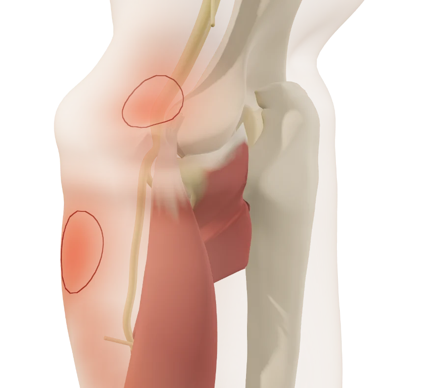
Explain the compression in 3D
The ulnar neuropathy model opens in your browser, at the grade you choose, with no sign-up.
Share this page with a colleague or a resident: