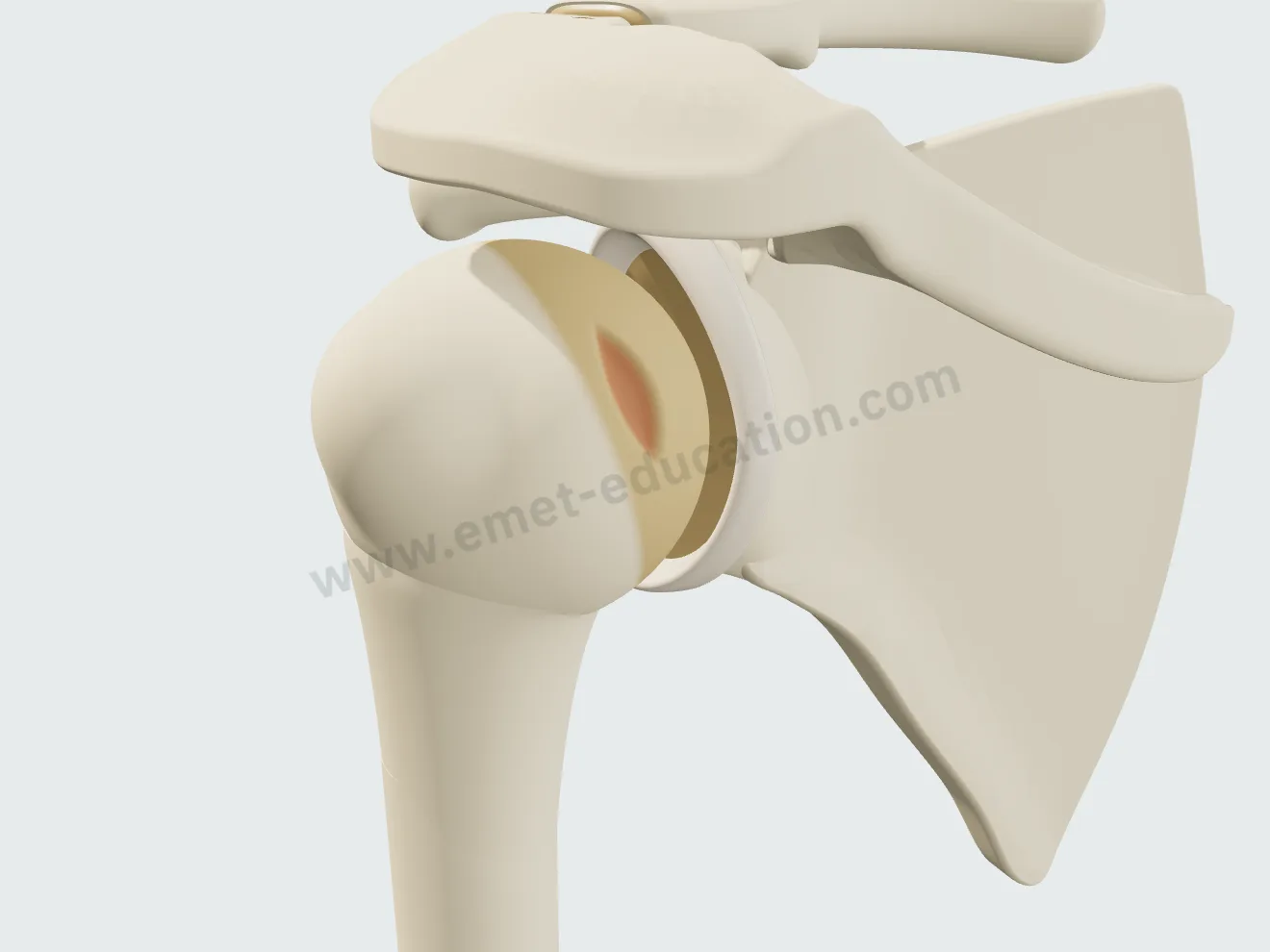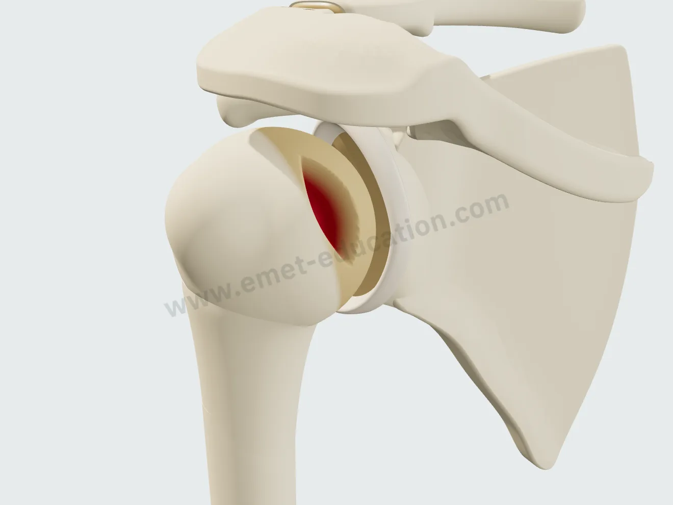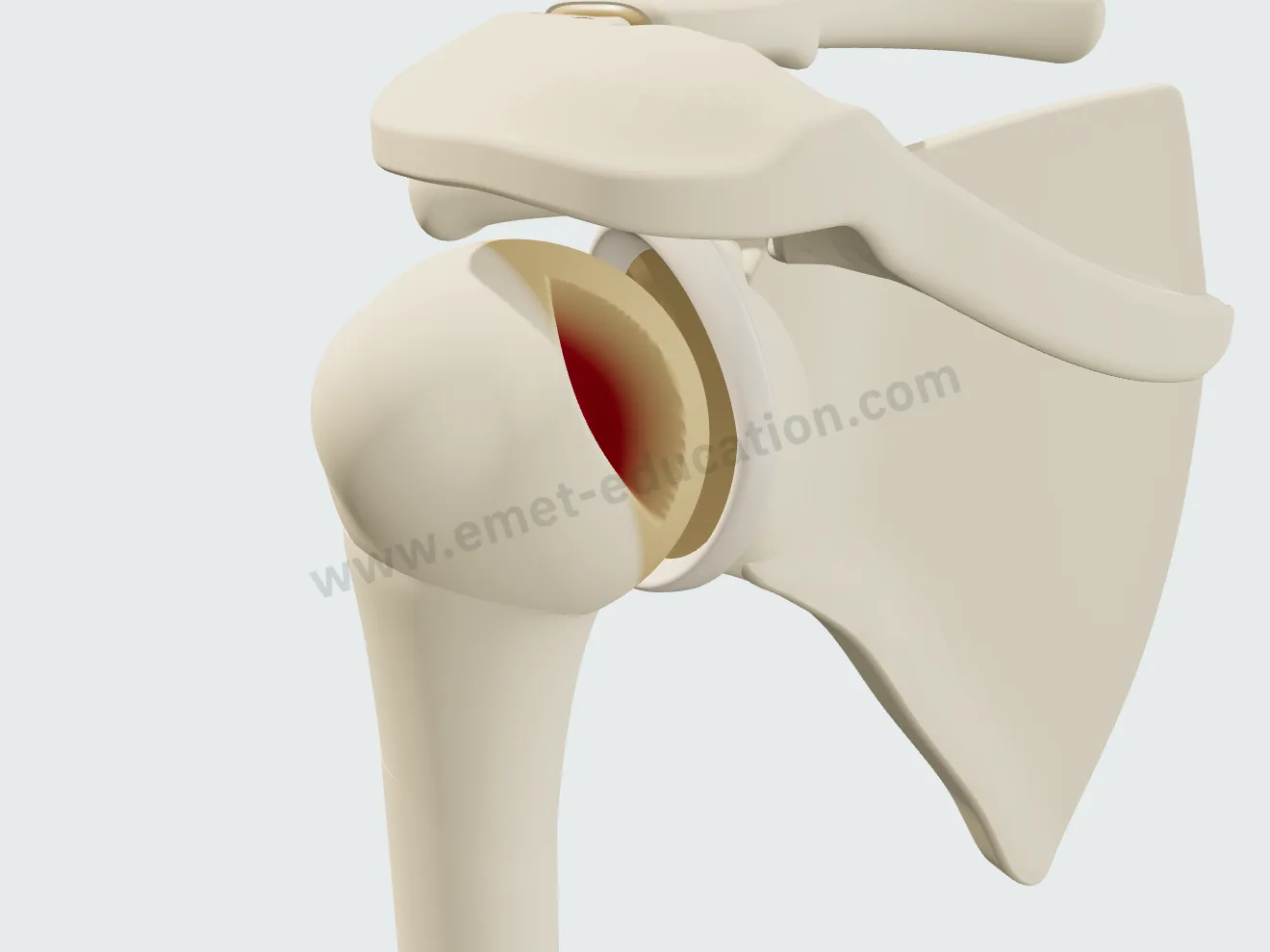
The dislocation and its lesions, in a few clicks
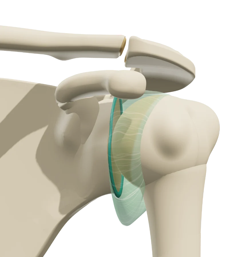
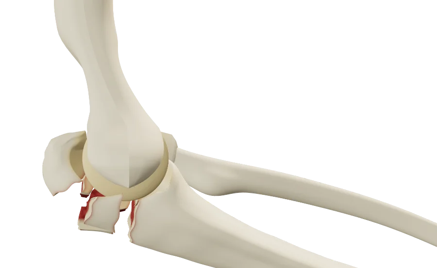
The “Anterior dislocation” button shows the injury, the “Pathology & lesions” panel what it leaves behind, on the glenoid and on the humeral head. Lesions add up, and each one changes the model in front of the patient.
Anterior dislocation, in motion
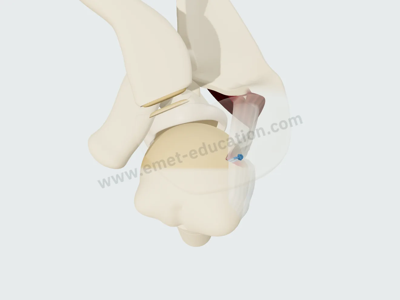
The arm moves into abduction and external rotation, then the humeral head slides over the anteroinferior rim and stops there, the Hill-Sachs lesion engaged on the rim. “Generate post-dislocation lesions” plays the dislocation and the lesions it leaves.
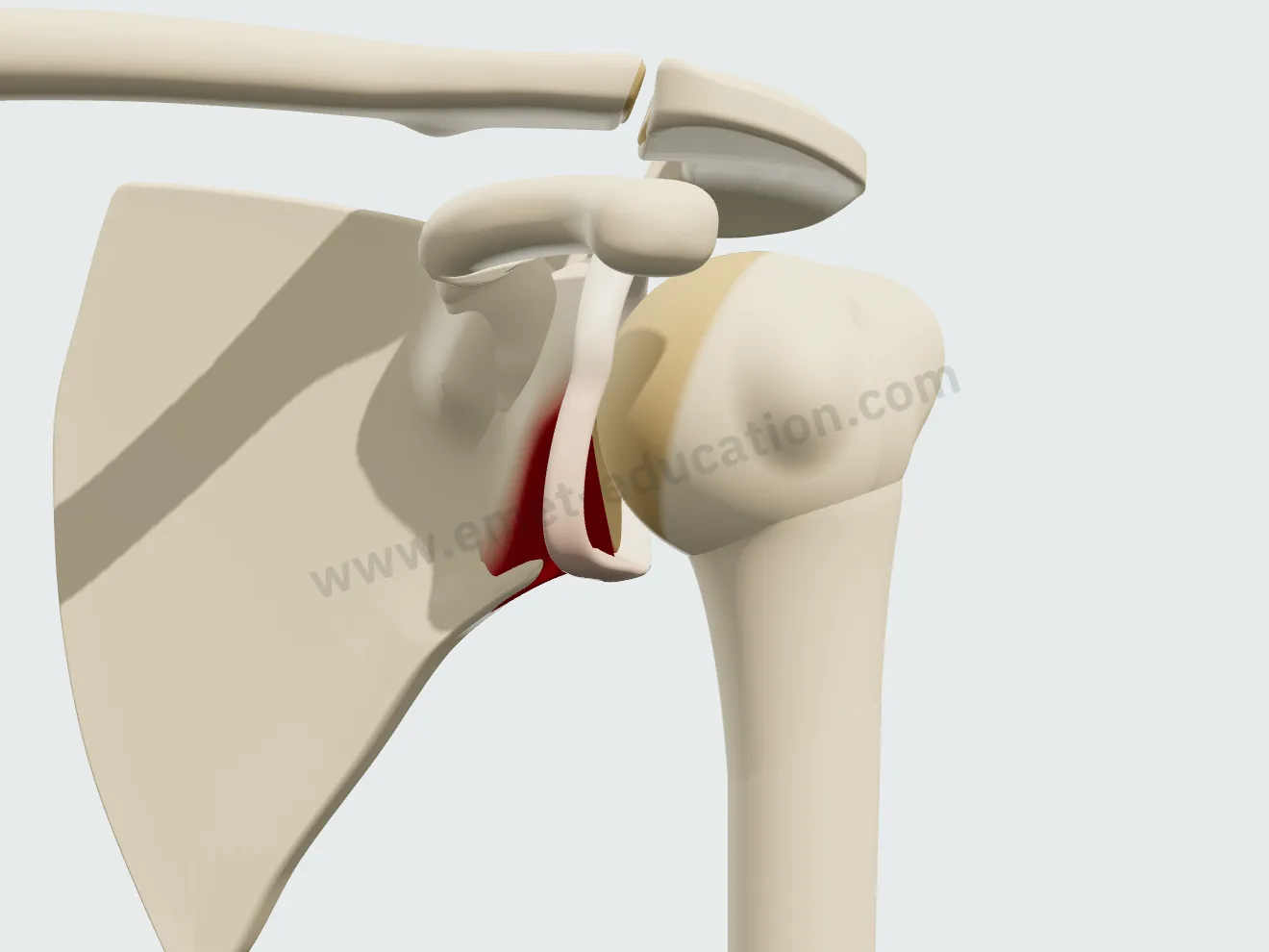
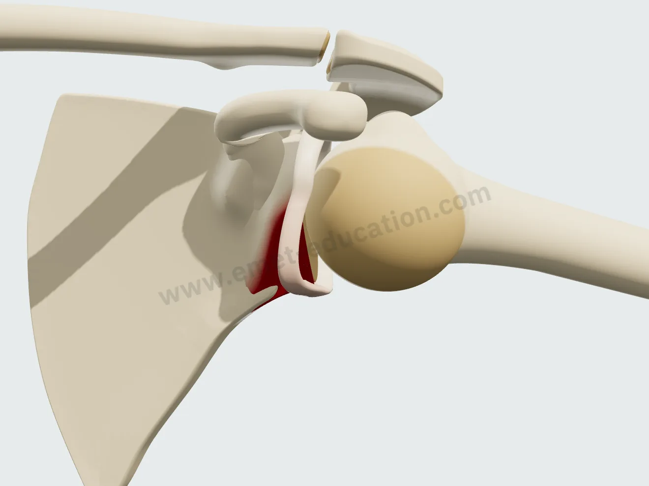
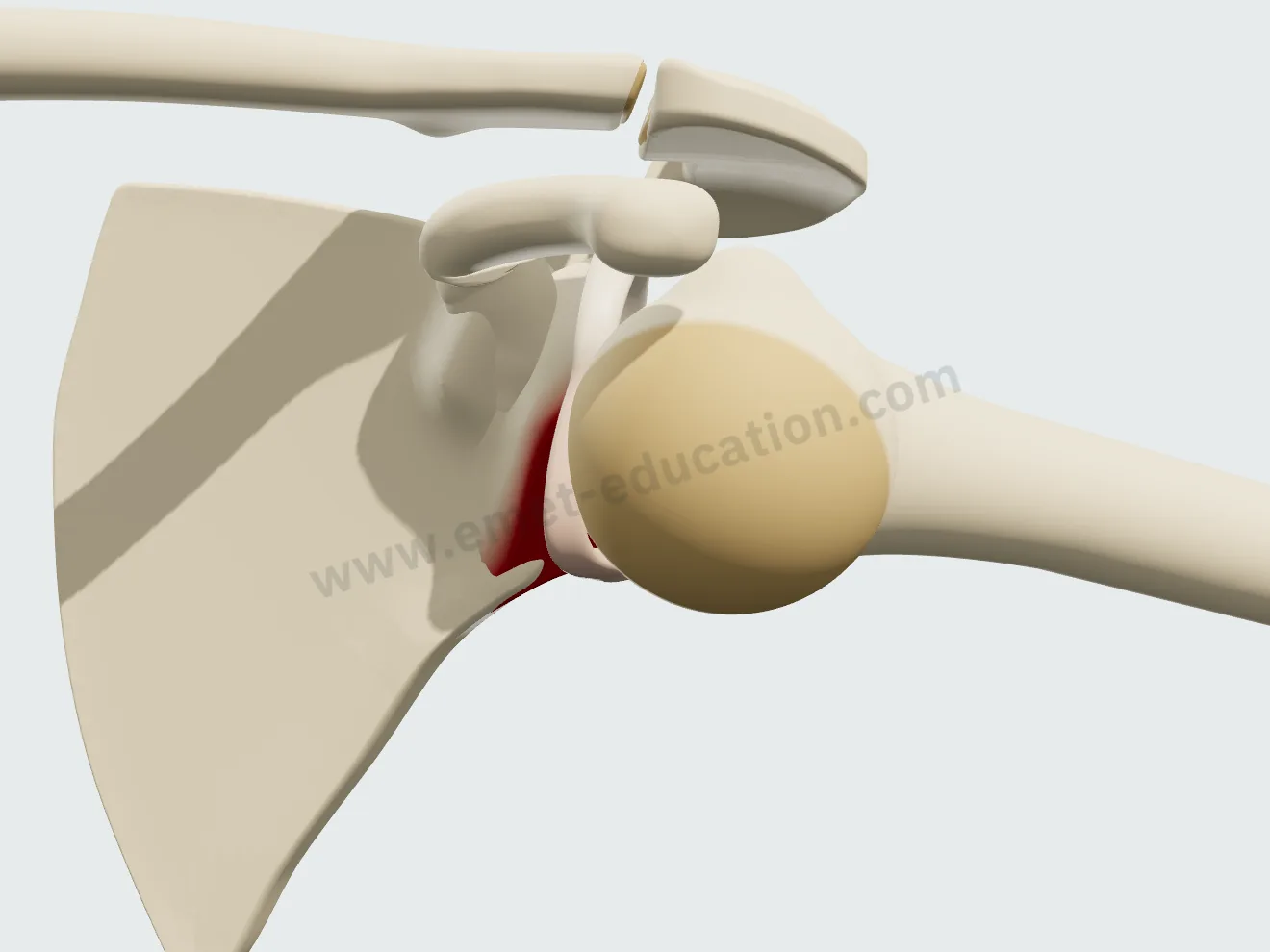
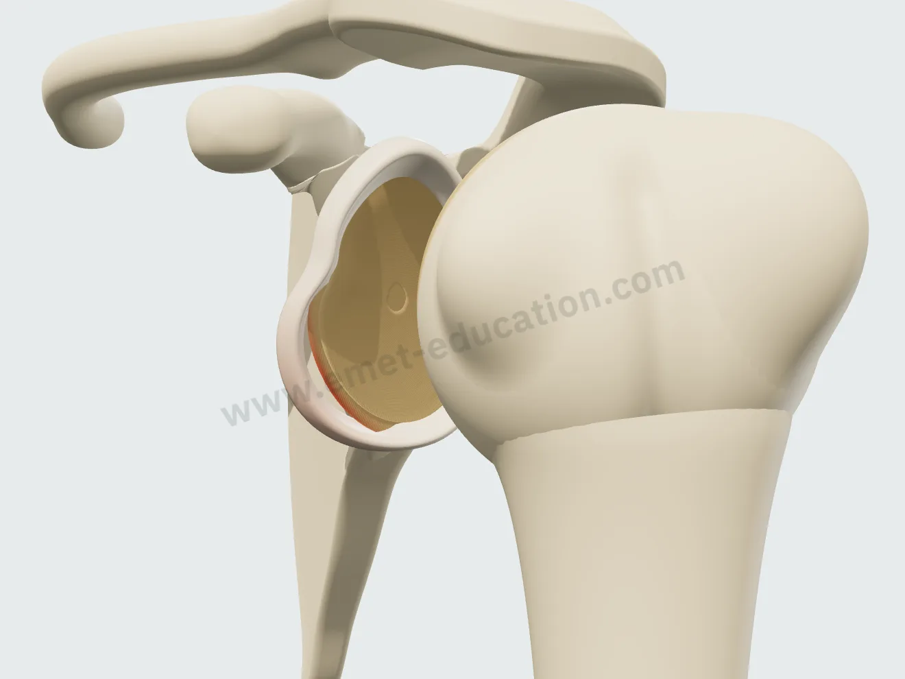
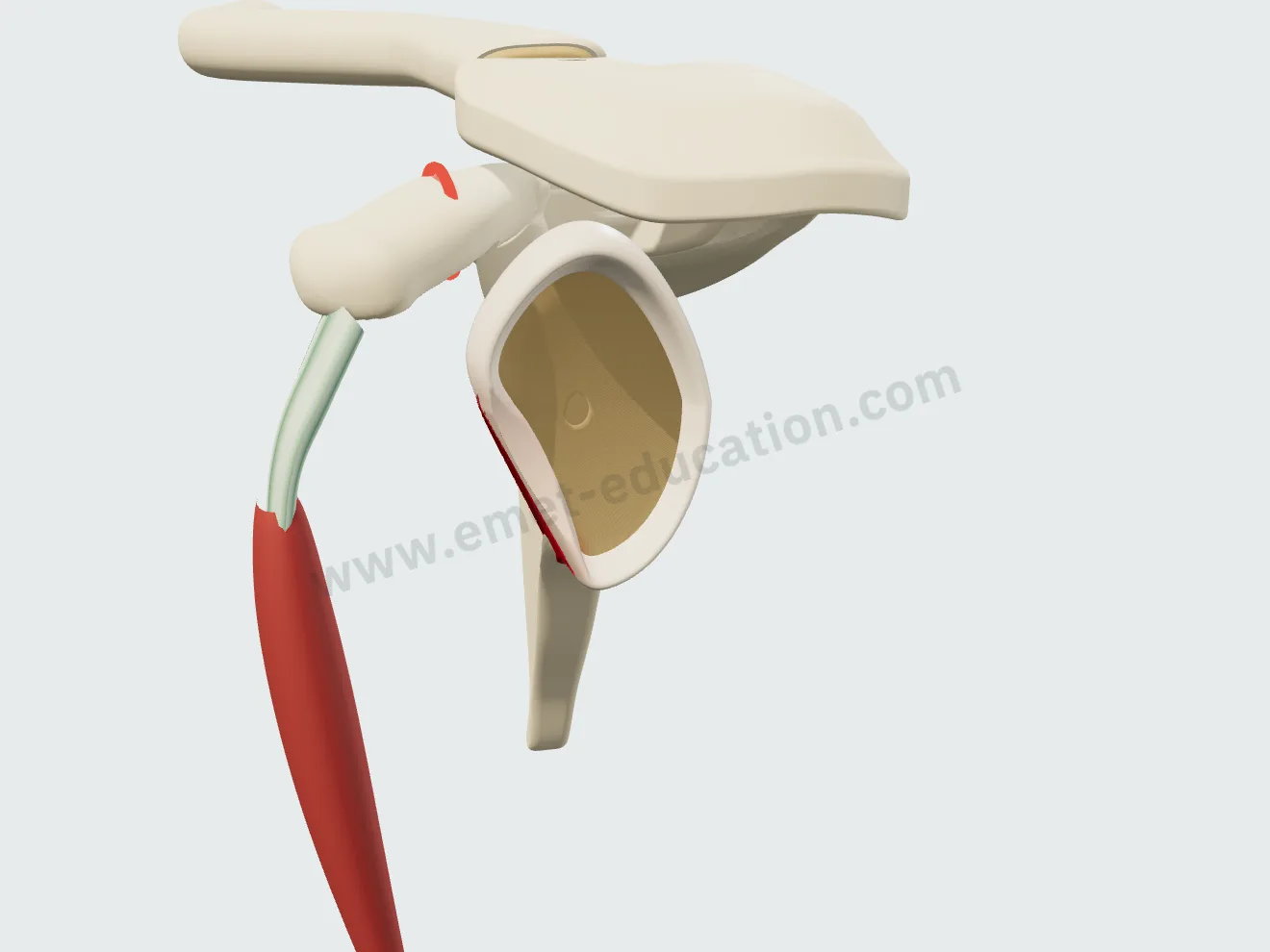
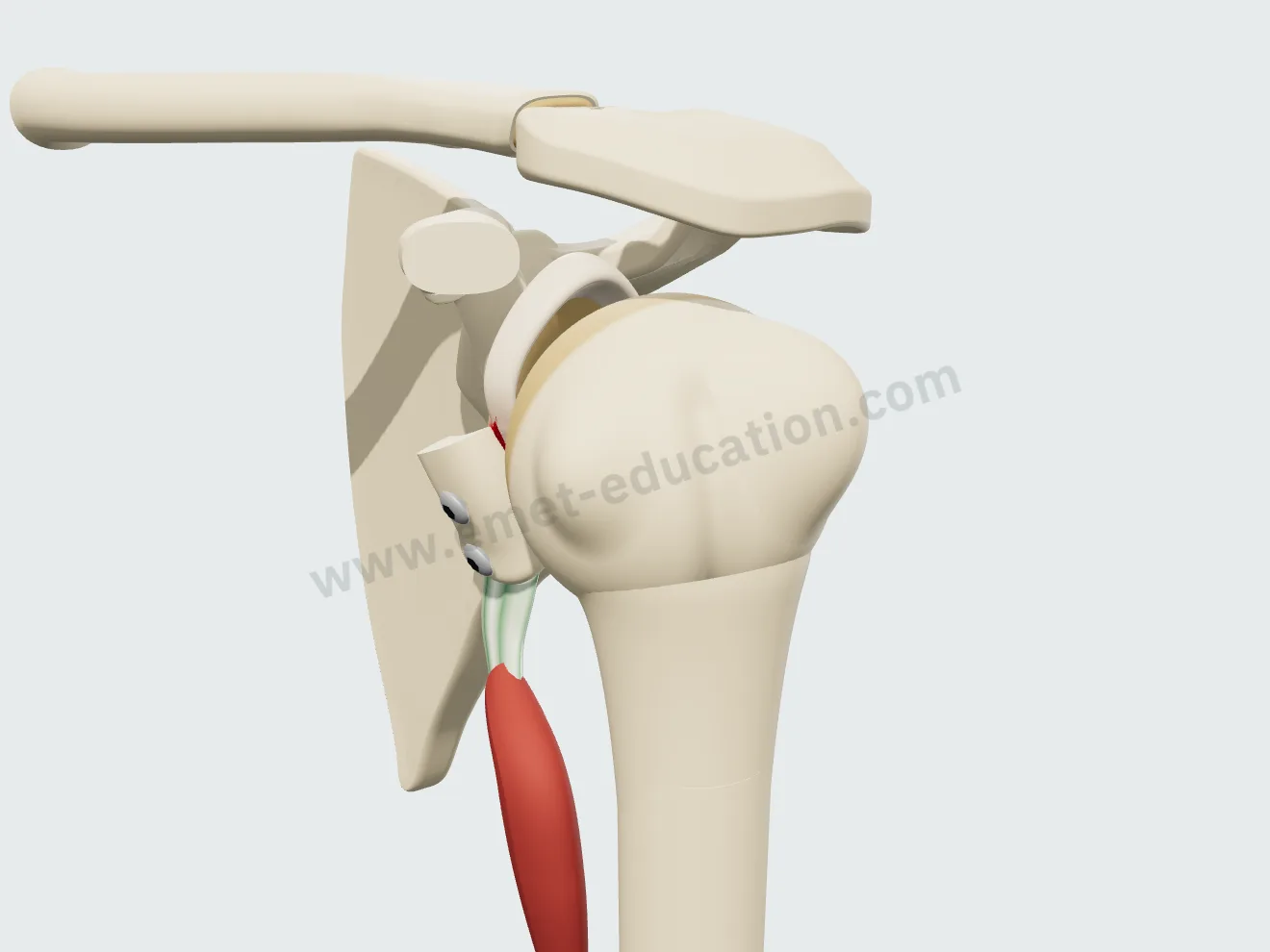
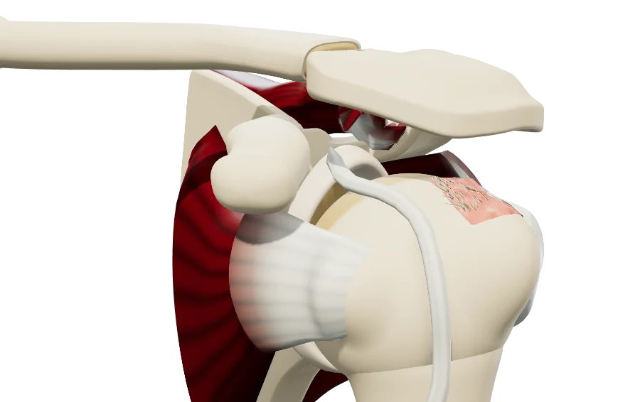
The Bankart lesion
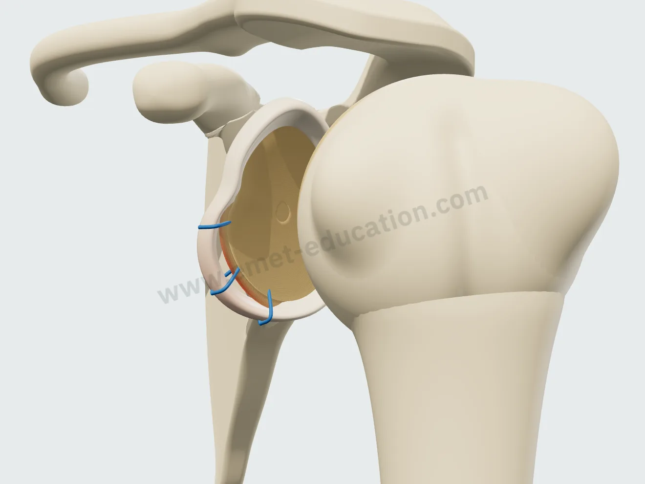
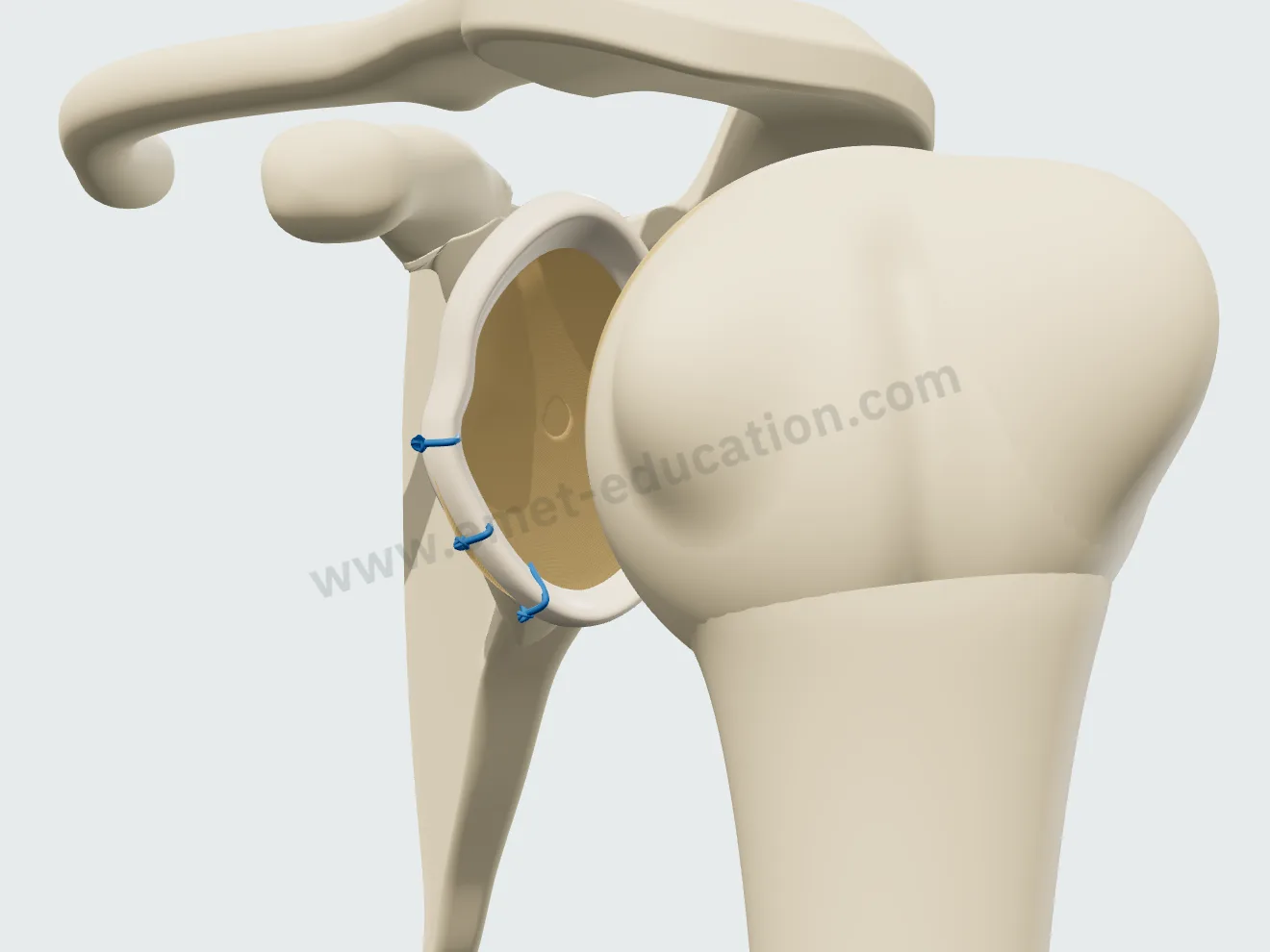
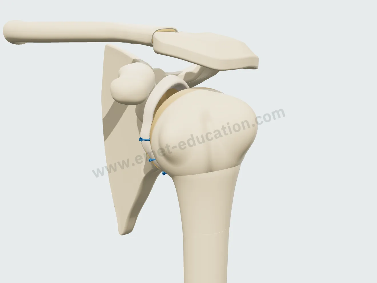
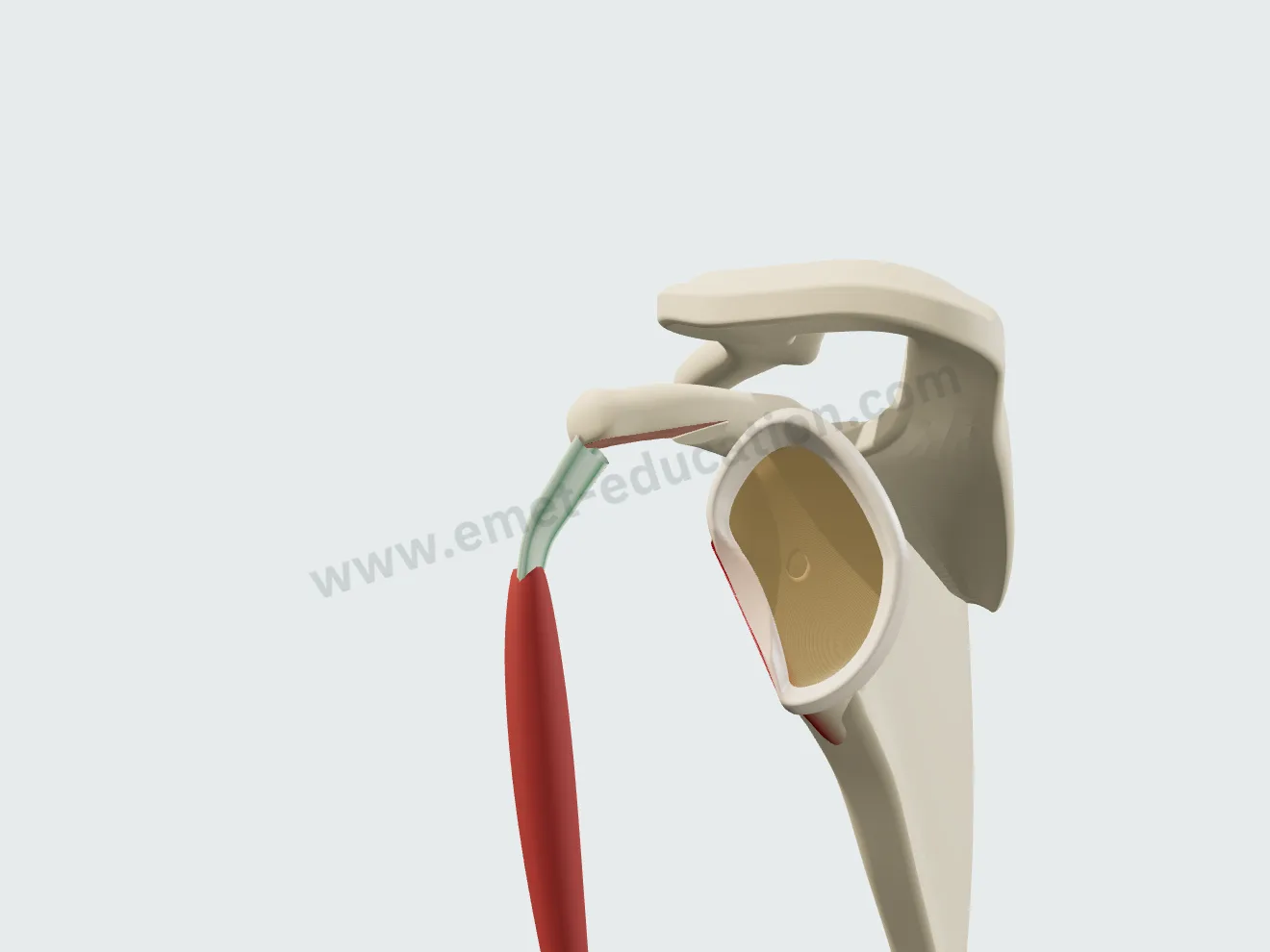
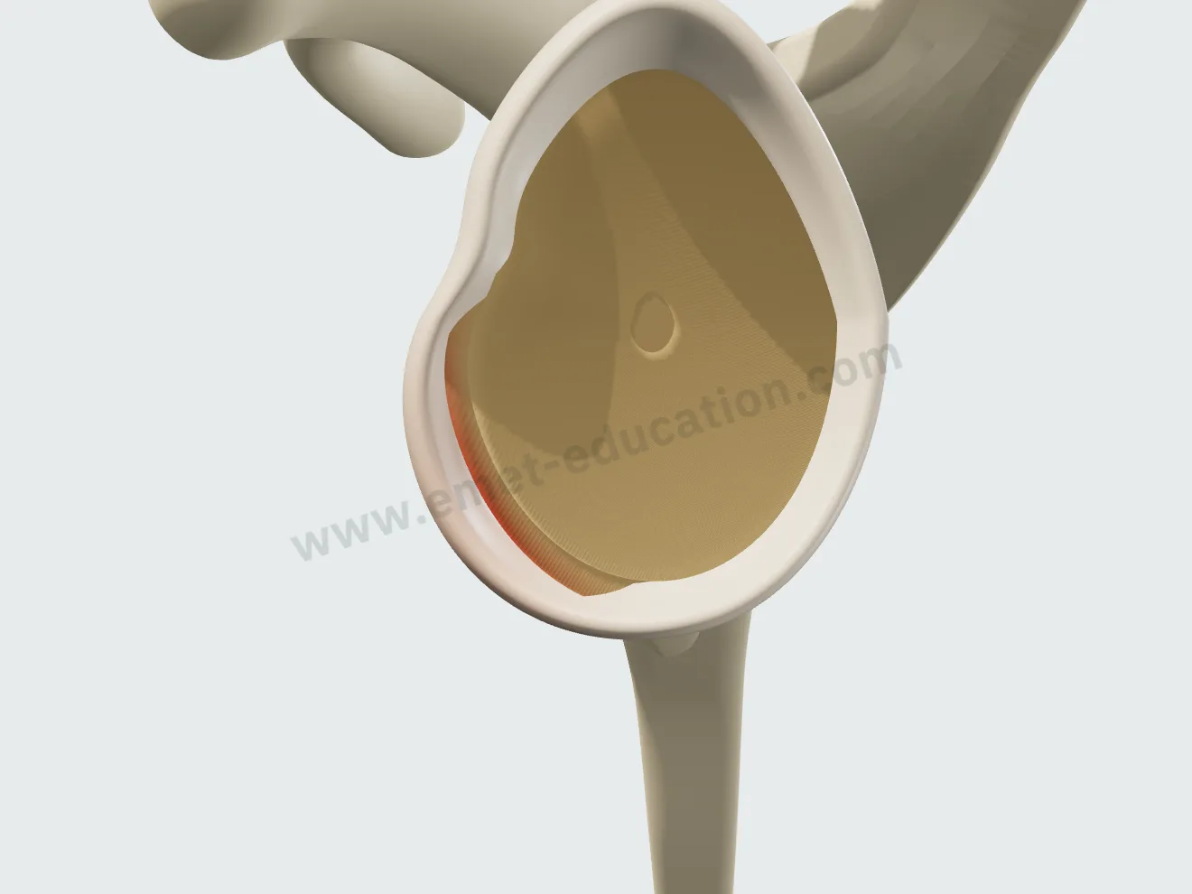
The anteroinferior labrum detaches from the rim, from 3 to 6 o'clock, slides medially along the neck and tilts; the bare bone of the rim turns red. The lesion is set independently of bone loss, and the two overlap without blending.

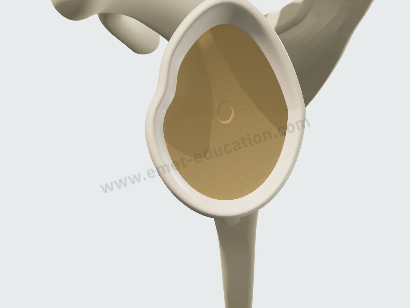
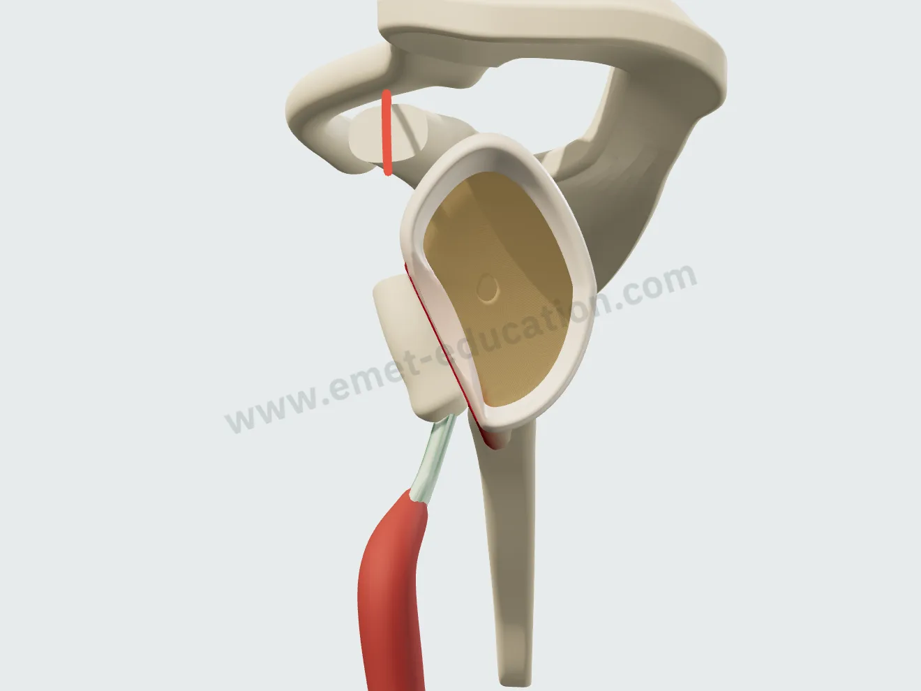
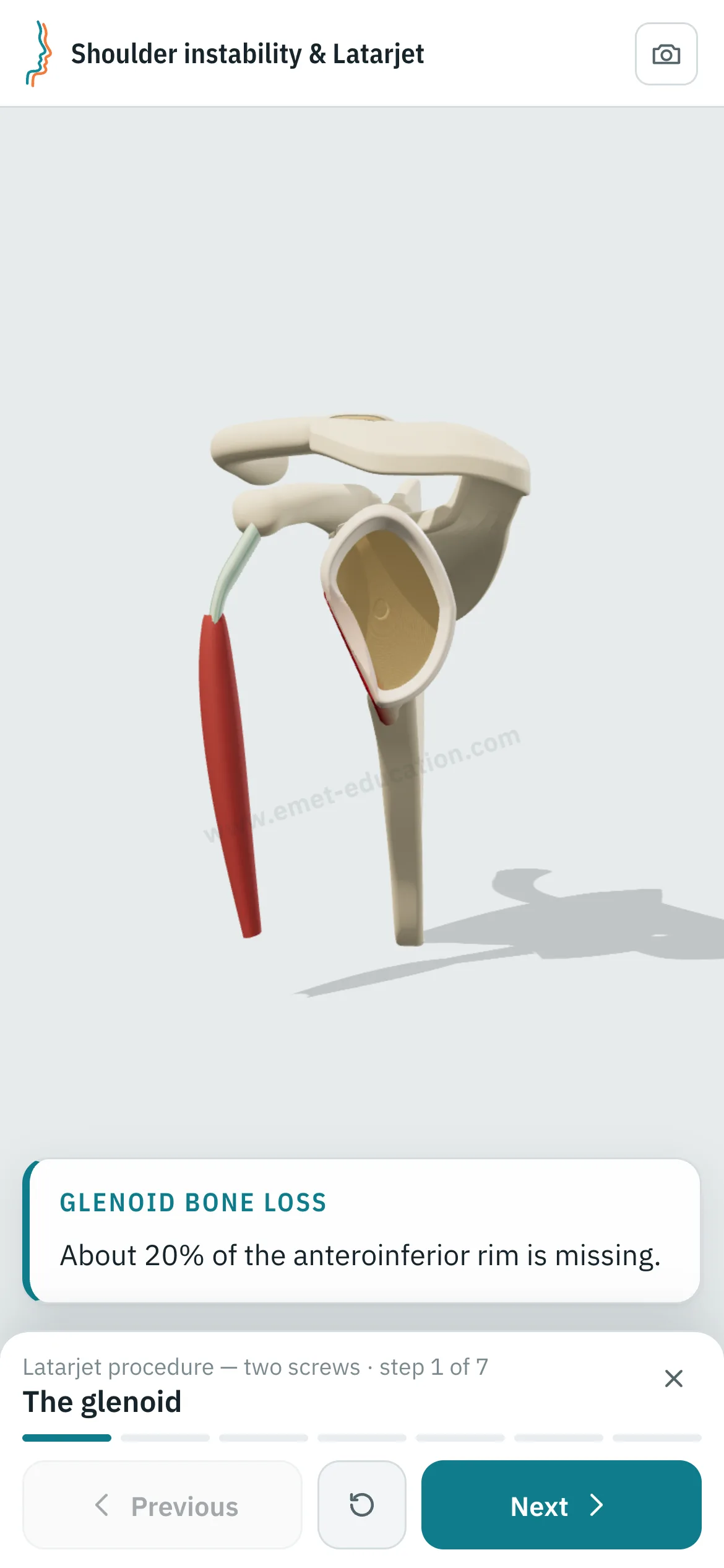
Glenoid bone loss, to the percent
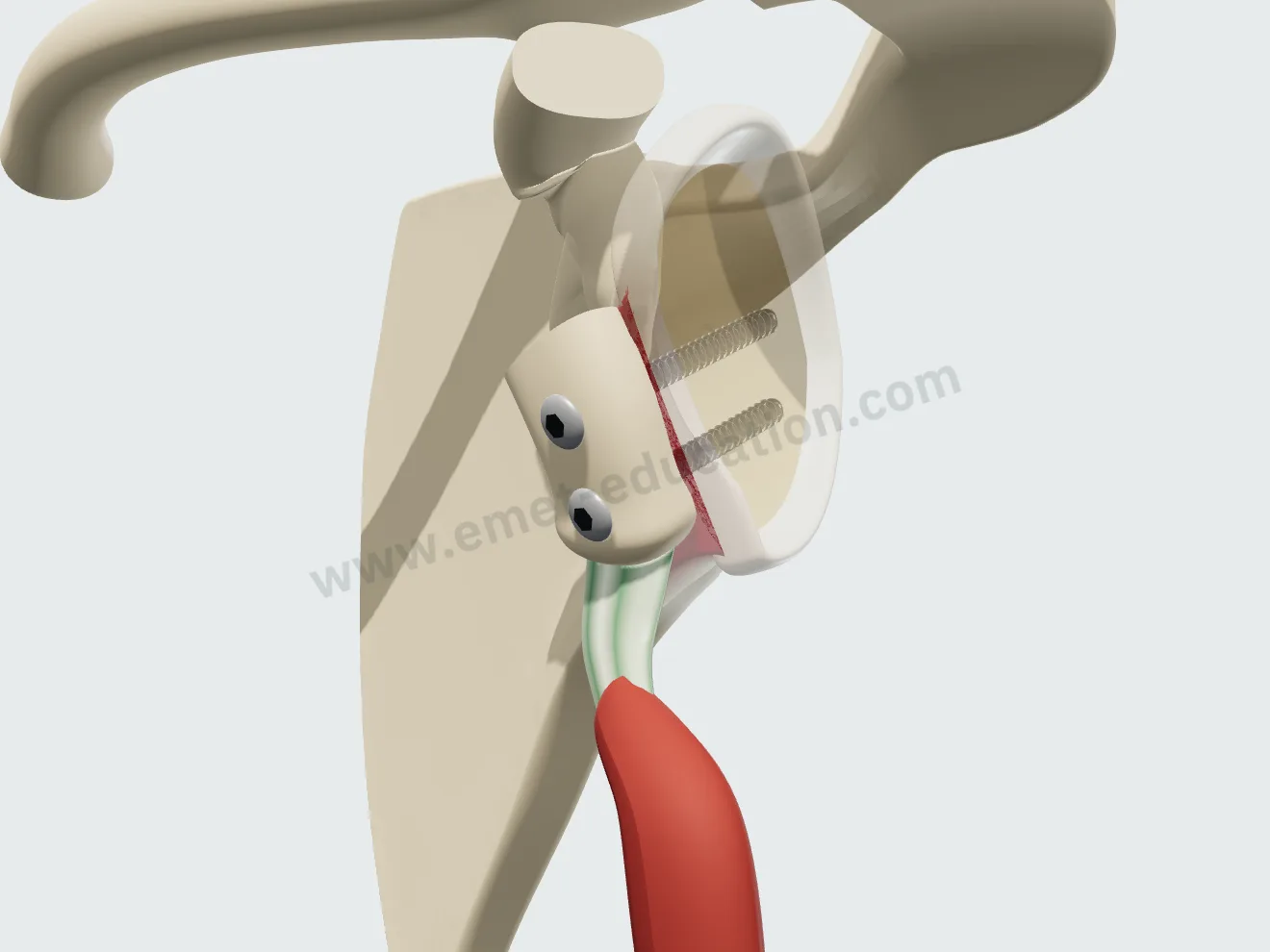
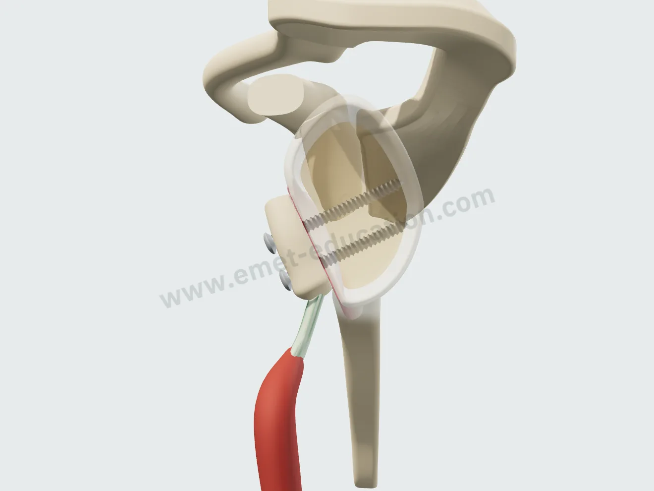
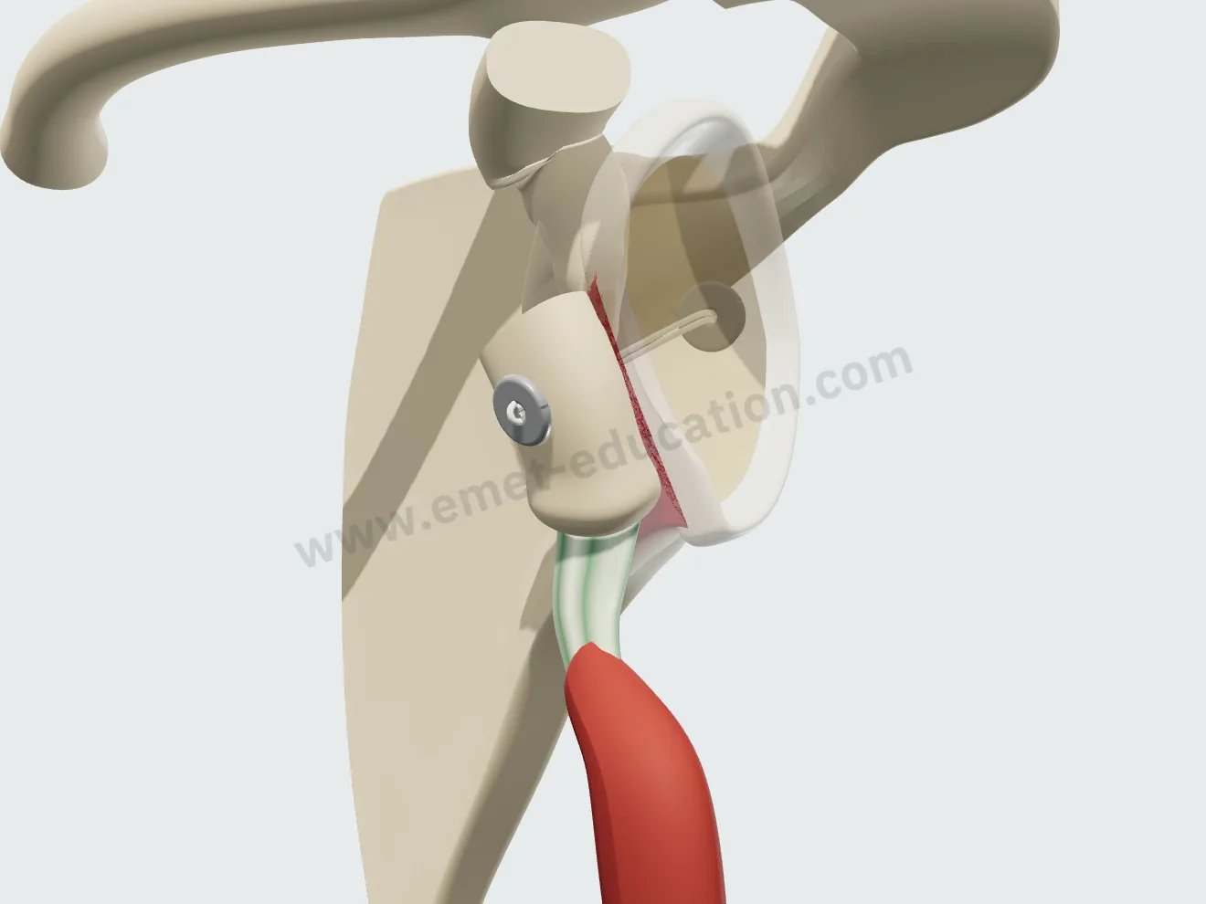
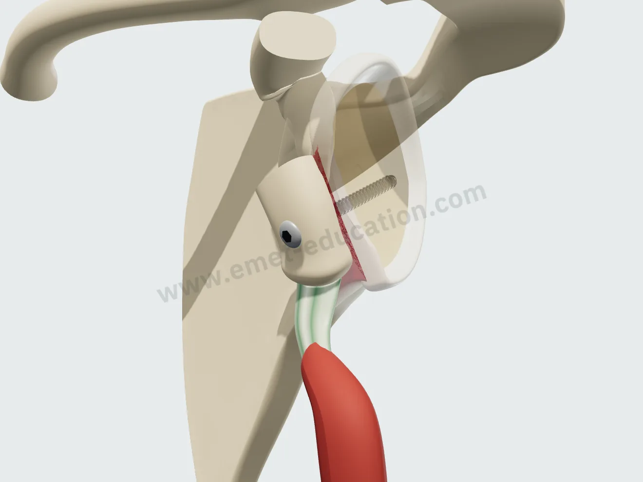
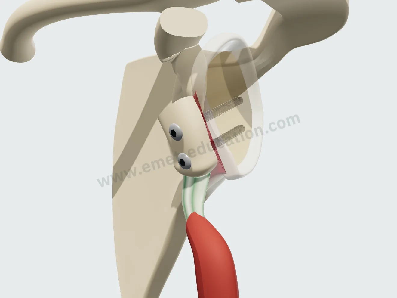
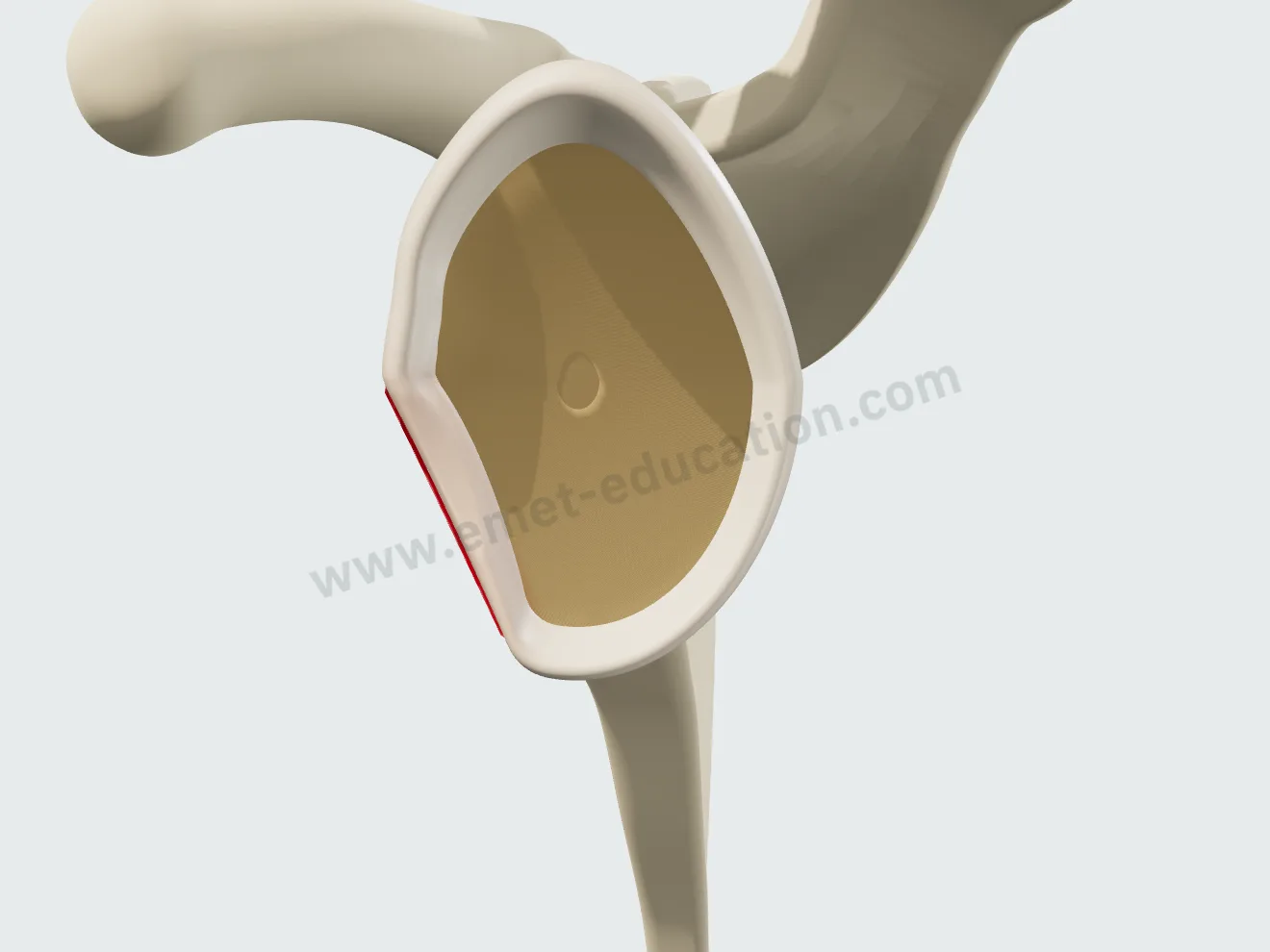
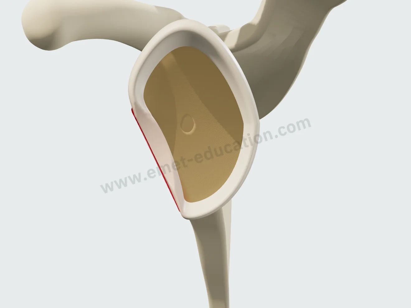
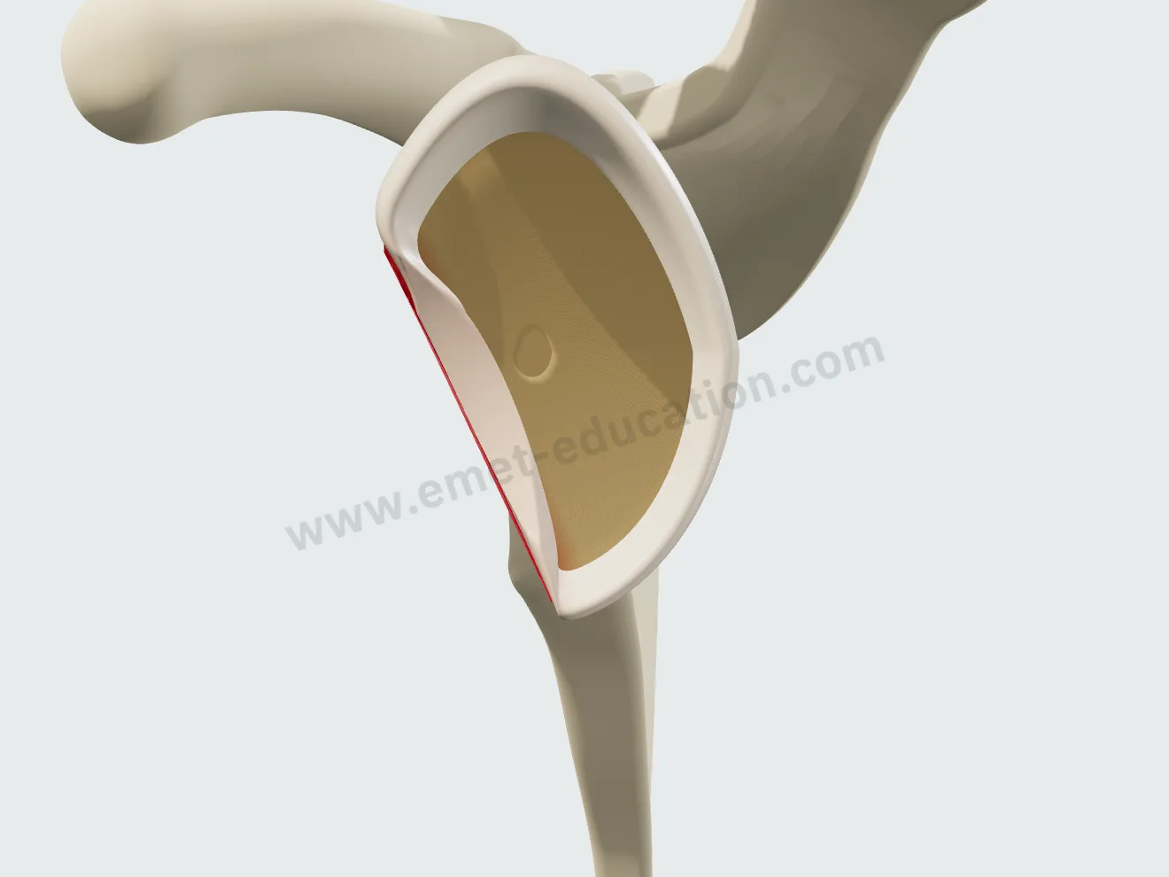
A slider from 0 to 30% truly removes the anteroinferior quadrant of the glenoid, along a nearly vertical line outlined in red. The loss persists after reduction, and the bone block is placed on that line.

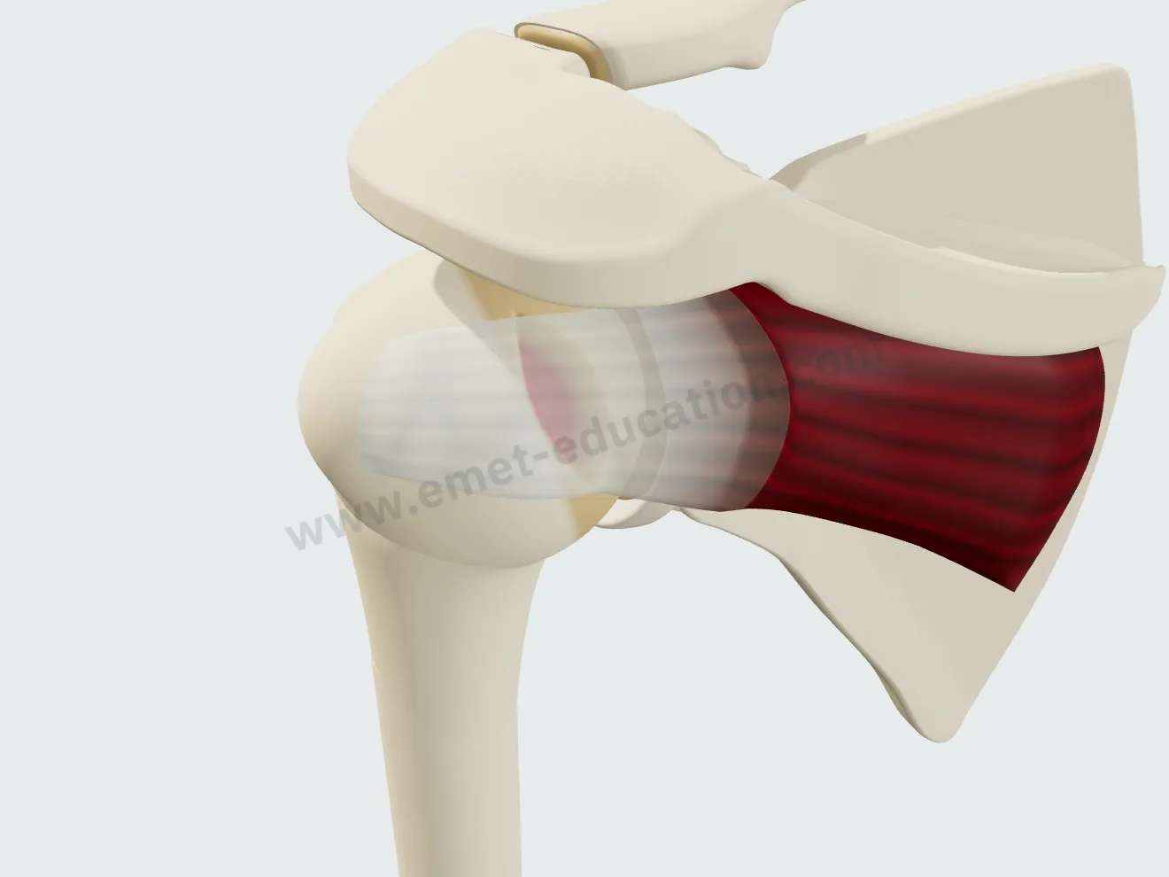
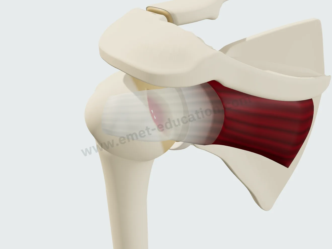
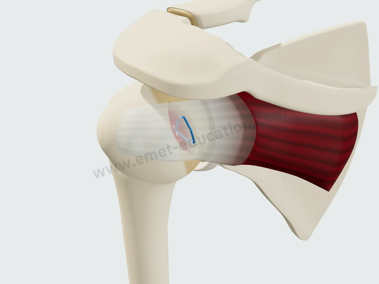
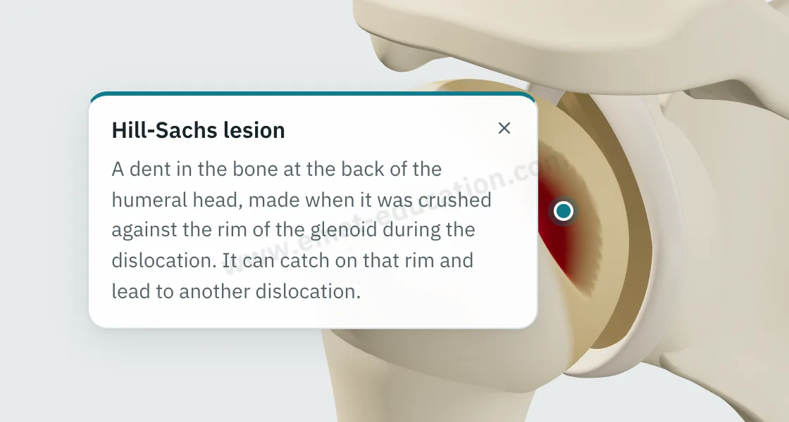
The Hill-Sachs lesion
A true impaction of the posterosuperolateral humeral head: a spindle-shaped defect along the cartilage edge, steep medial wall, gentler lateral slope. With glenoid bone loss, the model refers to bipolar lesions.