EMET Electronic Medical Twin
  1. EMET Education
  2. Musculoskeletal
  3. Elbow
  4. Olecranon fracture

3D model · Elbow

Olecranon Fracture 3D Model

Reproduce your patient's fracture during the visit, and show in 3D the treatment you recommend. They leave with their model on their phone.

Designed by an orthopedic surgeon

Opens in your browser, no sign-up. For orthopedic surgeons, emergency physicians, sports medicine physicians, physiatrists and primary care physicians.

How it works

In the visit, in three steps

Structured information in a few minutes, which the patient can review at home.

  1. 1

    Show

    Open the model and walk through the anatomy: ulnohumeral joint, olecranon, trochlea, triceps insertion.

  2. 2

    Reproduce

    Set the fracture from the radiographs: type, pattern, gap, coronoid. Then walk through the proposed fixation, step by step.

  3. 3

    Share

    Give the patient the QR code or an image: they find their own case, in their language.

Features

Explain in detail during the visit

Every setting updates the model live: you reproduce the fracture from your patient's radiographs in front of them, then walk through the fixation you recommend.

The fracture, described in a few clicks

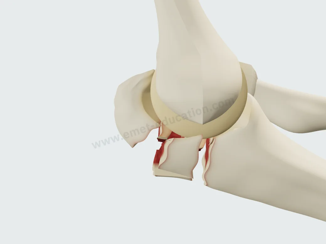
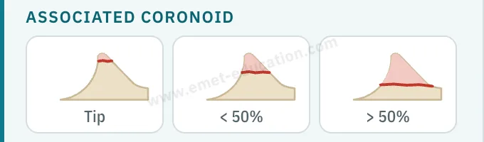
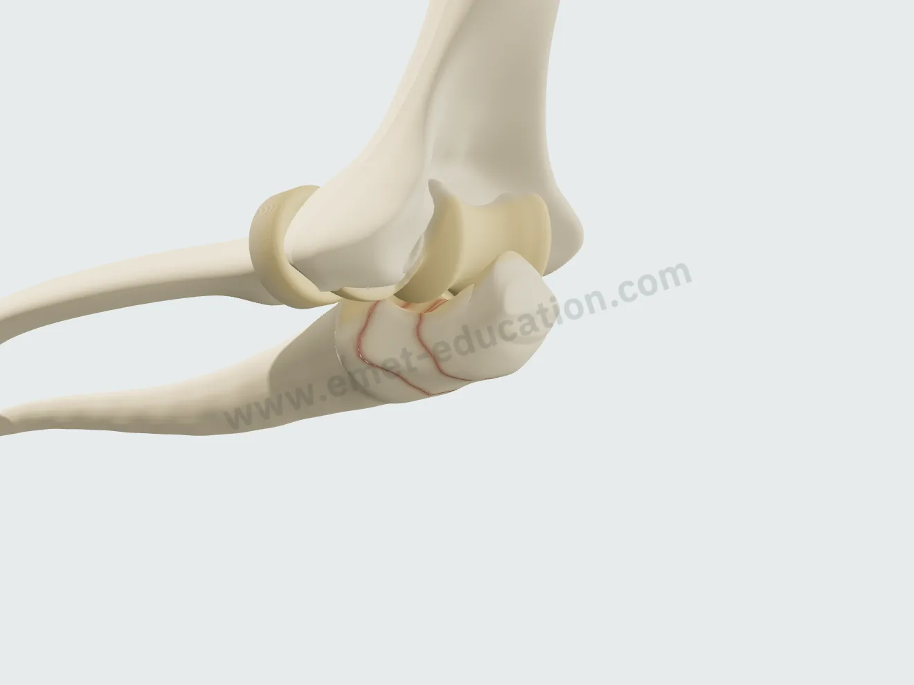
The “Pathology & lesions” panel describes the fracture in four settings: type, pattern, fracture gap, and associated coronoid fracture. Each one changes the model in front of the patient.

Three types, per the Mayo classification

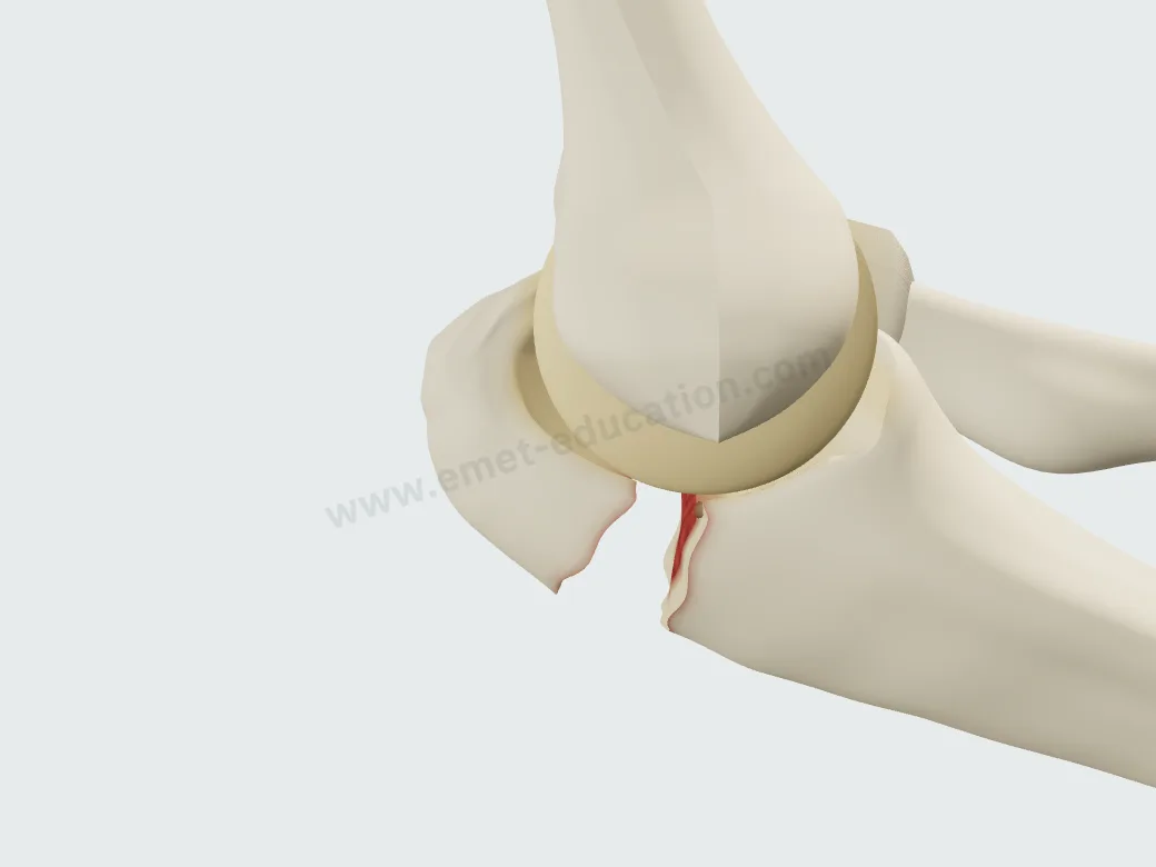
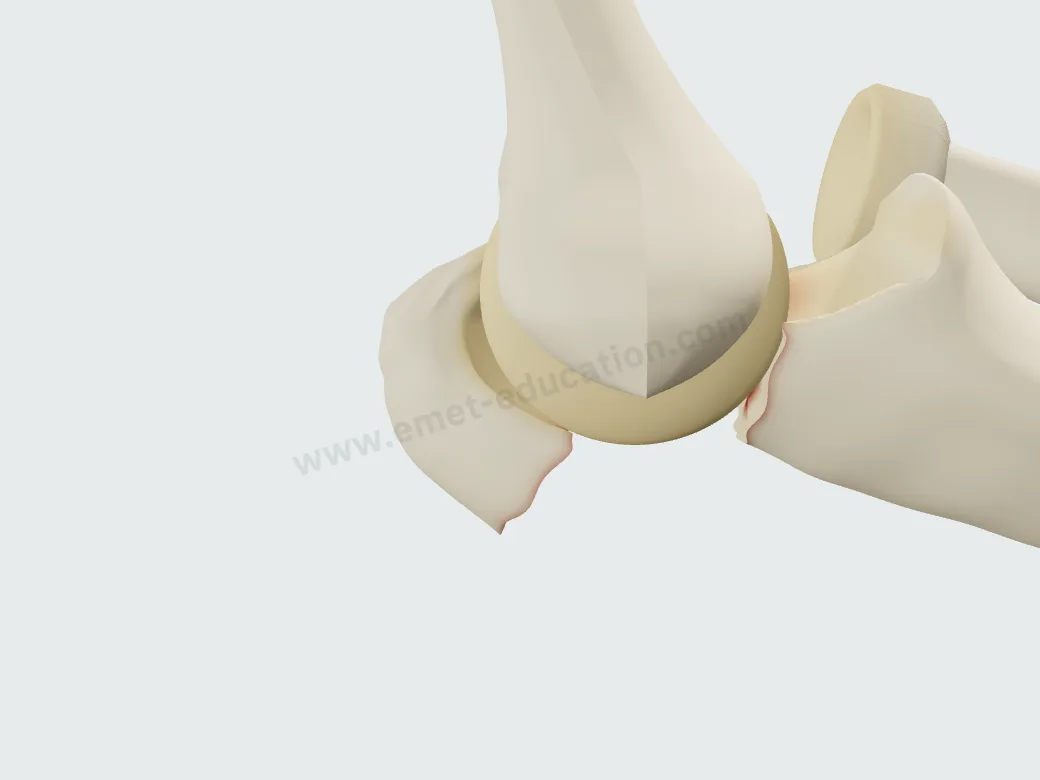
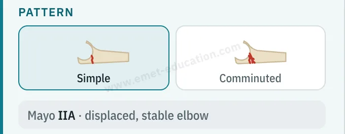
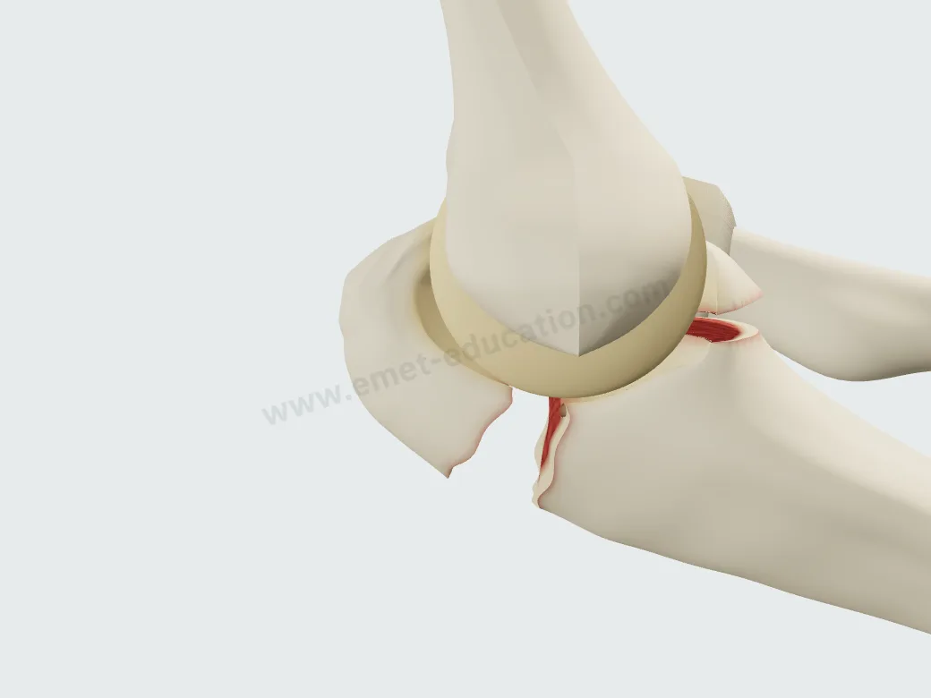
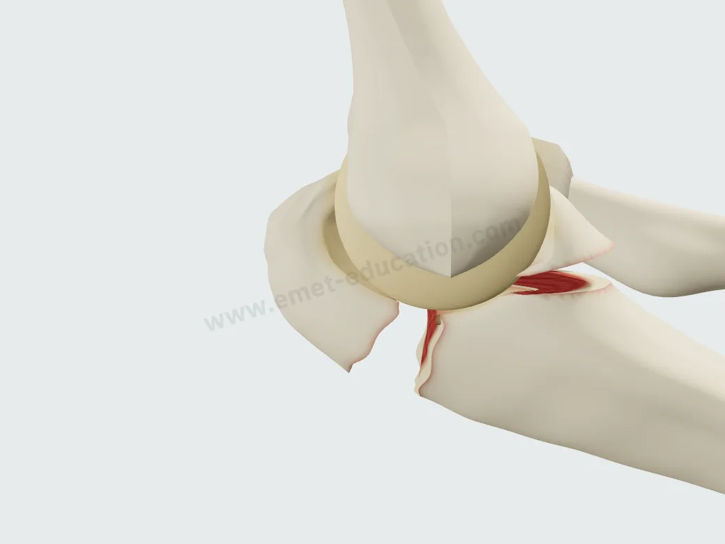
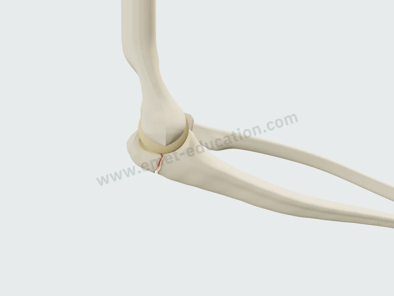
Undisplaced (I), displaced with a stable elbow (II), displaced with an unstable elbow and the forearm dislocated anteriorly (III). The classification updates live, for example “Mayo IIA · displaced, stable elbow,” and an explanatory card accompanies each choice.
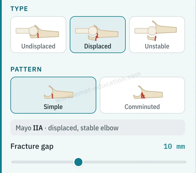
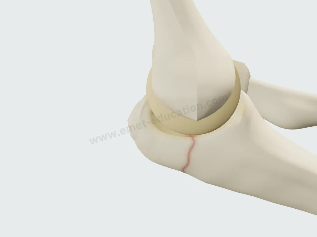
Model panel: fracture type (undisplaced, displaced, unstable), pattern (simple, comminuted), Mayo IIA classification, 10 mm fracture gap.
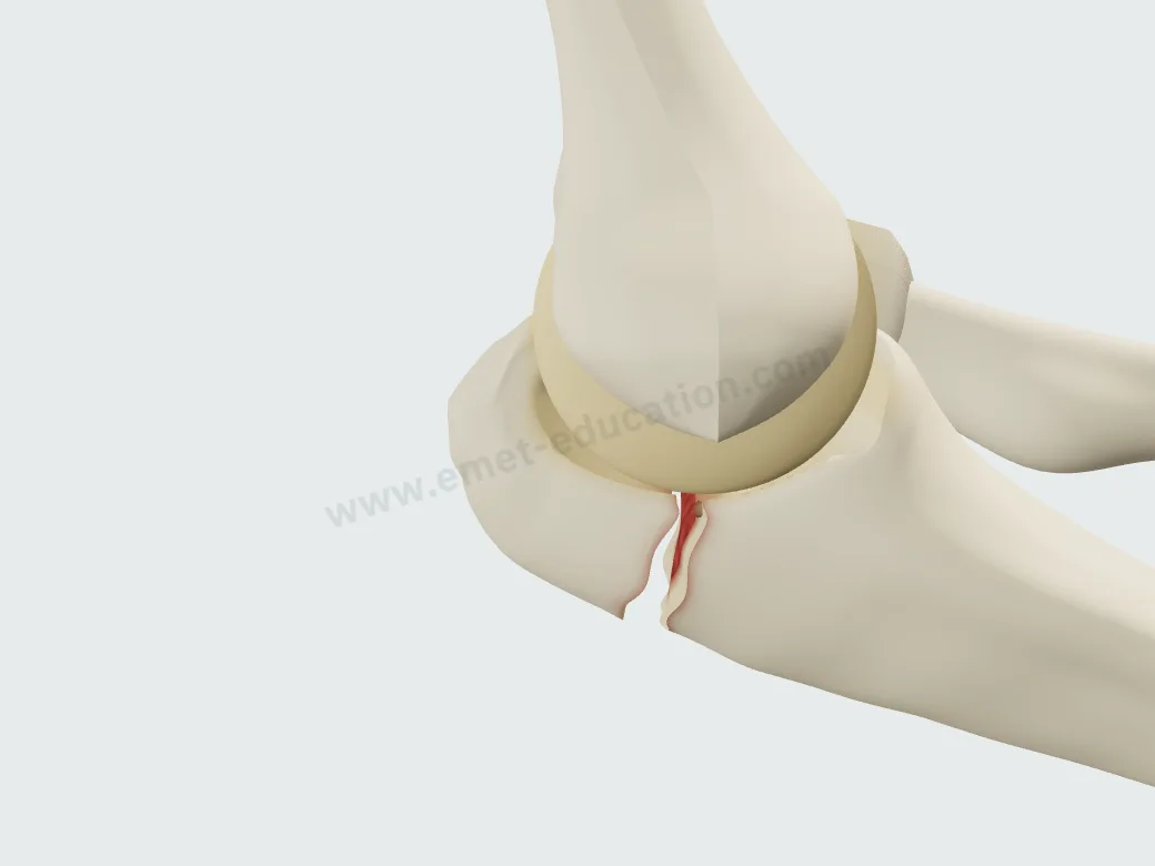
Undisplaced olecranon fracture (Mayo I): the fracture line shown by a thin red outline.
Mayo I · undisplaced
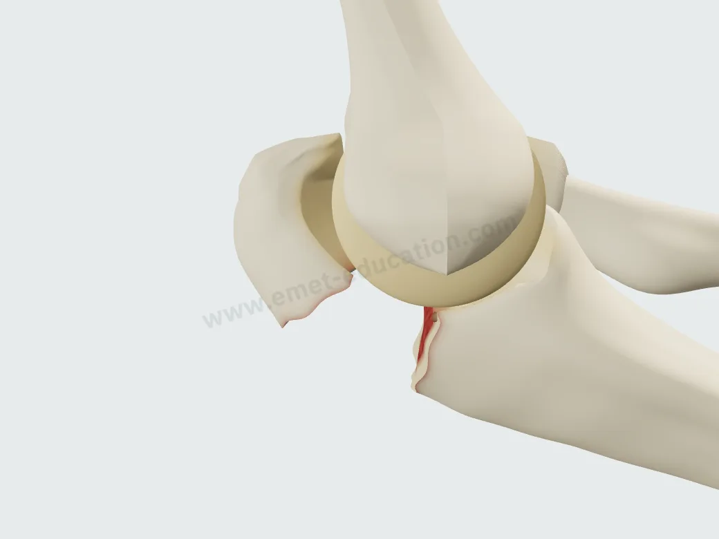
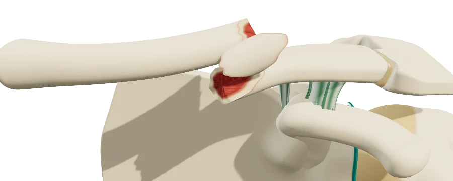
Displaced olecranon fracture, stable elbow (Mayo IIA): the proximal fragment pulled proximally.
Mayo II · displaced, stable elbow
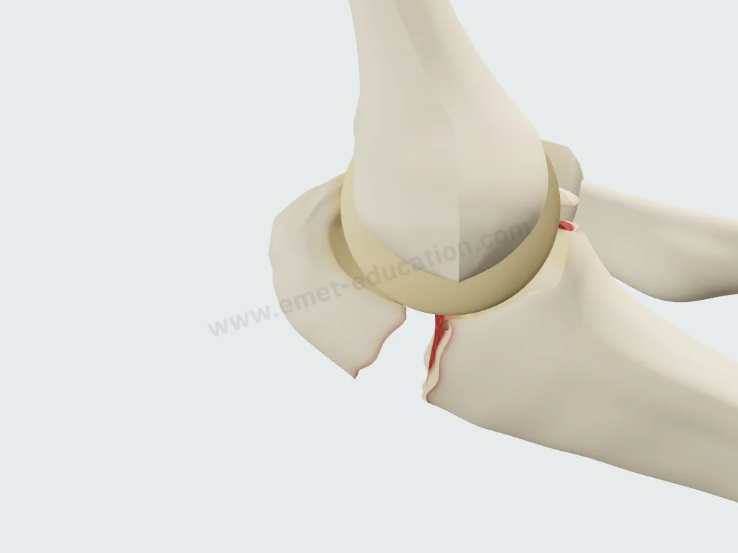
Transolecranon fracture-dislocation (Mayo III): the forearm dislocated anterior to the trochlea.
Mayo III · unstable elbow

The treatment, step by step

Two fixation techniques, played step by step, continuously or one step at a time. No procedure is locked: outside its usual indication, the button shows a warning that only you can see.

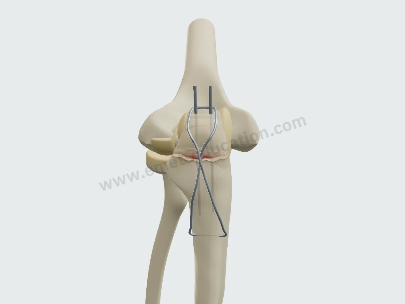
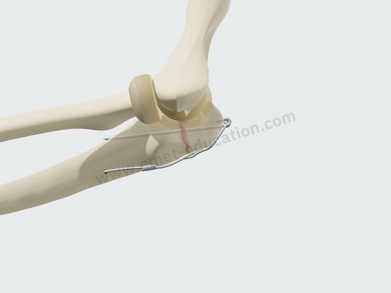
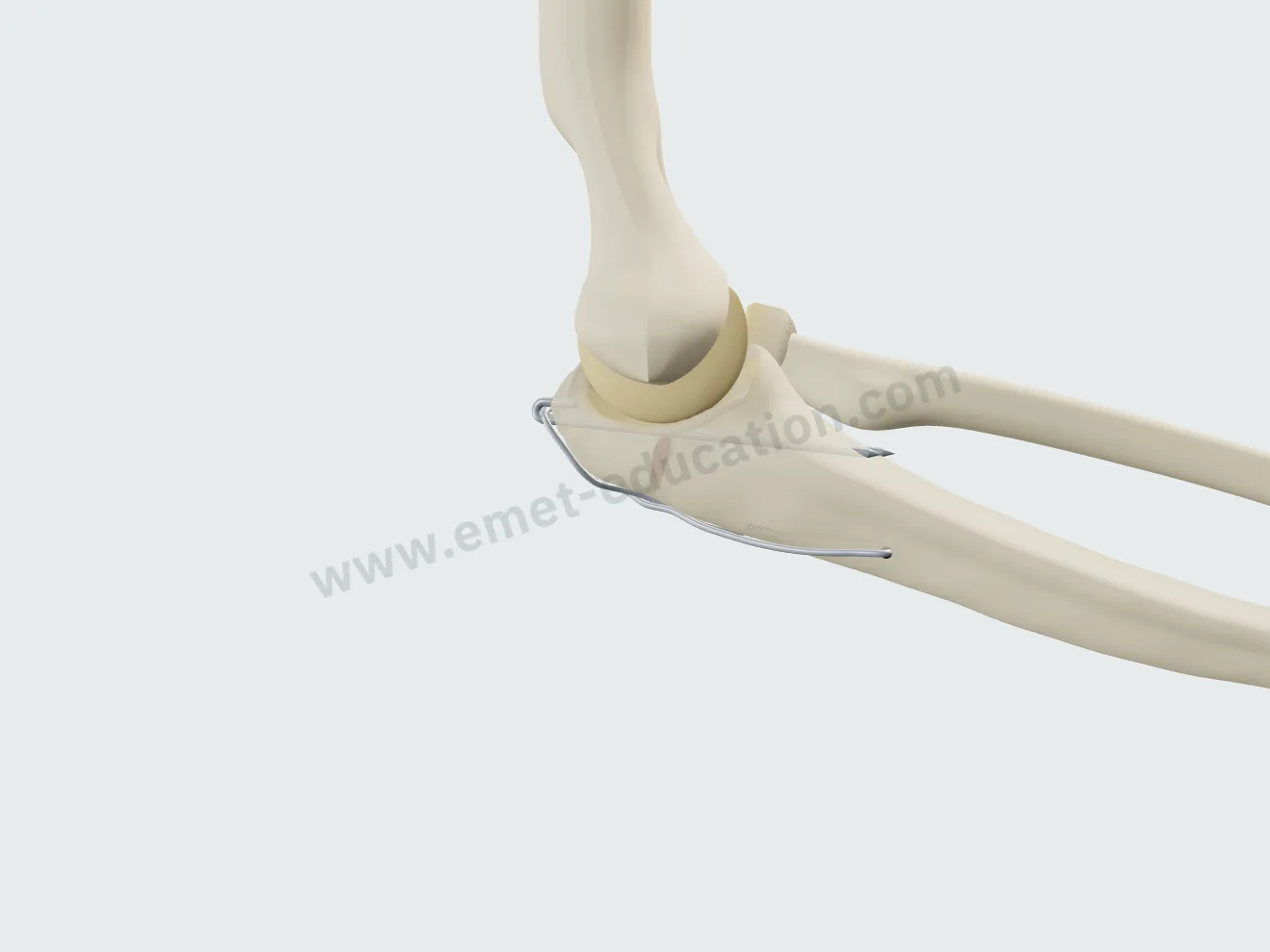
Tension-band wiring

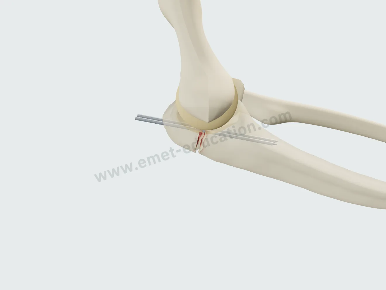
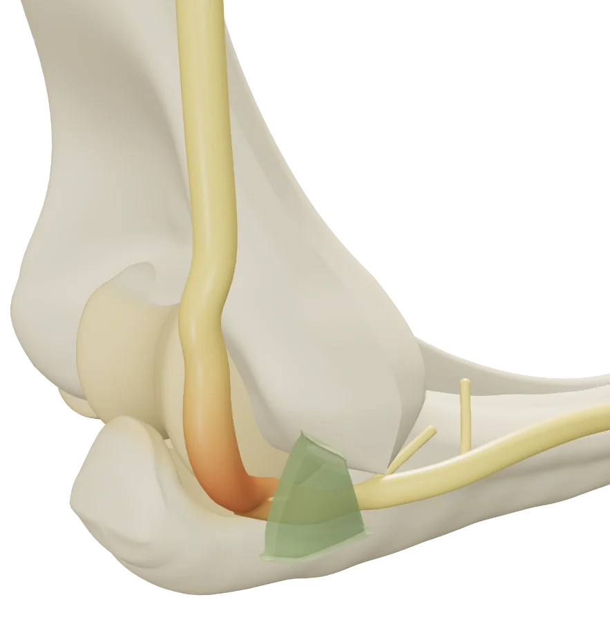
Two parallel K-wires, a figure-of-eight tension band, then twisting the wire to compress the fracture.

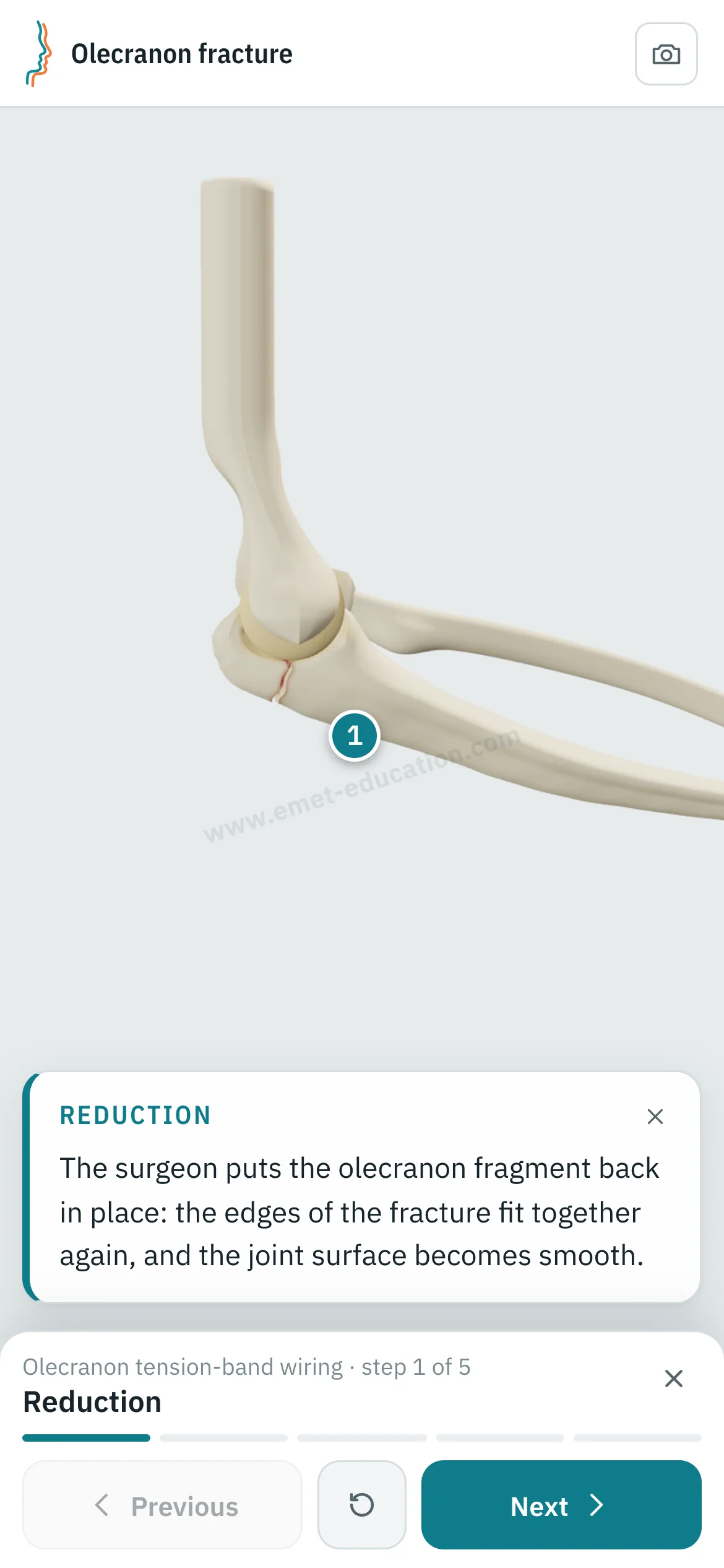
Tension-band wiring, step 1: reduction.
1Reduction
Tension-band wiring, step 2: K-wires.
2K-wires
Tension-band wiring, step 3: tension band.
3Tension band
Tension-band wiring, step 4: tightening.
4Tightening
Tension-band wiring, step 5: healing.
5Healing

Locking plate

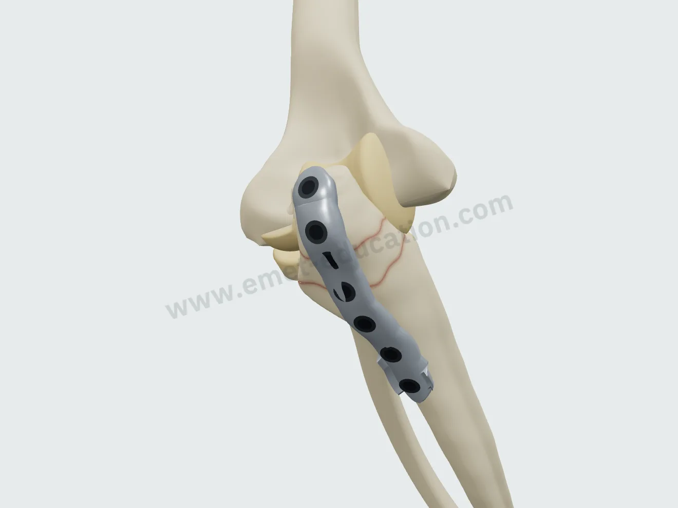
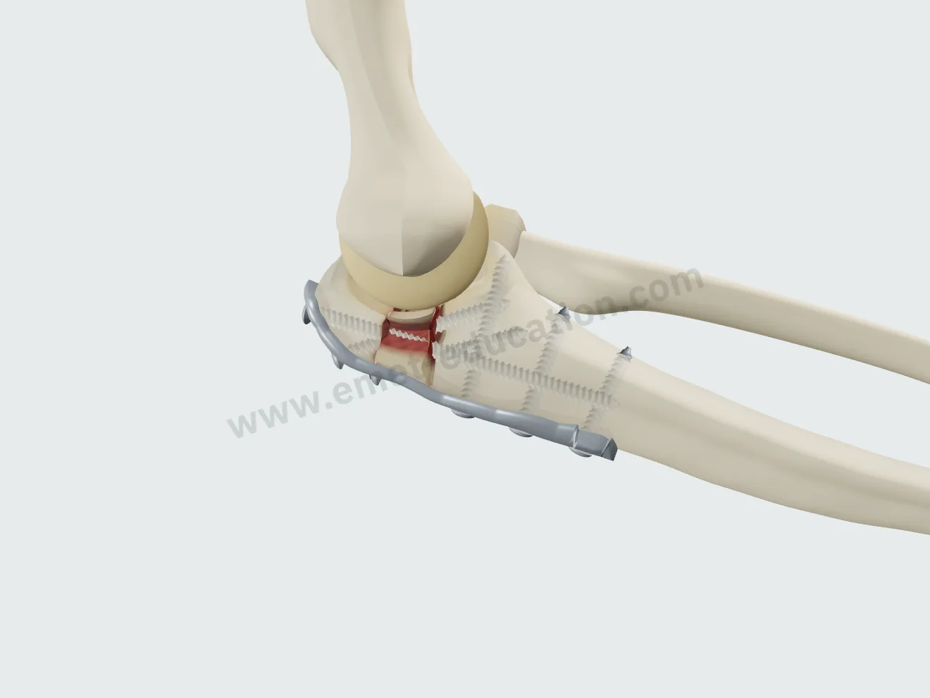
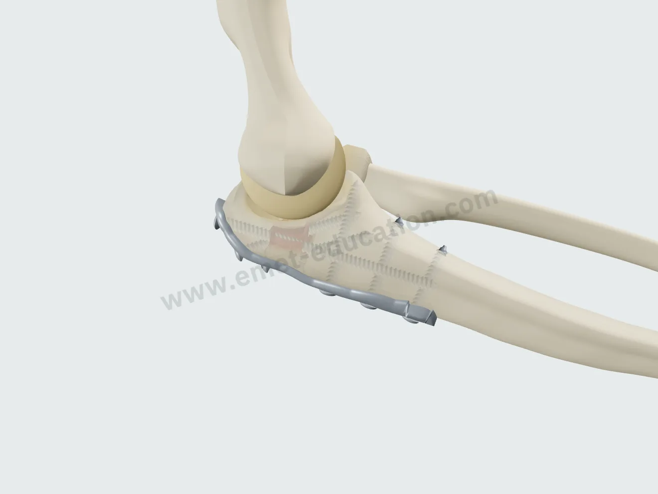
Precontoured posterior plate, locking screws, bridging a comminuted fracture.

Locking plate, step 1: reduction.
1Reduction
Locking plate, step 2: plate.
2Plate
Locking plate, step 3: screws.
3Screws
Locking plate, step 4: healing.
4Healing

After the visit

The patient leaves with their 3D model

They scan the QR code: the model, their findings and the proposed treatment follow them on their phone, in their language.

The model, exactly as you set it

At the end of the visit, the Consultation panel shows the anatomical findings, the proposed treatment and a QR code. The patient scans it and finds the model on their phone exactly as you set it, in their language.

  • Their fracture, and the proposed fixation to replay step by step.
  • Their anatomical findings and the explanatory card.
  • No identifying data in the link.
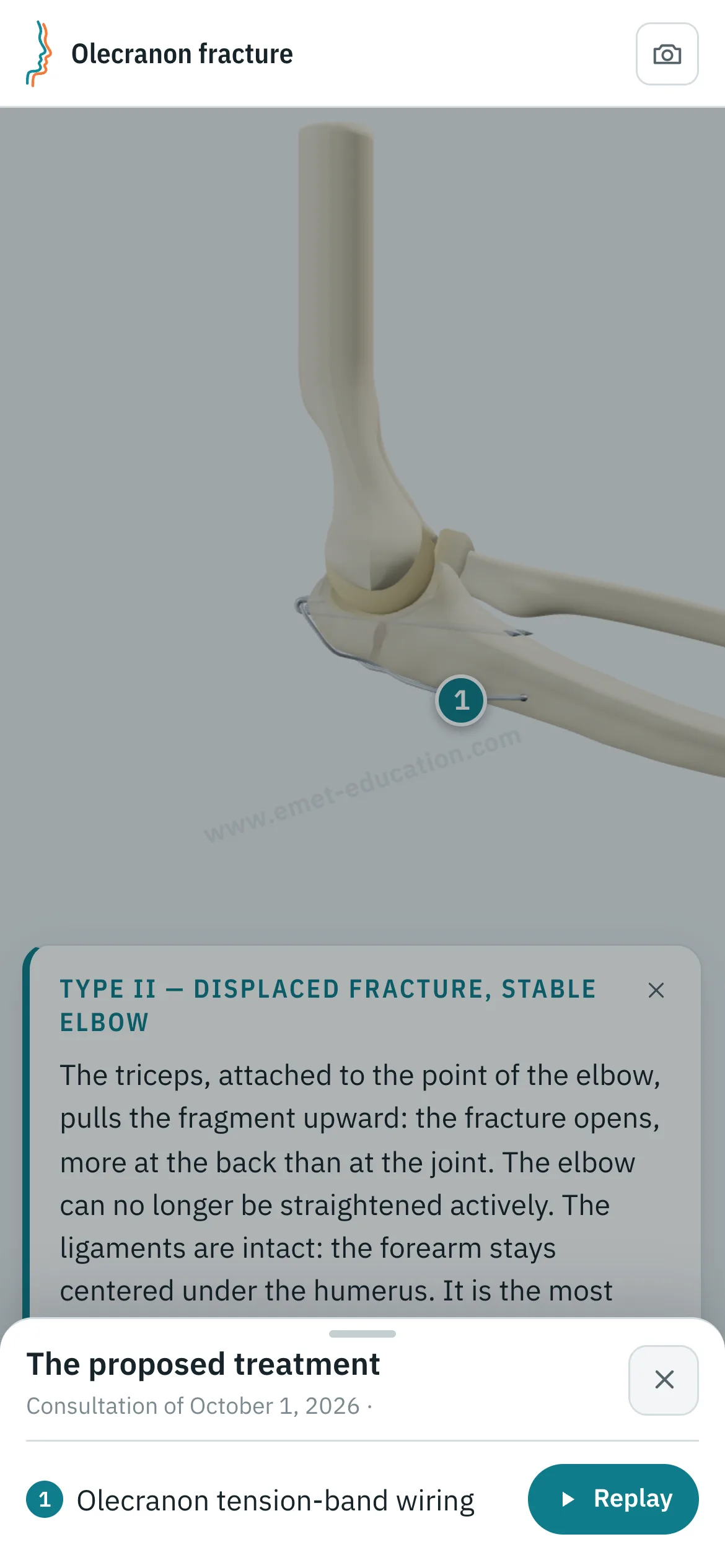
Patient view on a phone: the elbow after tension-band wiring, the card “Type II — displaced fracture, stable elbow,” and the “The proposed treatment” panel open: olecranon tension-band wiring, “Replay” button.
What the patient sees after scanning the QR code

Every structure explains itself on tap

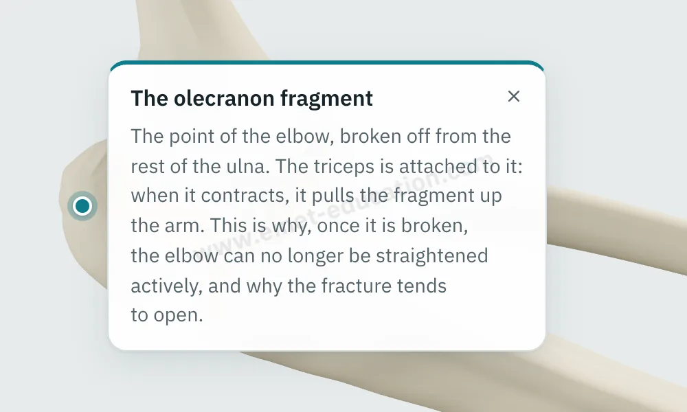
Tap a fragment, a bone, a K-wire or a screw: its name and an explanation appear.

  • One explanation per structure: bones, fragments, comminuted fragments, K-wires, tension band, plate, screws.
  • Written for the patient, in their language.
  • They can reread it on their own, on the tablet or at home.
Explanation bubble “The olecranon fragment” shown after tapping the bone.
“What is this?” on tap

They can replay the treatment step by step

From “The proposed treatment,” the patient replays the procedure: each step plays at their own pace, with its explanation, as often as they like.

Patient view on a phone: the tension-band wiring replayed step by step.
“Replay”: the procedure, step by step

Try it with your next case: the model opens in your browser, nothing to install.

Customize the fracture

Patient information

Quality information, documented

Only 12% of U.S. adults have proficient health literacy (National Assessment of Adult Literacy). A patient who sees their own fracture and the proposed procedure understands them, gives truly informed consent, and approaches surgery with confidence.

For the patient

They see their own fracture, reproduced from their imaging, and how the proposed fixation unfolds. They leave with the same material and can review it at home, with their family, as often as they like, before giving consent.

For the physician

Each visit created with an account keeps a record of the information provided: what was shown, when, and whether the patient reviewed it. The model image, with the anatomical findings, the proposed treatment and the QR code, downloads for the patient's chart and prints. Informed consent, as the Joint Commission puts it, is more than getting a signature.

  • Saved and datedThe exact state of the model: injuries shown, treatment proposed.
  • Identical, archivableThe patient receives what you showed; the image goes in the chart and prints.
  • TrackedEach time the patient opens the link is recorded.
  • AnonymousThe link given to the patient carries no identifying data.

“Document the informed consent conversation and the patient’s (or surrogate’s) decision in the medical record in some manner.”

AMA Code of Medical Ethics, Opinion 2.1.1, Informed Consent

The model supports the consent conversation and the written consent form; it does not replace them. Data and privacy: legal information (in French).

Questions

Questions from colleagues

Is the model free?

Yes: the fracture models are free. Treatments (fixation) and other features are paid options.

Do I need an account?

Not to open the model, set the fracture and walk through the treatment. After about fifteen adjustments, you will be asked to create a free account: it saves your visits, picks them up from one time to the next, and records when the patient opens the link.

What equipment do I need?

Just a web browser, nothing to install: computer, tablet or TV screen in the exam room; the patient opens it on their phone. One click saves an image, without the interface, for the chart.

What happens to patient data?

No patient names are stored. The link given to the patient carries only the state of the model: fracture, treatment, language and visit date. A name written on the image you hand out stays on that image.

Is it a medical device?

No: it is an educational tool. It does not diagnose, does not plan surgery, and does not replace the consent conversation or the written consent form. Among software functions that are not medical devices, the FDA lists those intended for general patient education and interactive anatomy diagrams used for training.

How long does it take in clinic?

Three adjustments and one procedure played: a few minutes, during the visit. The patient reviews it at home, as often as they like.

Can the elbow be moved?

Yes: flexion from −5° to 145°, plus pronation and supination. The proximal fragment stays under triceps pull at every angle, and the hardware moves with the bone.

Can I embed it on my website?

It is planned for upcoming plans: the model displayed on a practice or hospital website, credited to EMET Education.

Which languages?

English, Spanish, French and Russian. The patient receives the model in the language chosen during the visit, which helps with the roughly 28 million people in the U.S. with limited English proficiency, alongside a qualified interpreter, not instead of one.

Design

Who designed this model

Model designed by Dr. Mikaël Chelli, orthopedic surgeon, based on the reference classifications. Updated September 29, 2026.

  • Mayo Clinic classification (Morrey, 1995); Wiegand L. et al., Orthop Traumatol Surg Res, 2019.
  • Regan and Morrey classification of coronoid fractures (1989); Wiegand L. et al., 2019.

Try it with your next patient

The model opens in your browser, nothing to install, no sign-up.

Know a colleague who could use it? Email this page ·