EMET Electronic Medical Twin
  1. EMET Education
  2. Musculoskeletal
  3. Elbow

3D models · Elbow

Elbow 3D models

Elbow 3D models to show your patient their lesion and the treatment you recommend. They leave with their model on their phone.

Designed by an orthopedic surgeon

Open in your browser, no sign-up. For orthopedic surgeons, emergency physicians, sports medicine physicians, physiatrists, rheumatologists and primary care physicians.

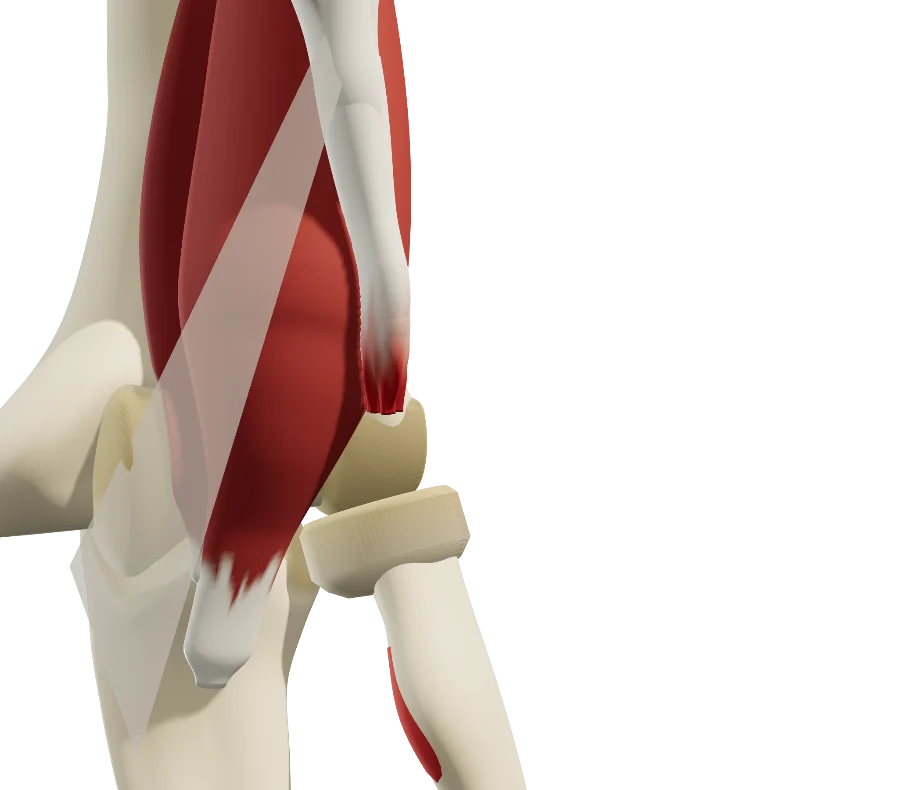
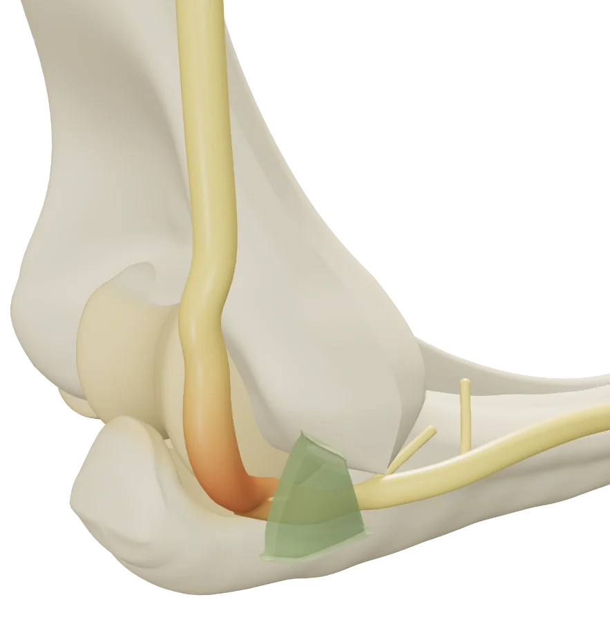
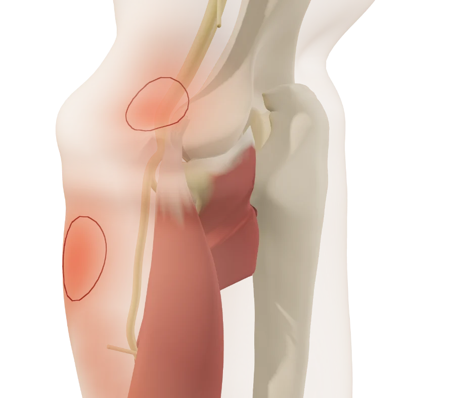
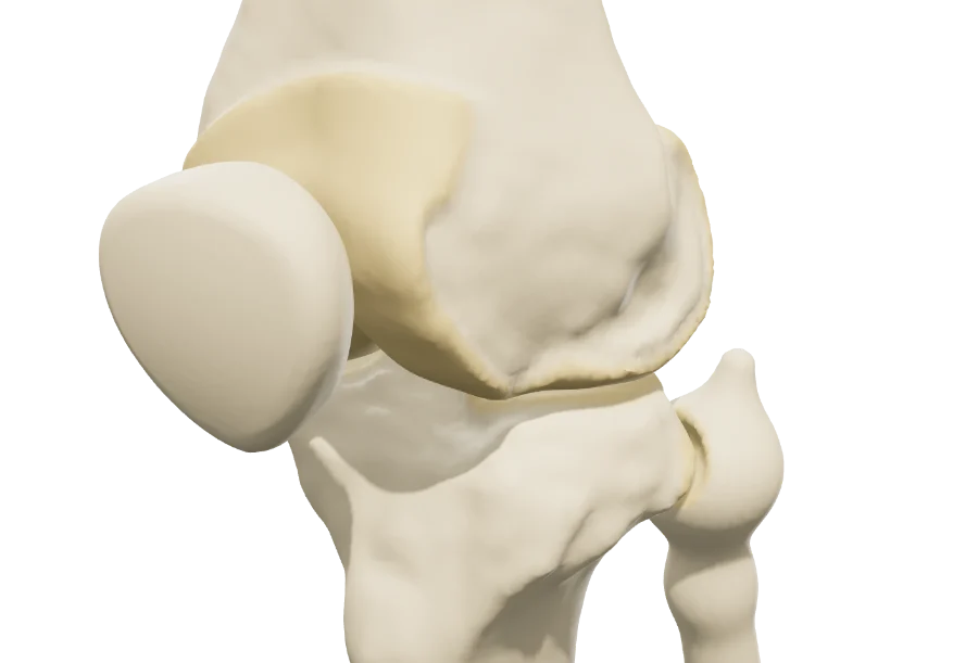
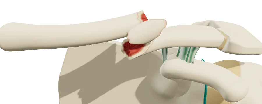
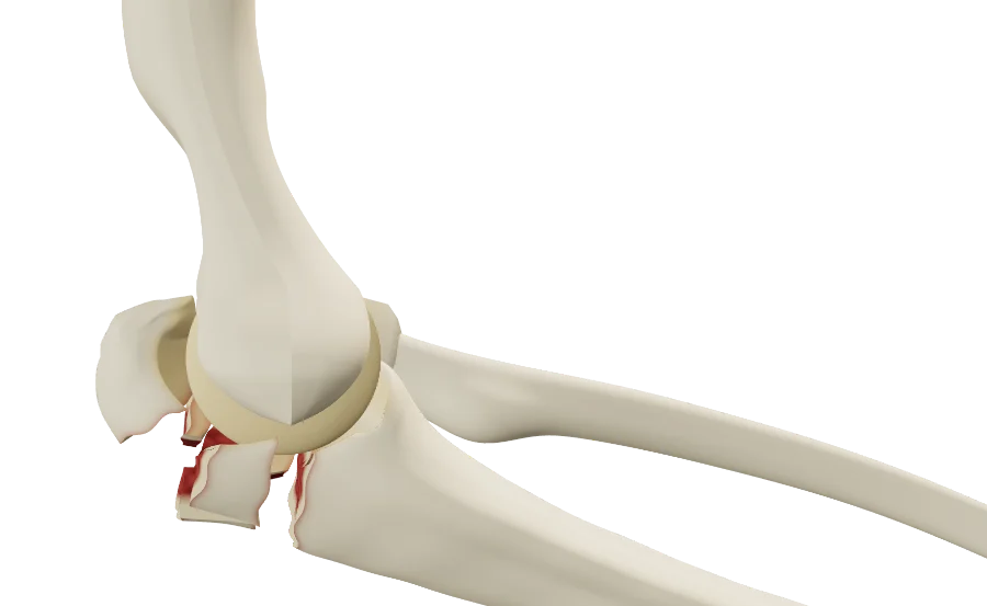
Elbow in 3D seen from the side and back: the fractured olecranon, its fragment pulled up by the triceps, the fracture open posteriorly. Elbow in 3D, front view: the ruptured distal biceps tendon, its frayed end retracted above the exposed radial tuberosity. Elbow in 3D seen from the medial side: the ulnar nerve compressed under Osborne's ligament, behind the medial epicondyle. Elbow and forearm in 3D from the lateral side, under translucent skin: the painful area of the tendon on the lateral epicondyle, and that of the posterior interosseous nerve a few centimeters lower.

The models

Elbow 3D models

The models share a common elbow anatomy: the three bones, flexion and pronation-supination, the tendons and the nerves. Fracture, tendon or nerve: the lesion is set from the workup, and the treatment plays step by step.

Olecranon fracture

The fracture set from the radiograph, triceps pull at every flexion angle, then fixation.

Lesions
Mayo classification (I to III, simple or comminuted), fracture gap from 4 to 20 mm, associated coronoid fracture (Regan and Morrey).
Treatments
Tension-band wiring, locking plate.

Ulnar neuropathy at the elbow

The nerve pressed against the bone as the elbow bends: the mechanism is shown by flexing the elbow, and ligament tension and nerve flattening are read on the model.

Lesions
Compression under Osborne's ligament (McGowan I to III), nerve instability (subluxation, dislocation), space-occupying lesion (ganglion cyst, heterotopic ossification, bone fragment).
Treatments
In situ neurolysis, anterior transposition, injection.

Distal biceps rupture

The rupture mechanism replayed, retraction to the millimeter, then nonoperative treatment or reattachment.

Lesions
Distal tendon rupture, retraction from 0 to 40 mm (up to 80 mm with a torn lacertus), torn lacertus fibrosus.
Treatments
Nonoperative treatment (healing onto the brachialis, strength and motion), reattachment to the radial tuberosity with four techniques.

Lateral epicondylalgia and posterior interosseous nerve

The two causes of lateral elbow pain, alone or combined, and where each one hurts, seen on translucent skin.

Lesions
Tendinopathy of the extensor carpi radialis brevis (tendinosis, deep fissure, partial tear), posterior interosseous nerve compression (arcade of Frohse, edge of the extensor carpi radialis brevis).
Treatments
Peritendinous injection, intratendinous PRP, open or arthroscopic debridement. Nerve treatments are in progress.

How it works

In the visit, in three steps

Structured information in a few minutes, which the patient can review at home.

  1. 1

    Show

    Open the model and walk through the anatomy: ulnohumeral joint, radial head, tendons and the course of the nerves.

  2. 2

    Reproduce

    Set the lesion from the exam and imaging: location, type, stage, classification. Then walk through the proposed treatment, step by step.

  3. 3

    Share

    Give the patient the QR code or an image: they find their own case, in their language.

Try it with your next case: the models open in your browser, nothing to install.

See the “Olecranon fracture” model

Questions

Questions from colleagues

Which elbow conditions?

Those in the list above: fractures, tendon ruptures, tendinopathies and nerve compressions, each with its reference classifications. A model marked “in progress” already opens, but is still being expanded.

Can the elbow be moved?

Yes: the shared elbow anatomy supports flexion from −5° to 145° and pronation-supination. Each model uses it for its condition: the ulnar nerve is compressed as the elbow bends, the olecranon fragment stays under triceps pull at every angle.

Are the models free?

Yes: the free plan opens every model, to show patients their diagnosis: most conditions and lesions. Treatments and other features are paid options.

Do I need an account?

Not to open a model, set the lesion and walk through the treatment. After about fifteen adjustments, you will be asked to create a free account: it saves your visits, picks them up from one time to the next, and records when the patient opens the link.

What equipment do I need?

Just a web browser, nothing to install: computer, tablet or TV screen in the exam room; the patient opens it on their phone. One click saves an image, without the interface, for the chart.

What happens to patient data?

No patient names are stored. The link given to the patient carries only the state of the model: lesion, treatment, language and visit date. A name written on the image you hand out stays on that image.

Which languages?

English, Spanish, French and Russian. The patient receives the model in the language chosen during the visit, which helps with the roughly 28 million people in the U.S. with limited English proficiency, alongside a qualified interpreter, not instead of one.
Design

Who designed these models

Models designed by Dr. Mikaël Chelli, orthopedic surgeon, based on the reference classifications of each condition. Updated October 1, 2026.

Try them with your next patient

The models open in your browser, nothing to install, no sign-up.

Know a colleague who could use them? Email this page ·