EMET Electronic Medical Twin
  1. EMET Education
  2. Musculoskeletal
  3. Shoulder

3D models · Shoulder

Shoulder 3D models

Shoulder 3D models to show your patient their lesion and the treatment you recommend. They leave with their model on their phone.

Designed by an orthopedic surgeon

Open in your browser, no sign-up. For orthopedic surgeons, emergency physicians, sports medicine physicians, physiatrists, rheumatologists and primary care physicians.

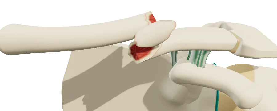
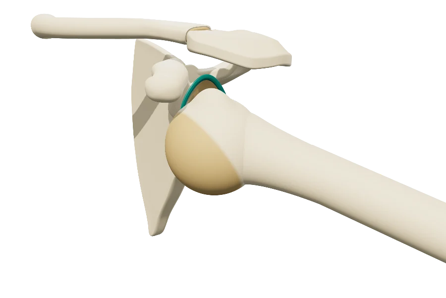
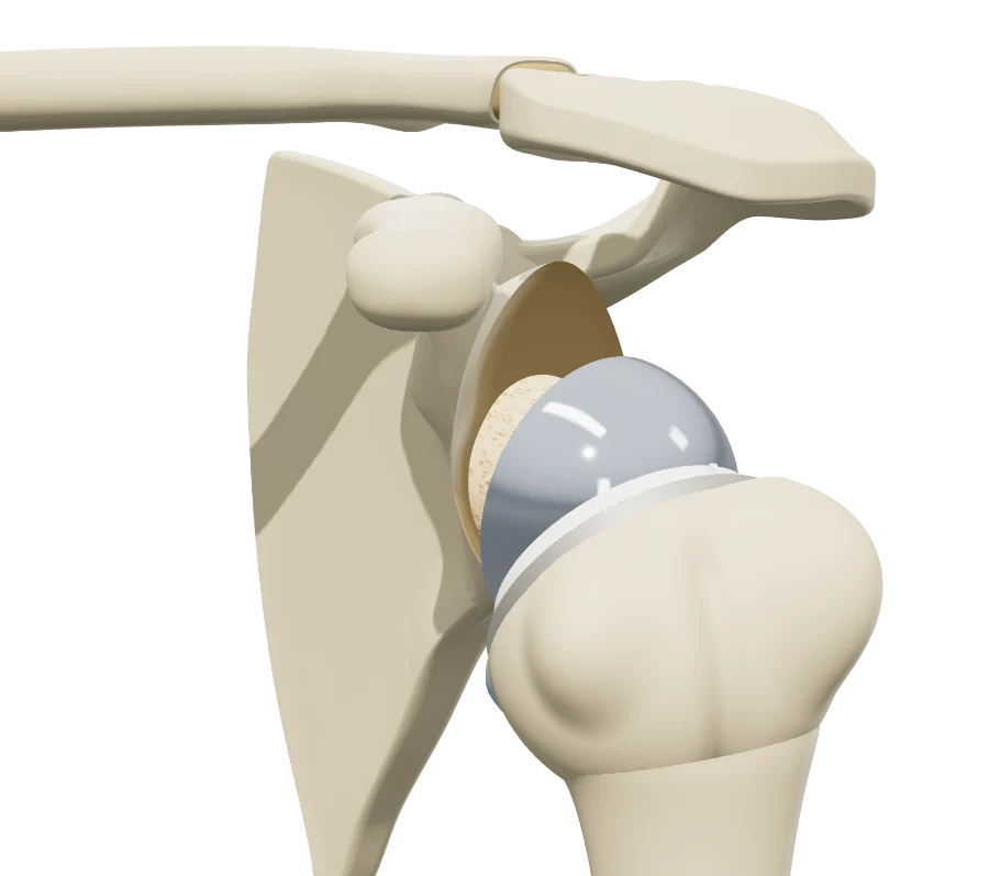
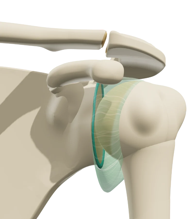
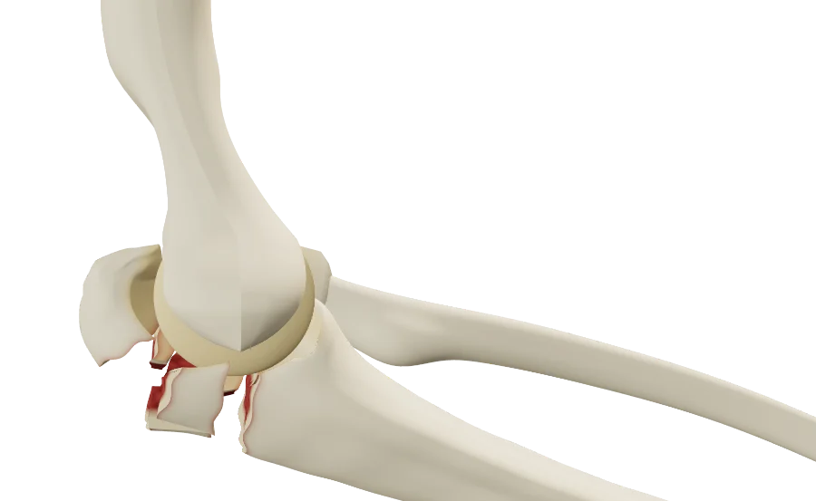
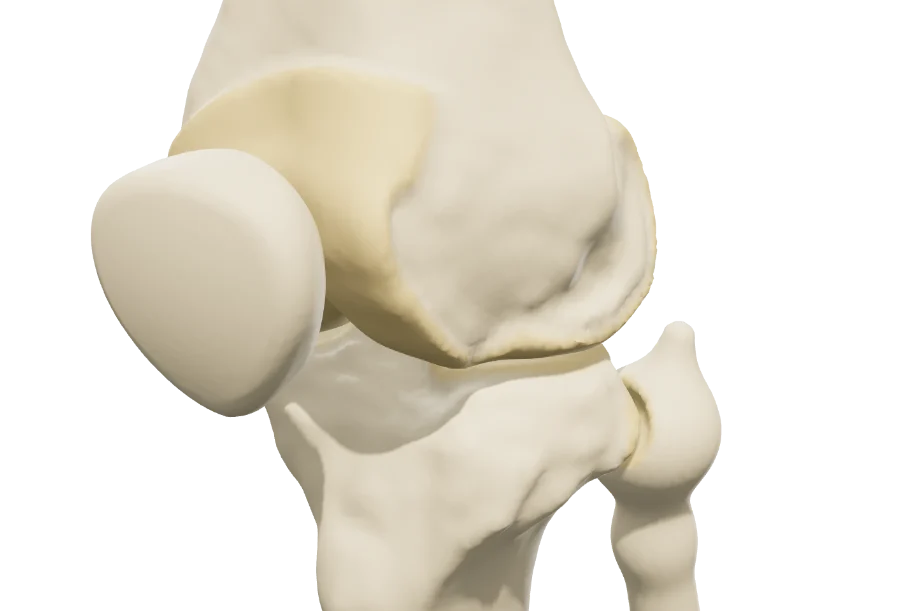
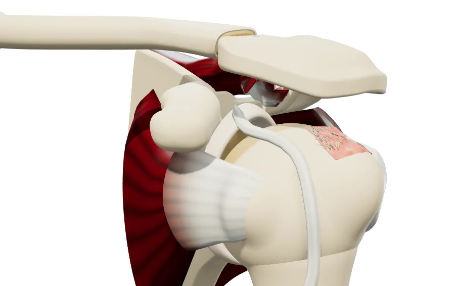
Rotator cuff in 3D: supraspinatus tear at the top of the humeral head. Clavicle in 3D fractured at its midpoint, with a third fragment: the medial fragment rises, the shoulder drops. Dislocated shoulder in 3D: the humeral head out of the glenoid, arm in abduction and external rotation. Reverse total shoulder arthroplasty in 3D: glenosphere on the scapula, cup on the humerus.

The models

Shoulder 3D models

The models share the same shoulder anatomy: scapula, humerus, clavicle, and depending on the model the rotator cuff, the capsule, the labrum or the coracoclavicular ligaments. You set the lesion from the exam and imaging, then walk through the proposed treatment: arthroscopic, arthroplasty, fracture fixation or nonoperative.

Rotator cuff

The cuff tendon by tendon, from calcific tendinitis to retracted tears, and the arthroscopic procedures.

Lesions
Full-thickness supraspinatus tear (retraction, stages 1 to 3), articular- or bursal-sided partial-thickness tear, infraspinatus, subscapularis (Lafosse), calcific tendinitis (types A, B, C), long head of the biceps (tenosynovitis, SLAP, rupture), bursitis, hooked acromion.
Treatments
Suture bridge repair, acromioplasty, biceps tenodesis, subacromial injection.

Shoulder instability

Anterior dislocation and its bony lesions, then the proposed stabilization, step by step.

Lesions
Animated anterior dislocation, Bankart lesion, glenoid bone loss, Hill-Sachs lesion.
Treatments
Latarjet procedure (double EndoButton, one or two screws), arthroscopic labral repair (Bankart), Hill-Sachs remplissage.

Painful unstable shoulder

Painful instability in young, often hyperlax patients, without true dislocation: capsule and labrum, alone or combined.

Lesions
Normal, lax or stretched capsule; labral lesions by sector, chosen on a glenoid diagram; painful translation of the humeral head.
Treatments
Labral repair, capsular plication with pants-over-vest stitches.

Shoulder osteoarthritis

How glenohumeral osteoarthritis develops, centered or eccentric, then the three arthroplasties.

Lesions
Centered osteoarthritis (Samilson and Prieto I to III), eccentric osteoarthritis (Hamada modified by Walch), osteonecrosis of the humeral head.
Treatments
Hemiarthroplasty, anatomic total shoulder arthroplasty, reverse shoulder arthroplasty and its effect on the deltoid.

Clavicle fracture

The shaft and the lateral quarter, fracture pattern and displacement set from the radiograph, then nonoperative or surgical treatment.

Lesions
Shaft: seven patterns (AO/OTA 15.2 A, B, C; Robinson), displacement and shortening. Lateral quarter: Neer I to V. Nonunion.
Treatments
Nonoperative treatment (soft callus, hard callus, remodeling), plate fixation, intramedullary screw, lateral locking plate, coracoclavicular button.

How it works

In the visit, in three steps

Structured information in a few minutes, which the patient can review at home.

  1. 1

    Show

    Open the model and walk through the anatomy: glenoid and humeral head, rotator cuff, capsule and labrum, clavicle and coracoid.

  2. 2

    Reproduce

    Set the lesion from the exam and imaging: location, type, stage, classification. Then walk through the proposed treatment, step by step.

  3. 3

    Share

    Give the patient the QR code or an image: they find their own case, in their language.

Try it with your next case: the models open in your browser, nothing to install.

See the “Rotator cuff” model

Questions

Questions from colleagues

Which shoulder conditions?

Those in the list above, each with its reference classifications: from the rotator cuff to fractures, including instability and osteoarthritis. All models open in your browser, nothing to install, and new ones join the catalog.

Are treatments shown?

Yes: each model plays its procedures step by step — rotator cuff repair, Latarjet procedure, capsular shift, arthroplasty, fracture fixation and nonoperative treatment of the clavicle. The patient reviews them at home, at their own pace.

Are the models free?

Yes: the free plan opens every model, to show patients their diagnosis: most conditions and lesions. Treatments and other features are paid options.

Do I need an account?

Not to open a model, set the lesion and walk through the treatment. After about fifteen adjustments, you will be asked to create a free account: it saves your visits, picks them up from one time to the next, and records when the patient opens the link.

What equipment do I need?

Just a web browser, nothing to install: computer, tablet or TV screen in the exam room; the patient opens it on their phone. One click saves an image, without the interface, for the chart.

What happens to patient data?

No patient names are stored. The link given to the patient carries only the state of the model: lesion, treatment, language and visit date. A name written on the image you hand out stays on that image.

Which languages?

English, Spanish, French and Russian. The patient receives the model in the language chosen during the visit, which helps with the roughly 28 million people in the U.S. with limited English proficiency, alongside a qualified interpreter, not instead of one.
Design

Who designed these models

Models designed by Dr. Mikaël Chelli, orthopedic surgeon, based on the reference classifications of each condition. Updated October 1, 2026.

Try them with your next patient

The models open in your browser, nothing to install, no sign-up.

Know a colleague who could use them? Email this page ·