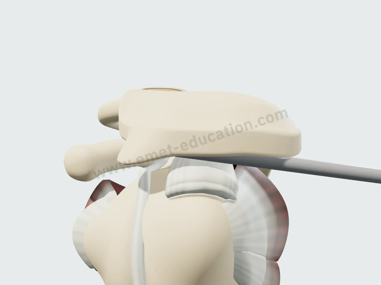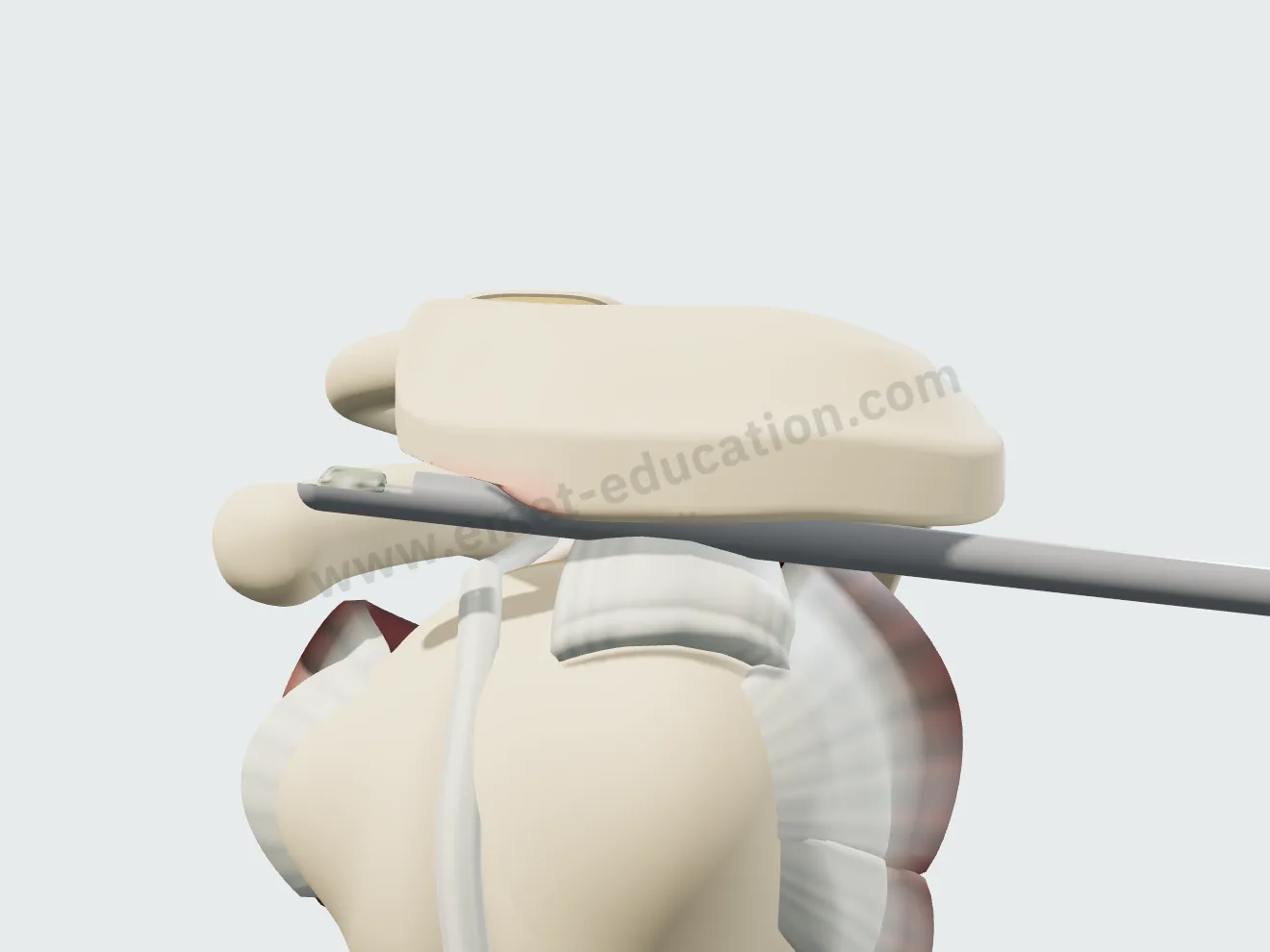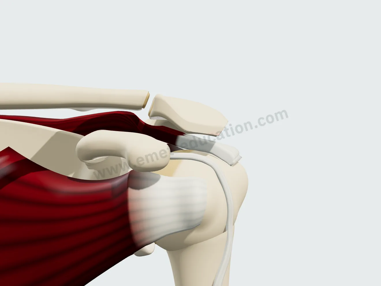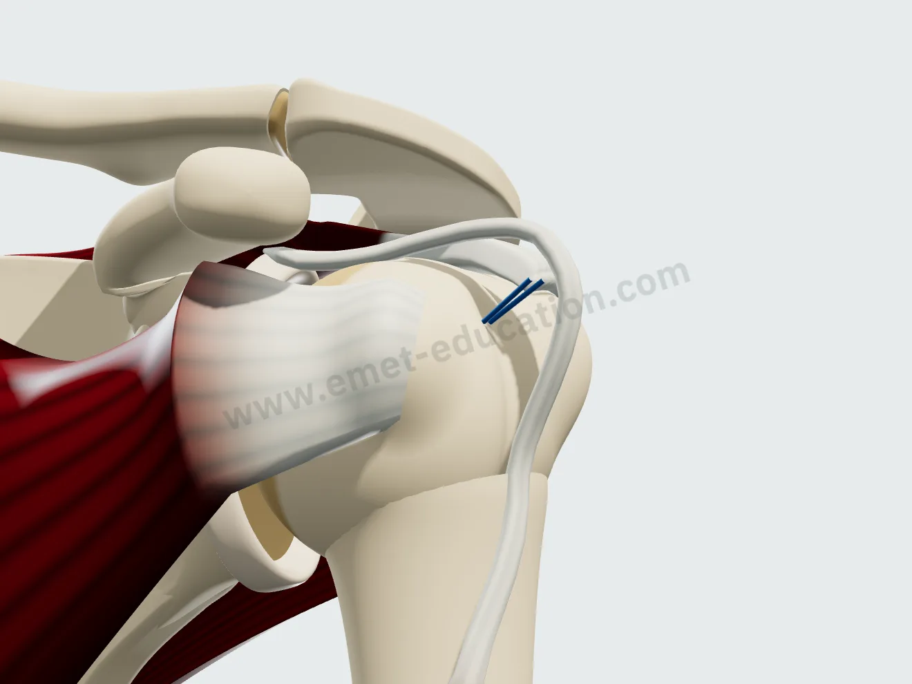
The lesions, described in a few clicks
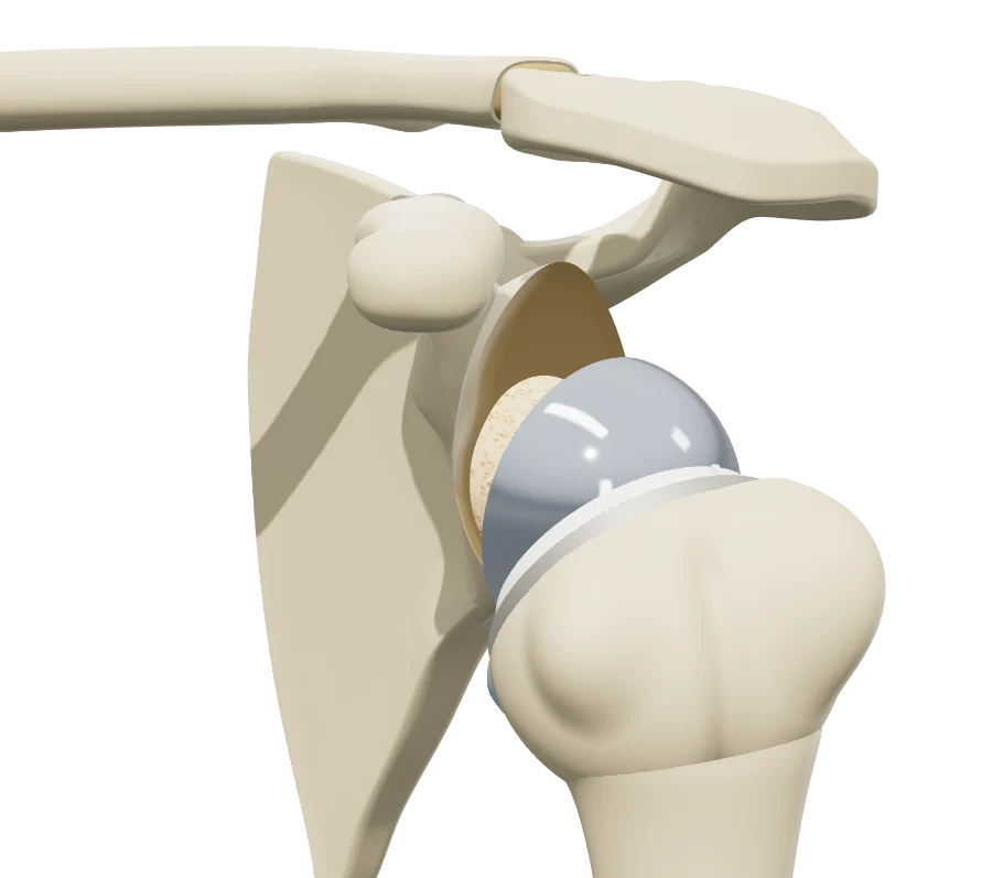
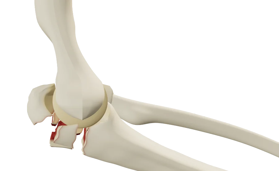
The “Pathology & lesions” panel describes the cuff tendon by tendon, the long head of the biceps and the subacromial space. Lesions add up, and each one changes the model in front of the patient.
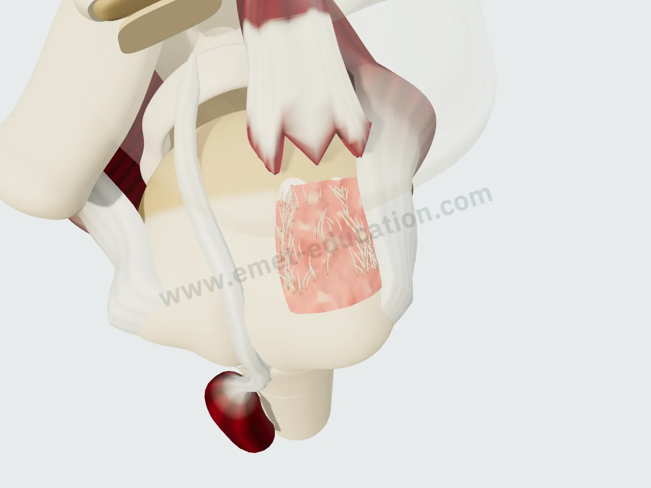
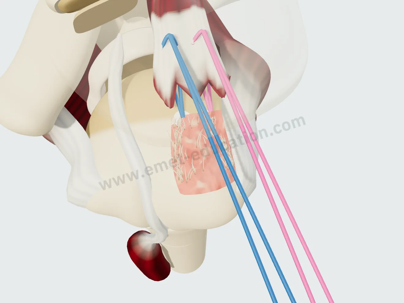
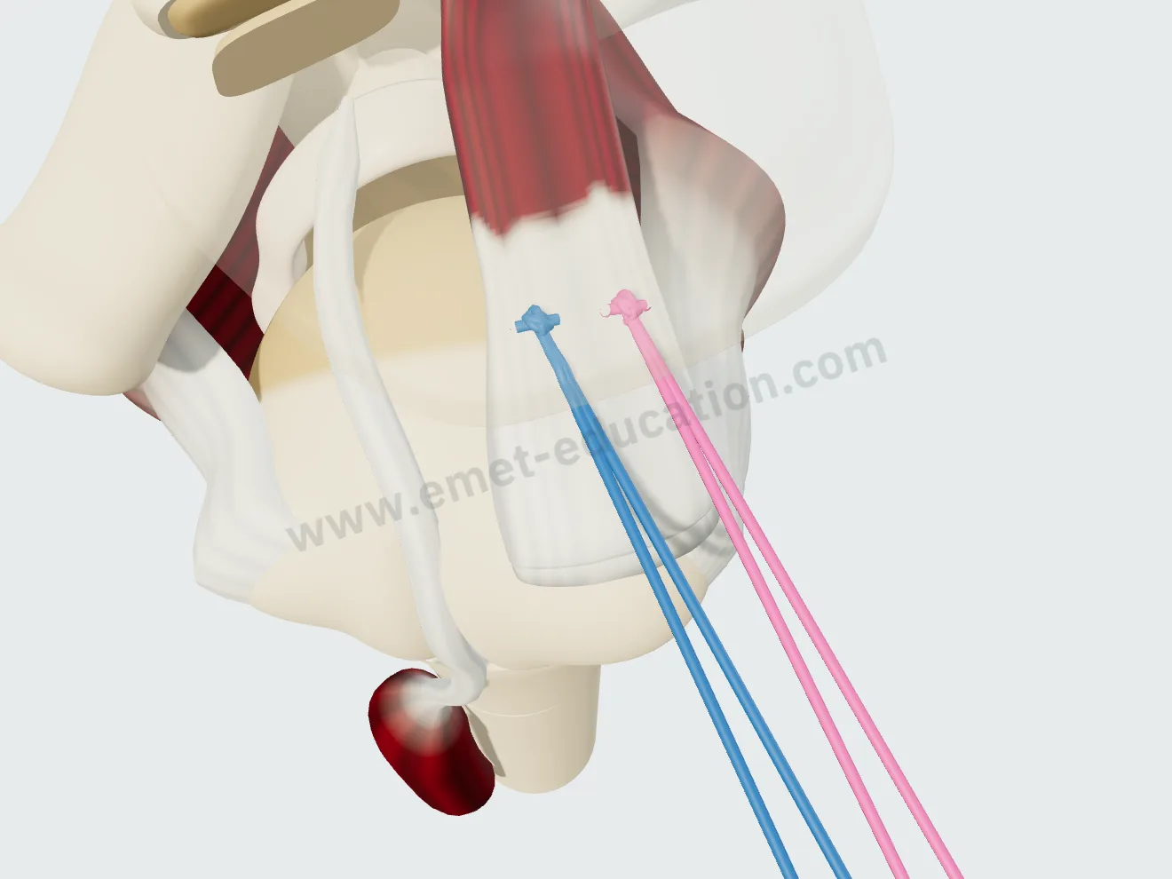
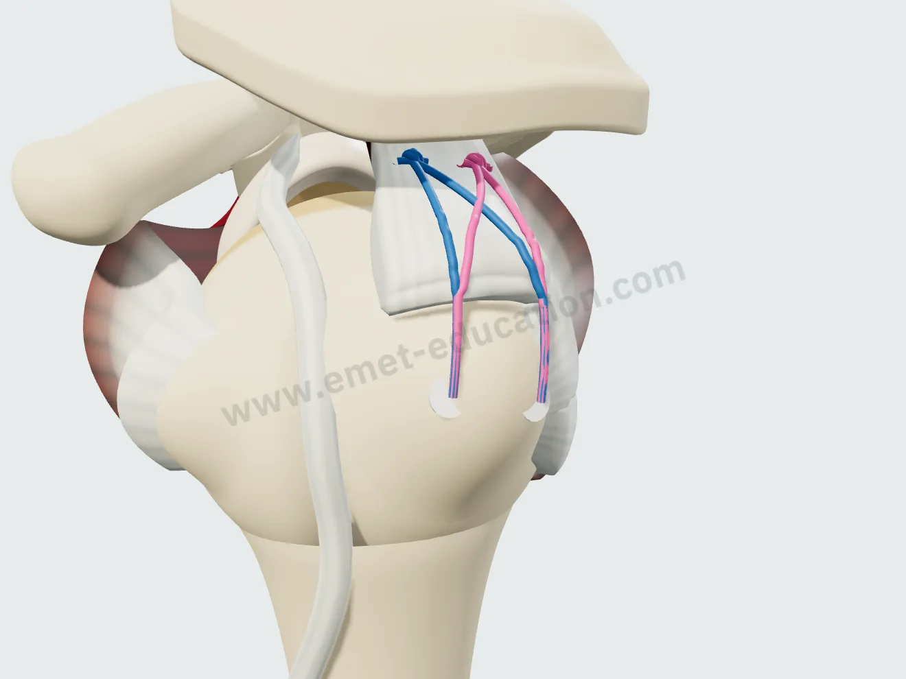
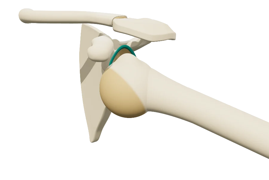
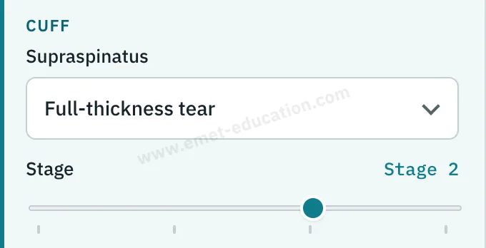
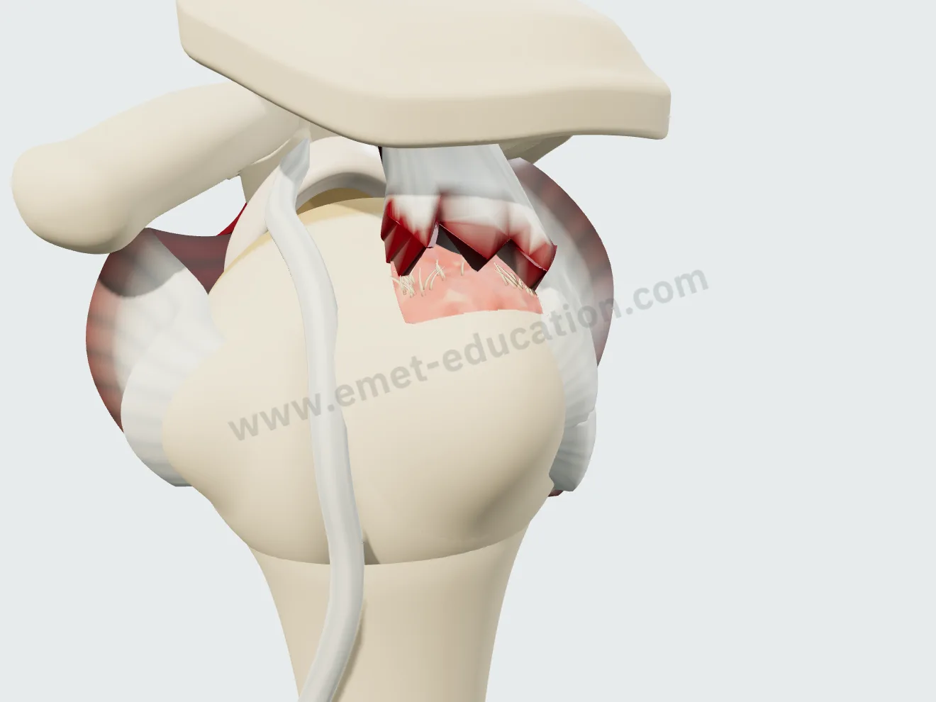
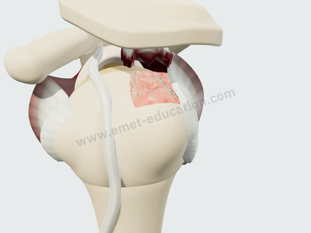
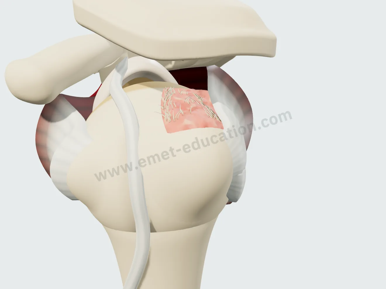
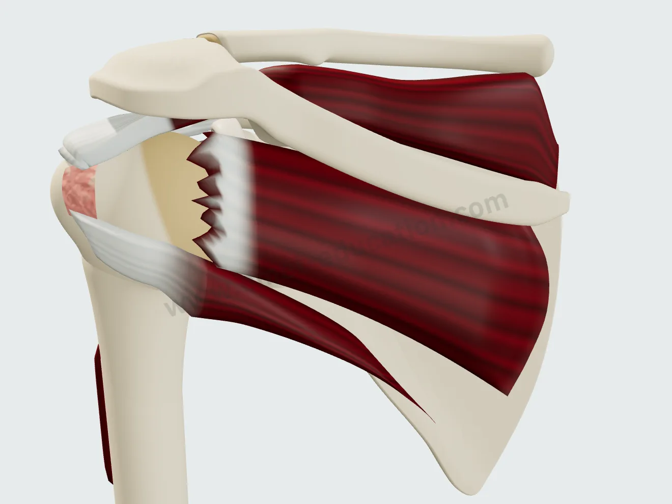
Supraspinatus retraction, in three stages
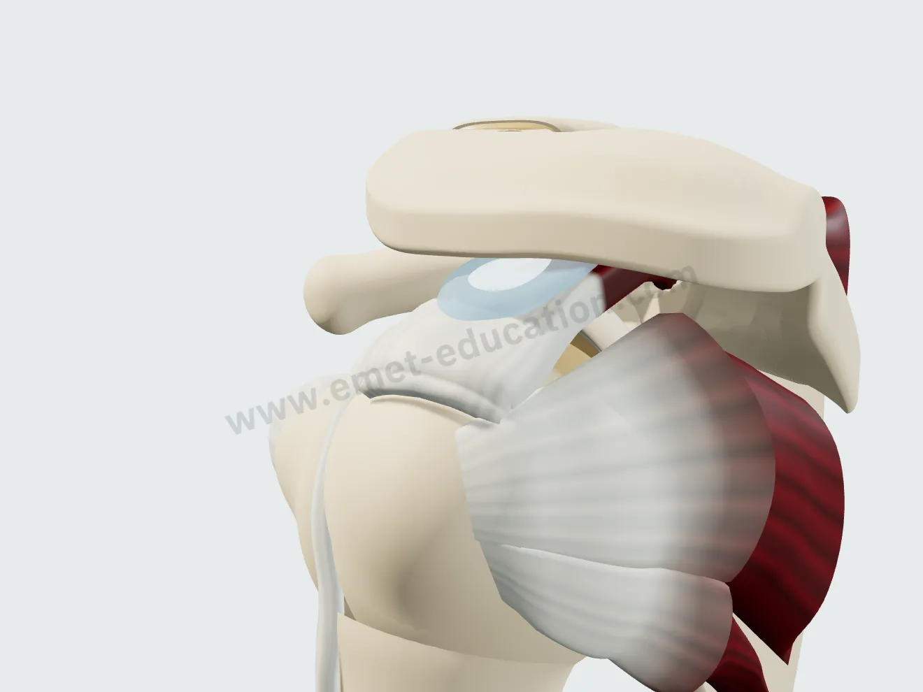
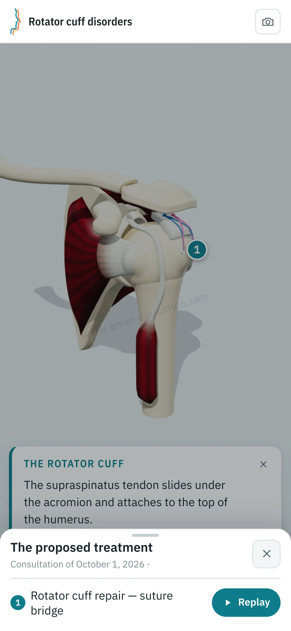
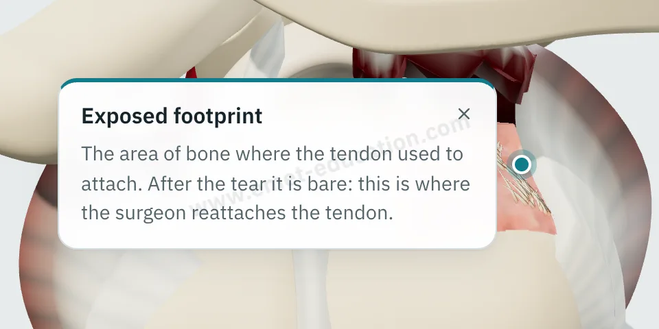
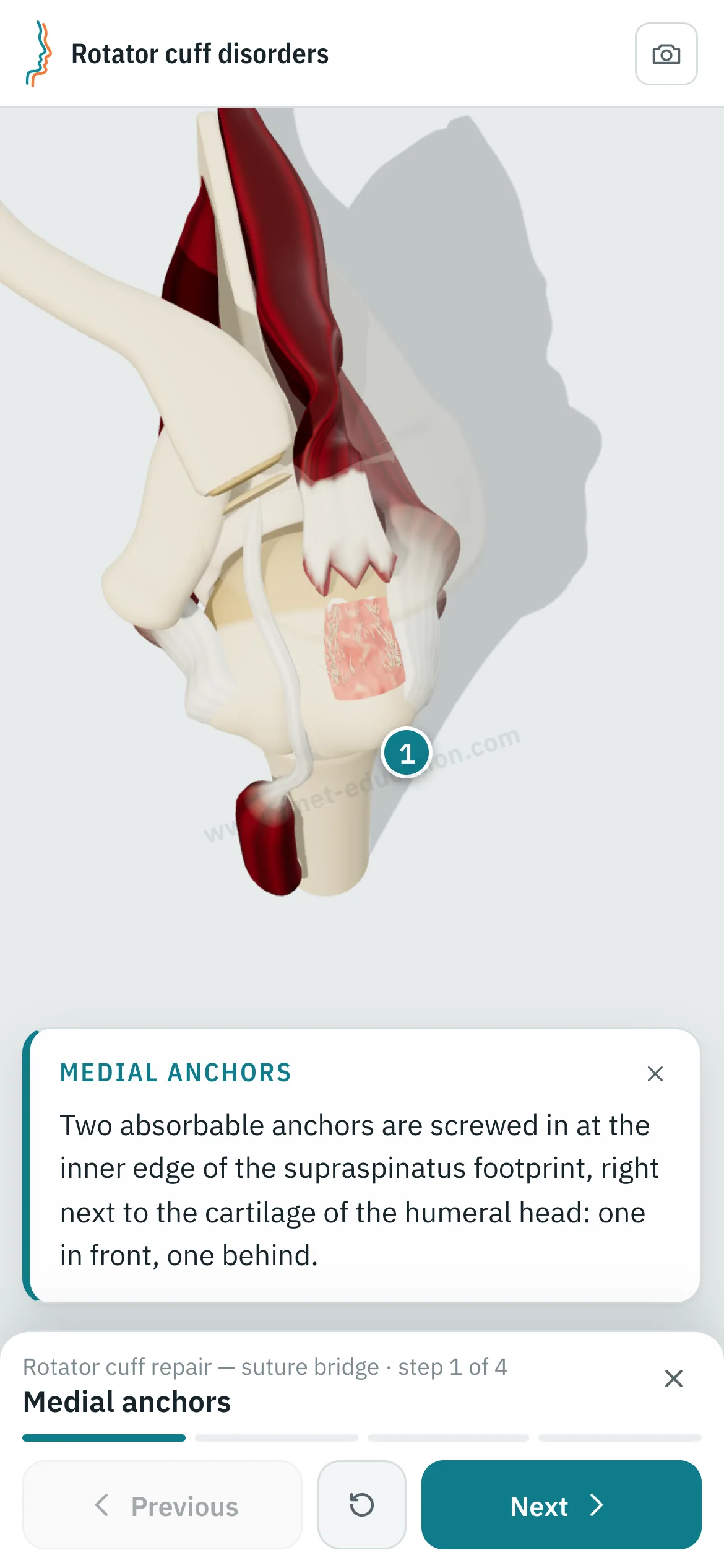
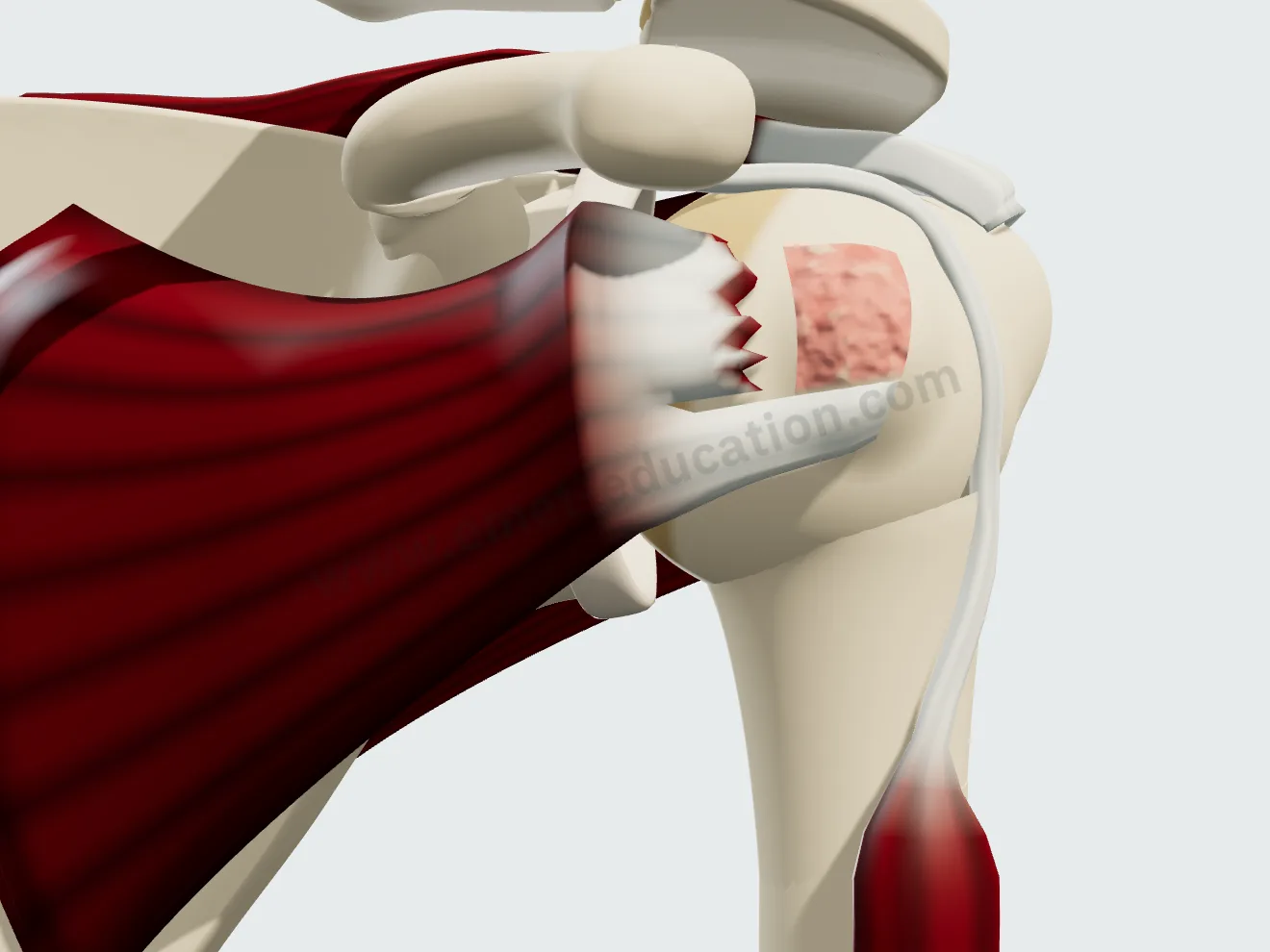
Non-retracted tear (1), stump at the top of the humeral head (2), retraction to the glenohumeral joint line (3). The enthesis tears with a jagged edge, then the tendon retracts; the exposed footprint appears on the greater tuberosity.
Partial-thickness tear, articular or bursal side
The tendon has its true thickness, 6 mm: the lesion erodes the articular or the bursal side over 10 to 90% of the thickness, above the footprint. At repair, it is first completed with the shaver.

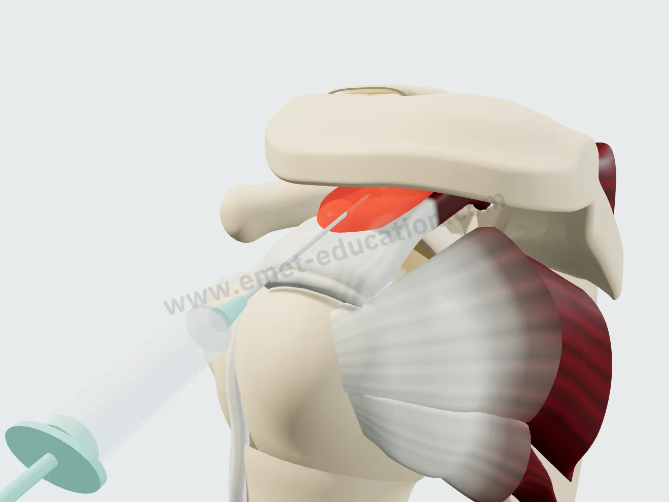
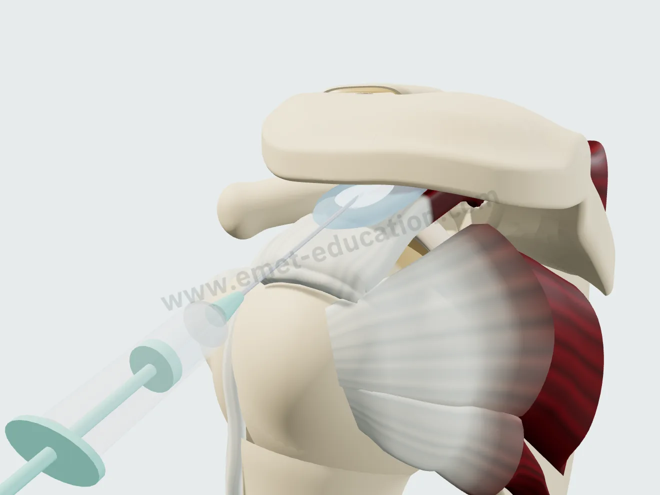
Calcific tendinitis
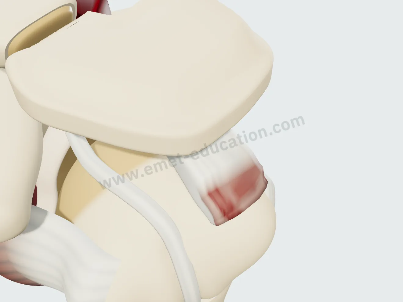
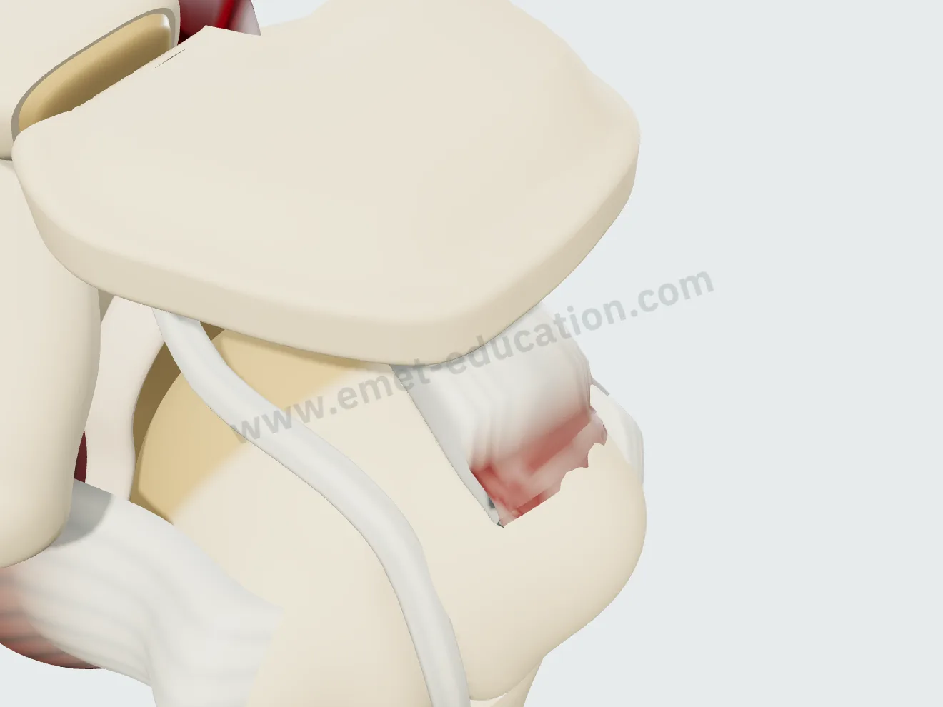
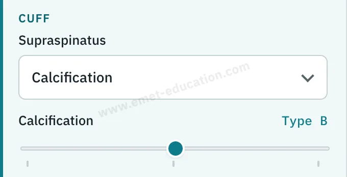
A chalky deposit within the supraspinatus, one centimeter medial to its insertion: single and well defined (A), larger and multilobulated (B), cloudy and infiltrating (C). The tendon bulges over the deposit.
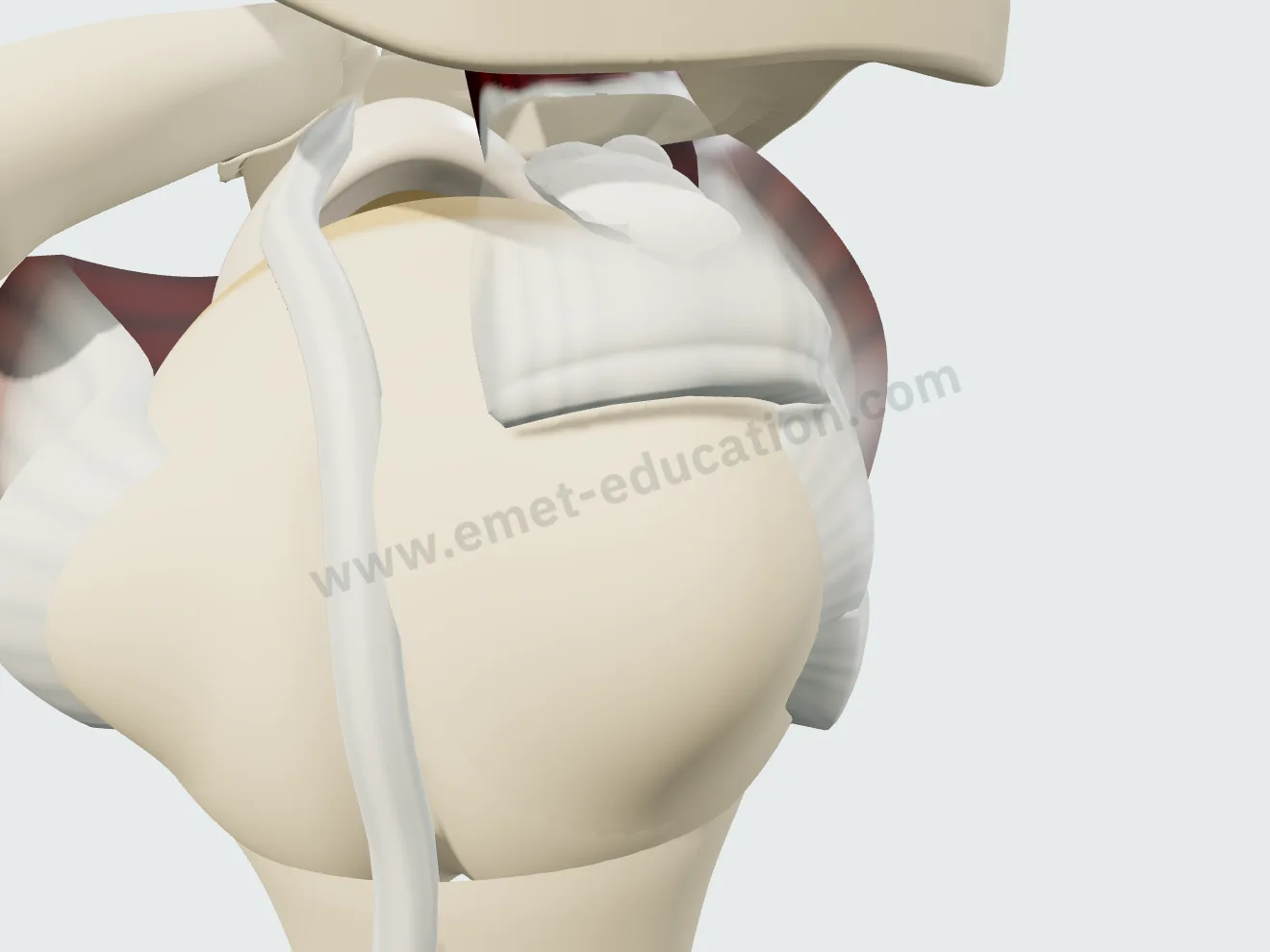
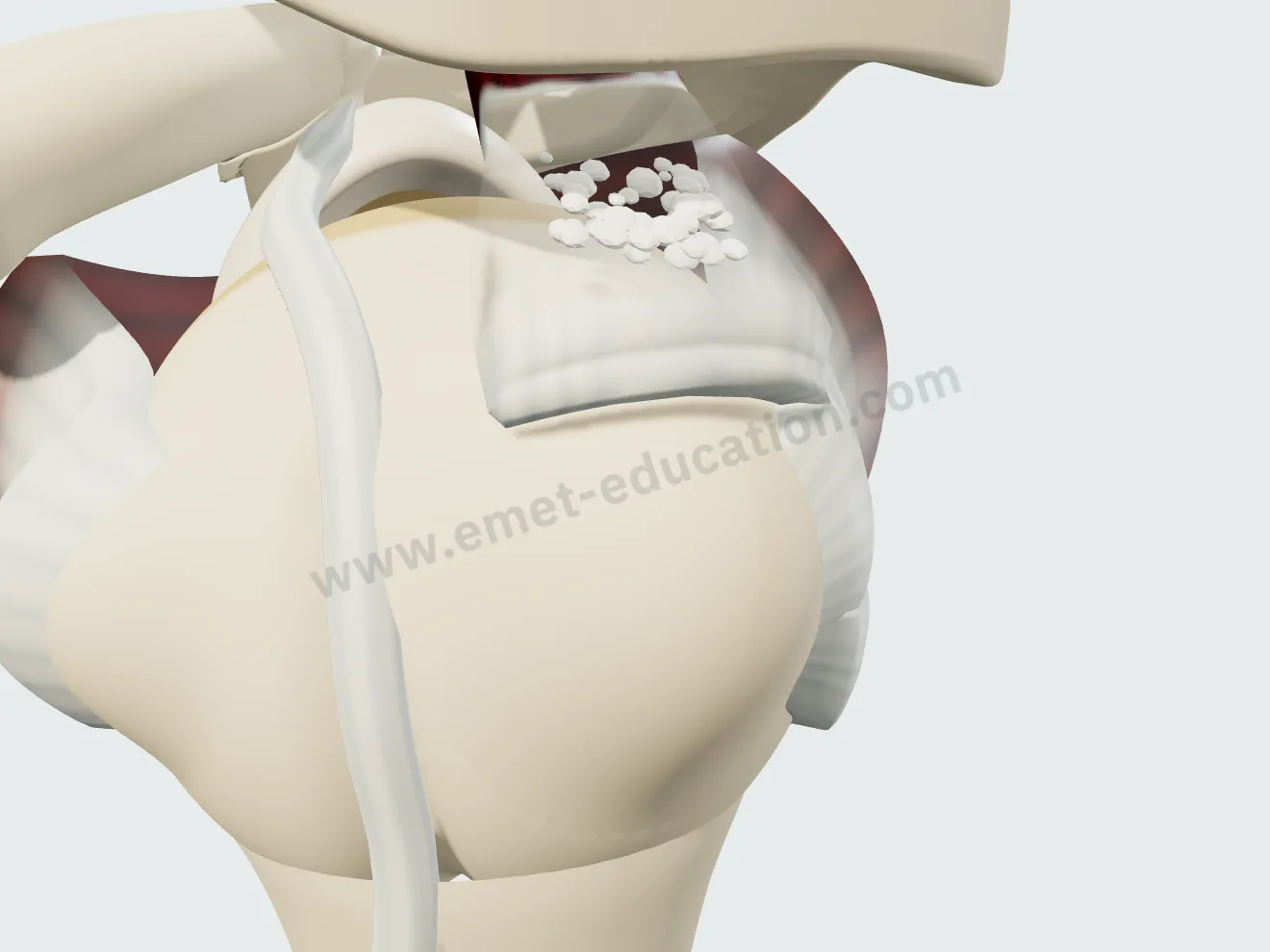
The whole cuff, tendon by tendon
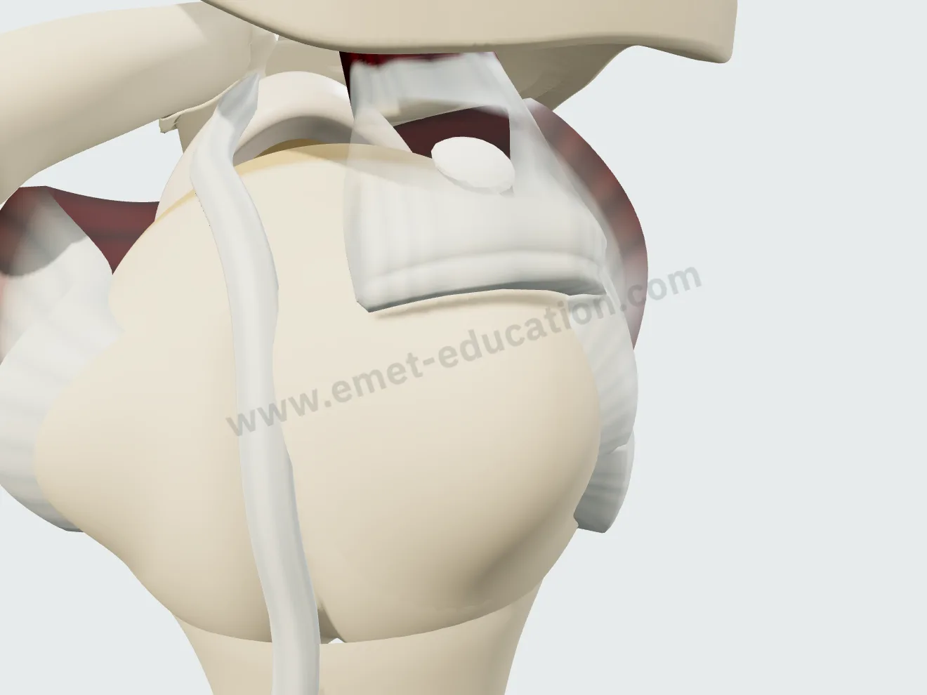
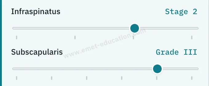
The infraspinatus has three stages of retraction, like the supraspinatus. The subscapularis follows the Lafosse classification: fraying (Ia), deep lesion of the upper third (Ib), tear of one third (II), of two thirds (III), complete retracted tear (IV).
The long head of the biceps
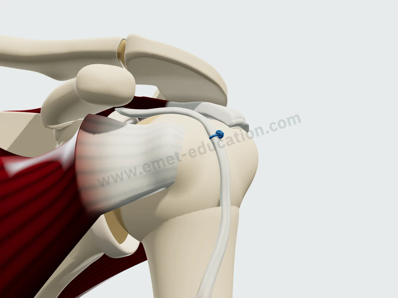
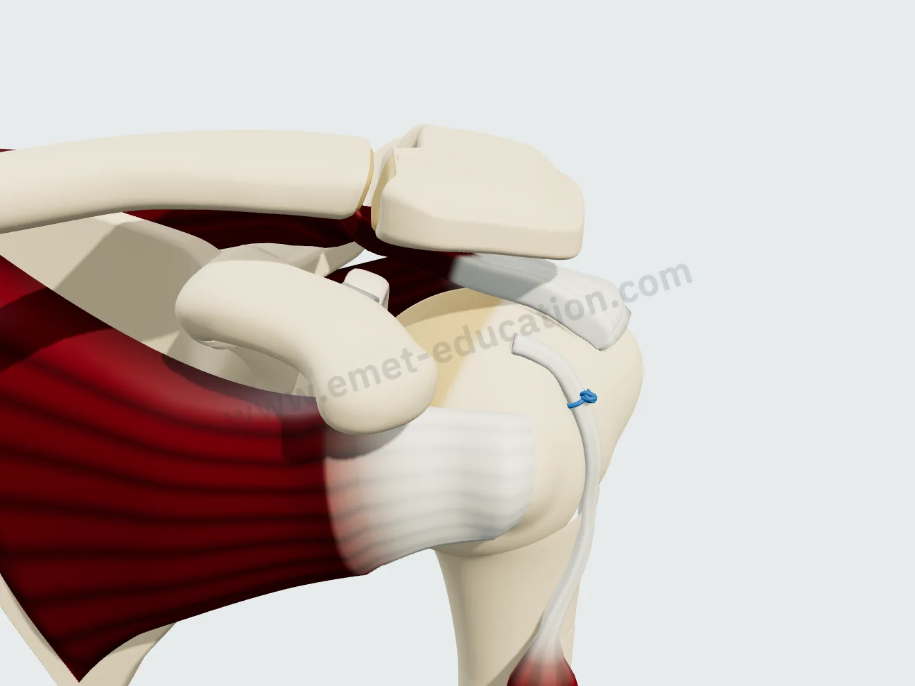
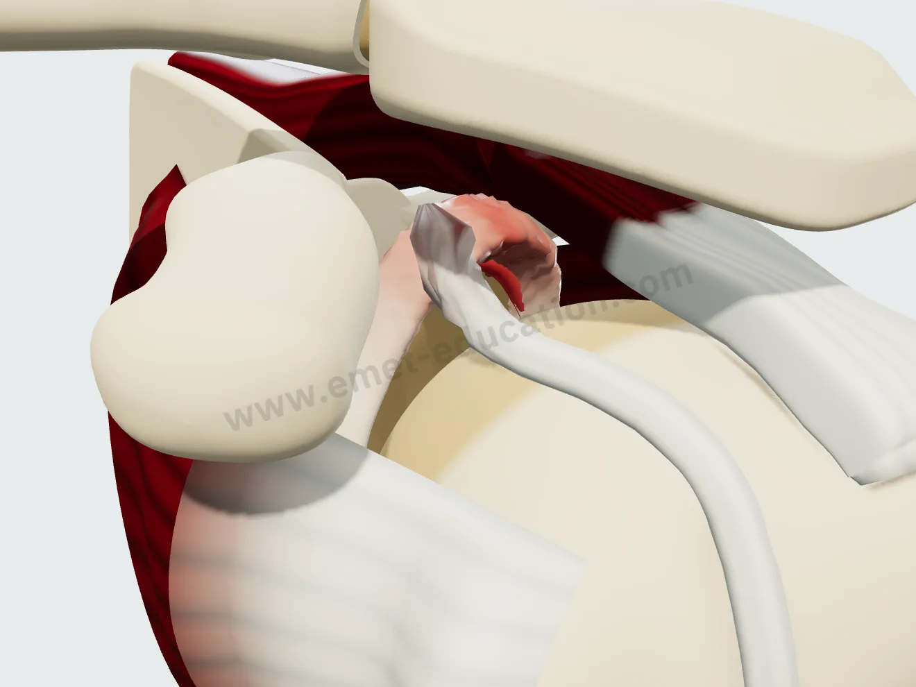
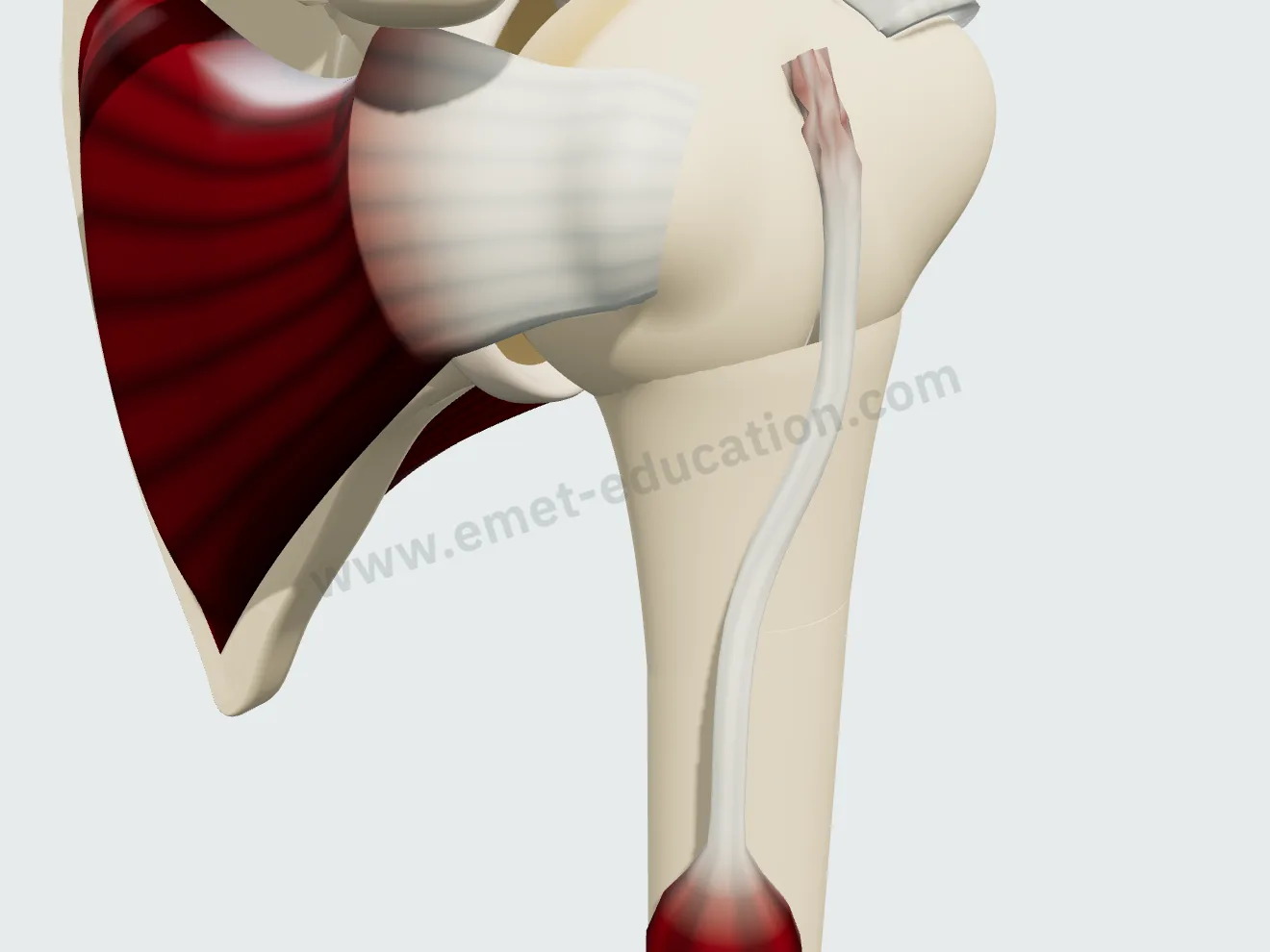
Tenosynovitis of the intra-articular portion, subluxation over the medial edge of the groove, SLAP lesion with detachment of the superior labrum, rupture one centimeter from the insertion with the tendon and muscle belly dropped.

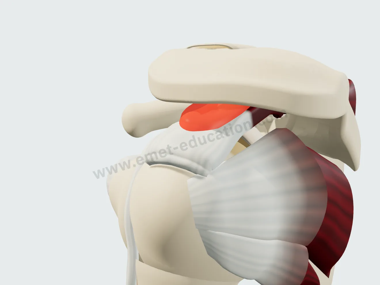
The subacromial space
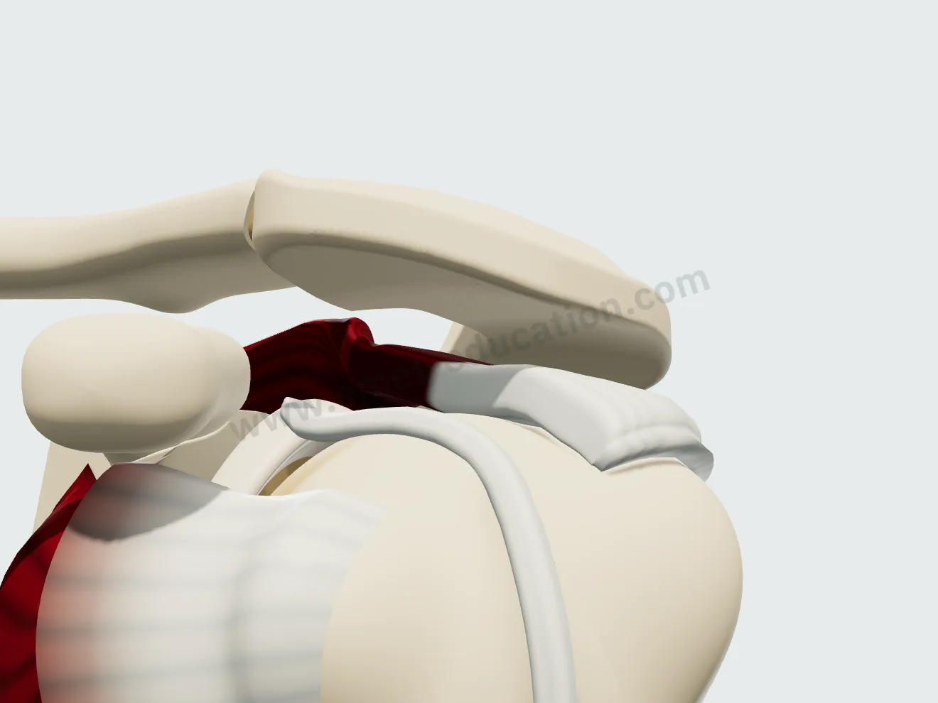
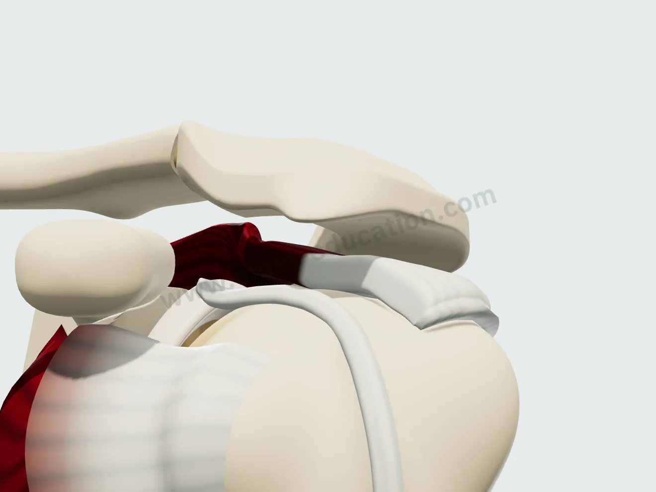
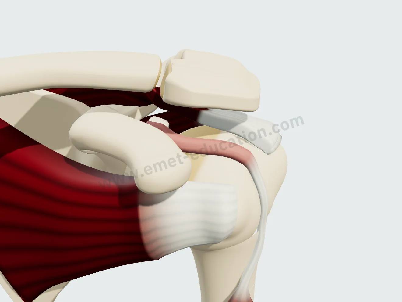
Subacromial bursitis, hooked acromion (Bigliani type III) with its osteophytic spur, increased critical shoulder angle (39° in the model, versus 32° normally). They add up, and impingement is shown with the arm moving.