- EMET Education
- 3D classifications
- Hamada classification
Classification · Massive rotator cuff tear
Hamada Classification: 3D Visualization
The radiographic stages of massive rotator cuff tears, from superior migration of the humeral head to cuff tear arthropathy, shown on a 3D model of the shoulder. With the original article, Walch's subdivision, reliability data and free infographics for teaching and publication.
Designed by an orthopedic surgeon
The 3D model opens in your browser, at the grade shown, with no sign-up. Infographics are free to reuse under CC BY 4.0.
- 1990Published by Hamada et al. in Clinical Orthopaedics and Related Research
- 1–5Five grades, from a preserved acromiohumeral interval to humeral head collapse
- 922Citations of the original article (OpenAlex, October 5, 2026)
- 4A · 4BWalch et al.'s subdivision of grade 4 (2005), by the presence of acetabularization
The six grades
From grade 1 to grade 5, in 3D
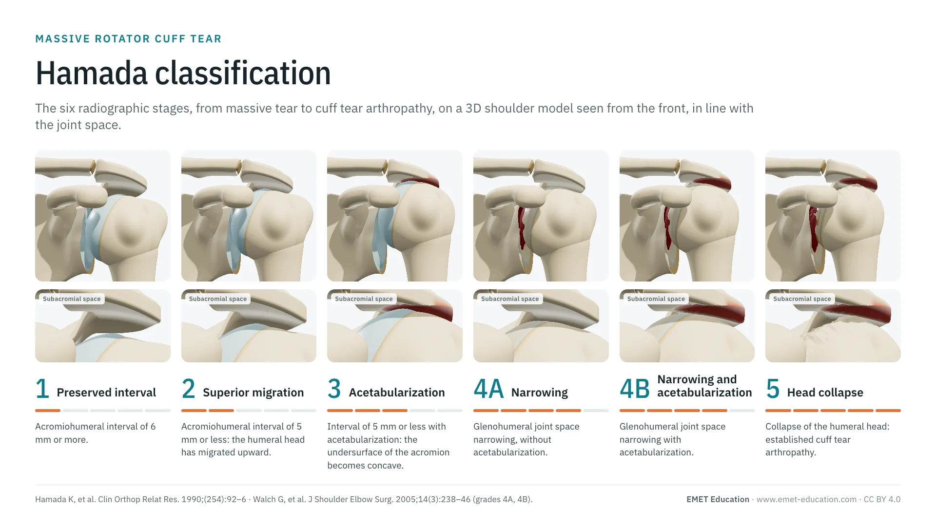
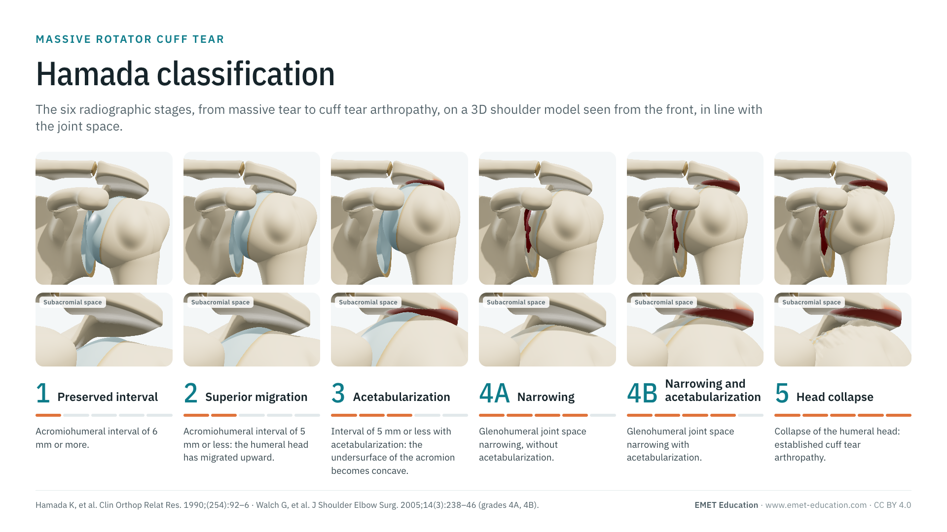
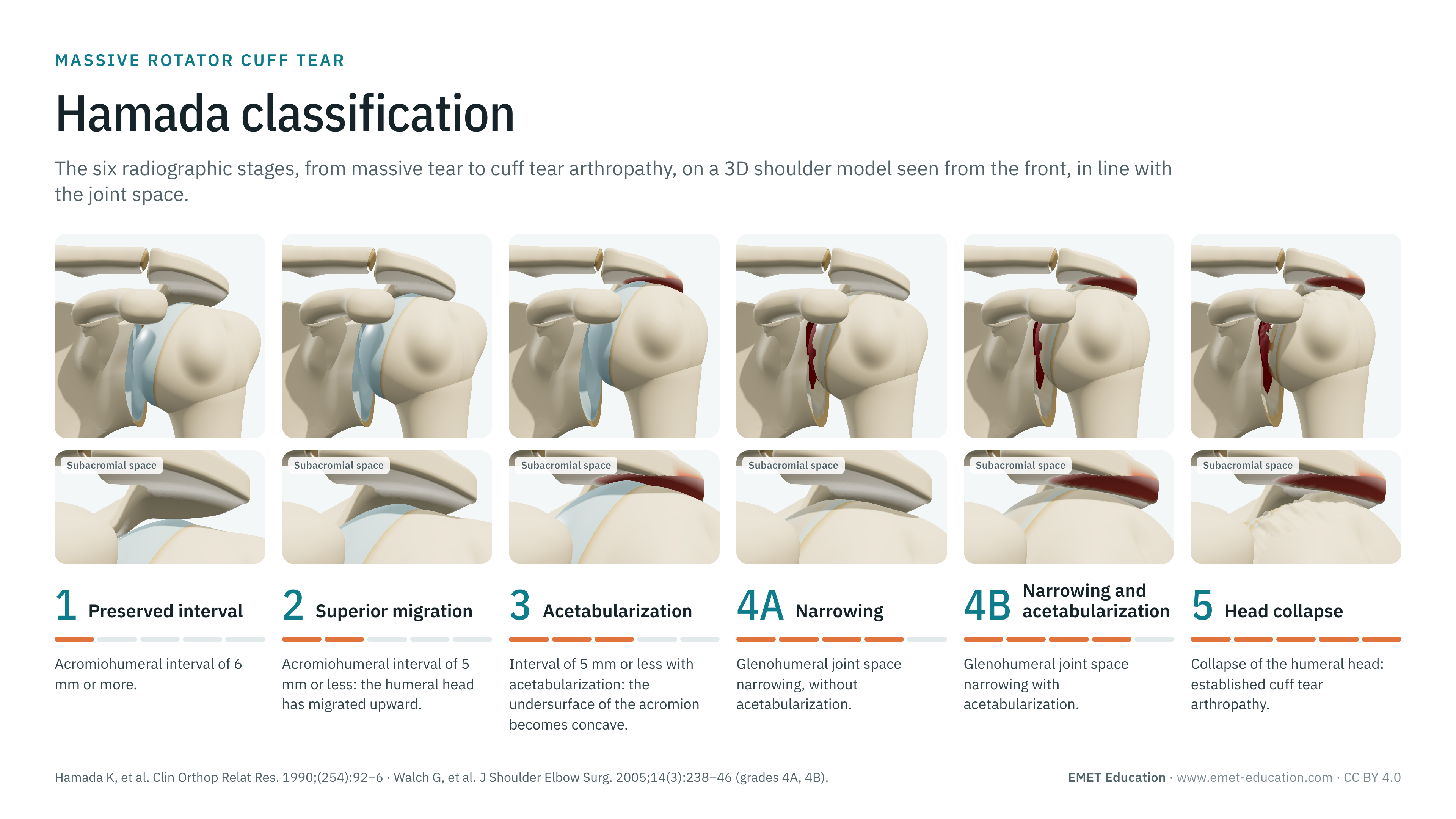
Each image is a rendering of the EMET Education shoulder osteoarthritis model in its eccentric form, at that grade: shoulder seen from the front, in line with the glenohumeral joint space, as on an anteroposterior radiograph; the coracoid process projects over the upper glenoid, as it does on the radiograph. The model sets the acromiohumeral interval measured vertically above the head (7, 4, 3, 3, 2, then 1.5 mm), hollows the acromion, rounds off the greater tuberosity, wears the upper glenoid, then collapses the humeral head.
-
1Preserved interval
- Radiographic criteria
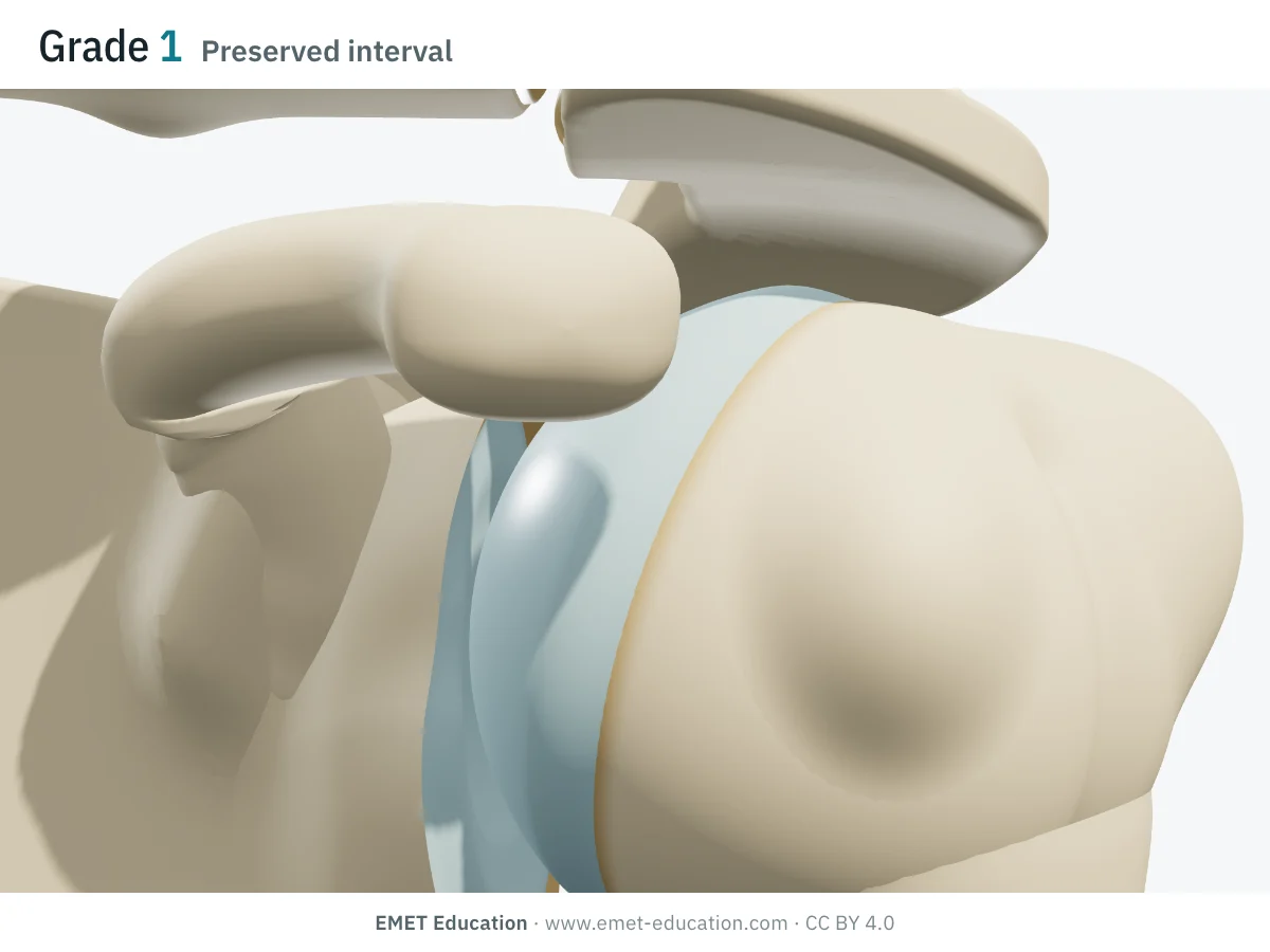
- Acromiohumeral interval of 6 mm or more.
- In the 3D model
- Acromiohumeral interval of 7 mm, normal acromion and greater tuberosity, normal glenohumeral joint space, round head.
-
2Superior migration
- Radiographic criteria
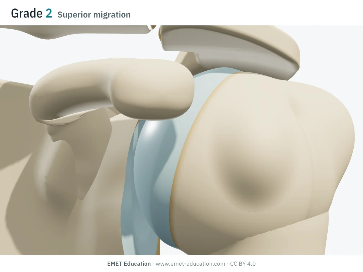
- Acromiohumeral interval of 5 mm or less: the humeral head has migrated upward.
- In the 3D model
- The head rises: acromiohumeral interval of 4 mm. Acromion, greater tuberosity and glenohumeral joint space still normal.
-
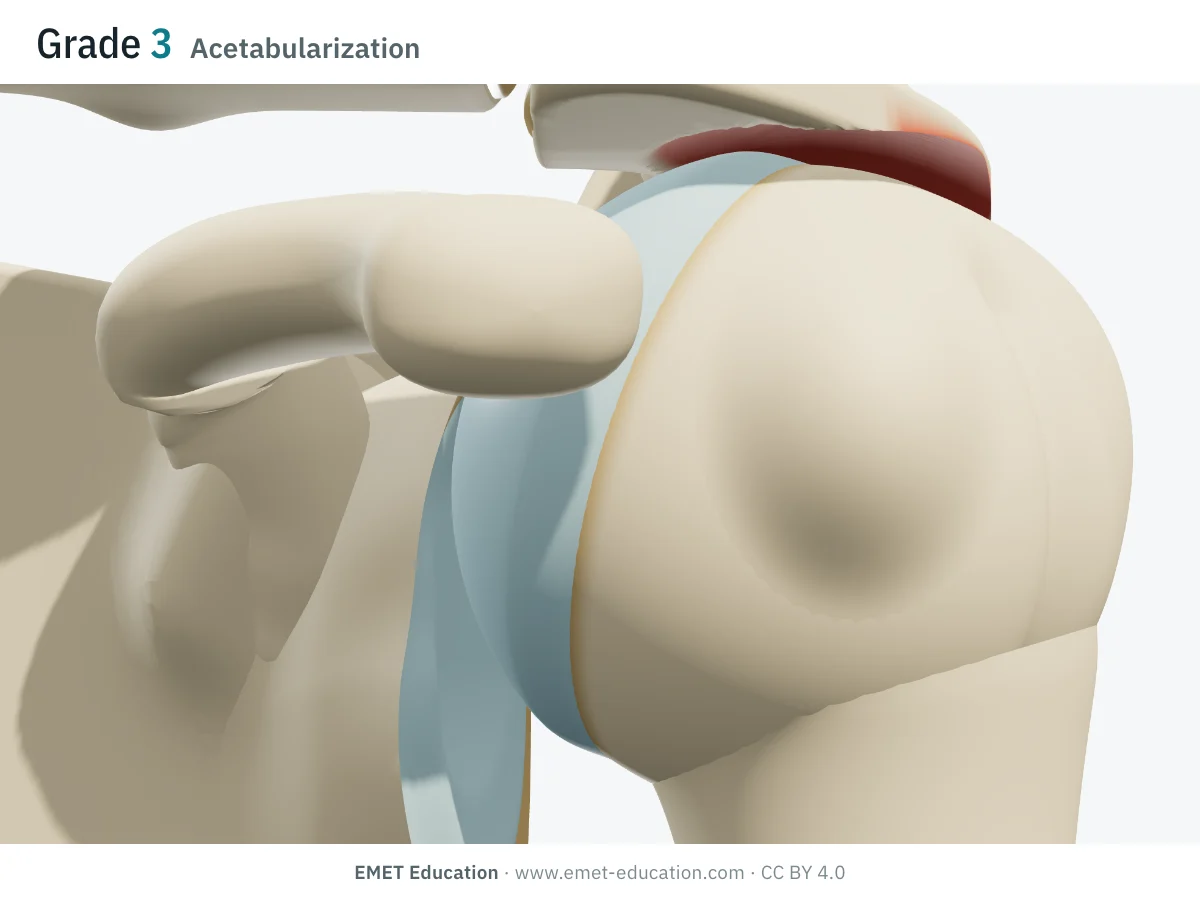
3Acetabularization
- Radiographic criteria
- Acromiohumeral interval of 5 mm or less with acetabularization: the undersurface of the acromion becomes concave where the head bears against it. Glenohumeral joint space preserved.
- In the 3D model
- Interval of 3 mm; a cup hollowed under the lateral border of the acromion, shaped by the head; greater tuberosity rounded off ("femoralization"); glenoid and cartilage intact.
-
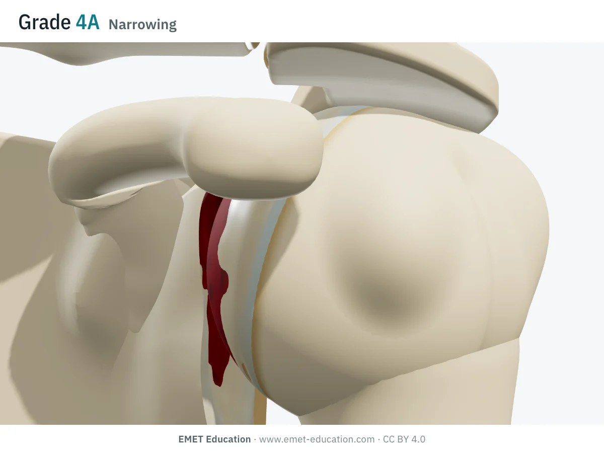
4ANarrowing
- Radiographic criteria
- Glenohumeral joint space narrowing, without acetabularization of the acromion.
- In the 3D model
- Interval of 3 mm; normal acromion; worn cartilage, upper half of the glenoid eroded (up to 4 mm at the top), bone exposed; the head settles into it.
-
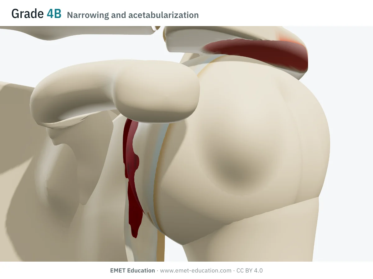
4BNarrowing and acetabularization
- Radiographic criteria
- Glenohumeral joint space narrowing with acetabularization of the acromion.
- In the 3D model
- Interval of 2 mm; hollowed acromion, rounded greater tuberosity; worn upper glenoid, bone exposed: the head articulates with both the glenoid and the acromion.
-
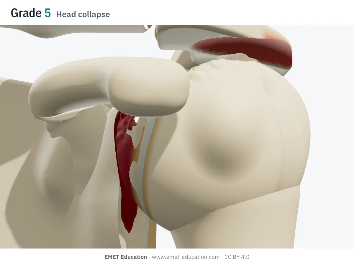
5Head collapse
- Radiographic criteria
- Collapse of the humeral head: established cuff tear arthropathy.
- In the 3D model
- Interval of 1.5 mm; top of the head collapsed by about 5 mm and irregular; hollowed acromion, rounded greater tuberosity, upper glenoid severely worn.
Schematic rendering, for teaching: the model illustrates each grade on a single shoulder, it does not reproduce a radiograph. Criteria reworded from the descriptions by Hamada (1990) and Walch (2005); their figures are not reproduced.
The original article
Hamada et al., 1990
K. Hamada, H. Fukuda, M. Mikasa and Y. Kobayashi (Tokai University, Japan) published Roentgenographic findings in massive rotator cuff tears. A long-term observation in Clinical Orthopaedics and Related Research in 1990 (254:92–6, doi:10.1097/00003086-199005000-00014). Before MRI, they had identified by an arthrographic technique 22 massive tears in patients treated without surgery, and analyzed their plain radiographs: narrowing of the acromiohumeral interval and changes of the humeral head, the tuberosities, the acromion, the acromioclavicular joint and the glenohumeral joint. They derived five grades from these findings. In seven patients followed for more than eight years, the grade had progressed in five, and one shoulder had progressed to cuff tear arthropathy.
In their view, a massive tear progresses to arthropathy step by step, each step with its radiographic signature. They proposed four mechanisms:
- arm elevation in activities of daily living;
- rupture of the long head of the biceps, which had still been depressing the head;
- the abnormal fulcrum of the head against the acromion and the coracoacromial ligament;
- weakness of external rotation.
In 2005, Walch et al. observed, in 307 biceps tenotomies, glenohumeral narrowing without acetabularization of the acromion: they split grade 4 into 4A (without acetabularization) and 4B (with) (doi:10.1016/j.jse.2004.07.008). Hamada adopted this subdivision in 2011 (doi:10.1007/s11999-011-1896-9). As of October 5, 2026, OpenAlex counts 922 citations of the original article, 678 of Walch's and 305 of the 2011 article. The term cuff tear arthropathy comes from Neer, Craig and Fukuda (1983).
Reliability
How reproducible is the grade?
Moderately. A reader agrees with themselves better than two readers agree with each other, and radiograph quality weighs heavily: agreement is similar to that of other cuff tear arthropathy classifications, but remains low overall.
| Study | Measure | Value |
|---|---|---|
| Kappe et al., 2011 | Intraobserver kappa, two readers, 52 shoulders | 1.00 and 0.49 |
| Kappe et al., 2011 | Interobserver kappa | 0.41 |
| Schumaier et al., 2019 | Intraobserver ICC, five shoulder surgeons, 108 simulated cases | 0.65 |
| Schumaier et al., 2019 | Interobserver ICC | 0.58 |
| Iannotti et al., 2010 | Interobserver agreement on good-quality radiographs (as reported by Brolin et al.) | 0.43 |
In the study by Kappe et al., which compared six classifications on the same radiographs, interobserver agreement for the Hamada grade (0.41) was close to that of Samilson-Prieto (0.51) and Kellgren-Lawrence (0.43); the Sirveaux classification, which grades only the glenoid, was the most reproducible (0.60). In Iannotti et al., agreement rose from 0.18 on poor radiographs to 0.43 on good ones: the grade is only as good as the view.
Variants and related classifications
What Hamada grades, and what it does not
The Hamada grade follows the superior migration of the head and its consequences for the acromion and the joint space. It does not describe glenoid wear in the coronal plane, loss of anterosuperior containment of the head, or the quality of the cuff muscles, which MRI or CT assess: other classifications cover these, and are often combined with it.
When reporting a grade 4, say whether it is 4A or 4B. Related scales:
| Classification | What it grades | Typical use |
|---|---|---|
| Hamada as modified by Walch (2005) | The Hamada grades, grade 4 split into 4A (narrowing without acetabularization) and 4B (with). | The version on this page; adopted by Hamada in 2011 and in reverse arthroplasty series. |
| Seebauer (2004) | Centering and stability of the head: types 1A and 1B (centered head), 2A and 2B (head decentered upward, up to anterosuperior escape). | Choosing the prosthetic treatment in established arthropathy. |
| Sirveaux (2004) | Glenoid wear in the coronal plane, from E0 (no erosion) to E3 (erosion extending to the lower glenoid). | Reverse arthroplasty: planning the glenoid component; the most reproducible in Kappe et al. |
| Samilson-Prieto (1983) | Size of the inferior osteophyte: under 3 mm, 3 to 7 mm, over 7 mm. | Centered osteoarthritis with an intact cuff: the other form in the 3D model (the 3D page). |
| Cuff tear arthropathy (Neer, 1983) | The description of the end stage: massive tear, superior migration and collapse of the head, wear of the acromion and glenoid. | The disease framework behind grade 5. |
Use
In research and in clinical practice
In research
The grade describes the stage of massive tears in natural history studies and surgical series. After biceps tenotomy, Walch et al. measured a mean loss of 1.3 mm of acromiohumeral interval over 57 months, and the proportion of arthritic shoulders rose from 38% to 67%. Hamada et al. (2011, 75 patients) found more fatty degeneration of the subscapularis at grades 3 to 5, more retears of the repaired supraspinatus at grade 2 than at grade 1, and no progression to grades 3 to 5 in operated patients: they concluded that repair should come before the acromiohumeral interval narrows. In a series of 211 reverse shoulder arthroplasties, outcomes beyond 30 months did not differ between grades 1 to 3 and grades 4A to 5 (Kääb et al., 2022).
In clinical practice
The grade places the shoulder along the path to arthropathy. Grades 1 and 2 point rather toward joint-preserving surgery (repair, partial repair, palliative procedures), arthritis at grades 4 and 5 rather toward arthroplasty, most often reverse. But the grade alone does not decide: age, function (pseudoparalysis), retraction and tendon and muscle quality on MRI weigh as much. In a survey of 108 simulated cases, the Hamada grade was associated with the treatment surgeons chose, but their agreement on treatment remained fair (0.44).
The acromiohumeral interval, on which grades 1 and 2 rest, reflects the extent of the tear. In Nové-Josserand et al. (264 operated shoulders), it averaged 9.5 mm when only the supraspinatus was torn, 7.5 mm with the infraspinatus, 5.4 mm with an associated anterior lesion, and 2.2 mm when the infraspinatus muscle had degenerated: narrowing below 6 to 7 mm suggests a tear extending into the infraspinatus.
Grading pitfalls
Five pitfalls when grading a shoulder
- Grade on a true anteroposterior view. The acromiohumeral interval and glenohumeral narrowing depend on the projection and arm position. The anteroposterior view in neutral rotation, with the beam in line with the joint space, is the reference; the interval is measured at its shortest, between the inferior cortex of the acromion and the head.
- Measure, do not estimate. One millimeter separates grade 1 (6 mm or more) from grade 2 (5 mm or less): measure on a calibrated image.
- Do not assume a sequence. A grade 4A shoulder has not necessarily been grade 3: the joint space can narrow without acetabularization, and progression is not always linear. Grade what you see, and say 4A or 4B.
- A collapsed head is not always grade 5. Grade 5 implies a massive tear, with an upward-migrated head; a collapsed but centered head, under a normal acromiohumeral interval, points to another diagnosis, such as osteonecrosis.
- Complete the grade. The Hamada grade says nothing about glenoid wear (Sirveaux), anterosuperior escape (Seebauer) or muscle quality; before a surgical decision, it is not enough.
Self-test
Which grade?
Six views of the same shoulder from the front and side, slightly from below, in random order. Find the grade of each one; the answer comes with the criteria.
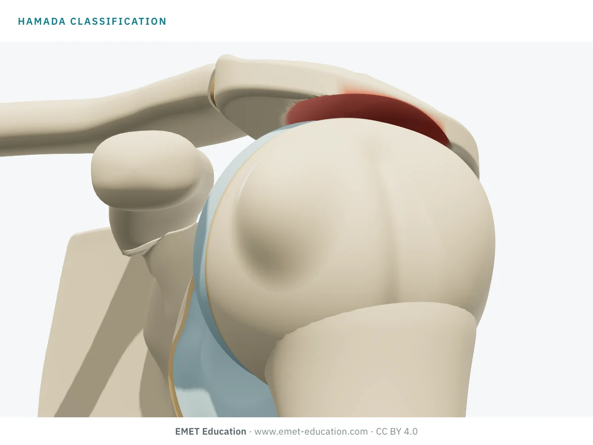
Which Hamada grade is this?
Download and cite
Free infographics, for teaching and publication
Use them in a course, a presentation, a social media post or a scientific article, under the Creative Commons Attribution 4.0 license: free reuse, including commercial, provided EMET Education is credited with a link to this page.
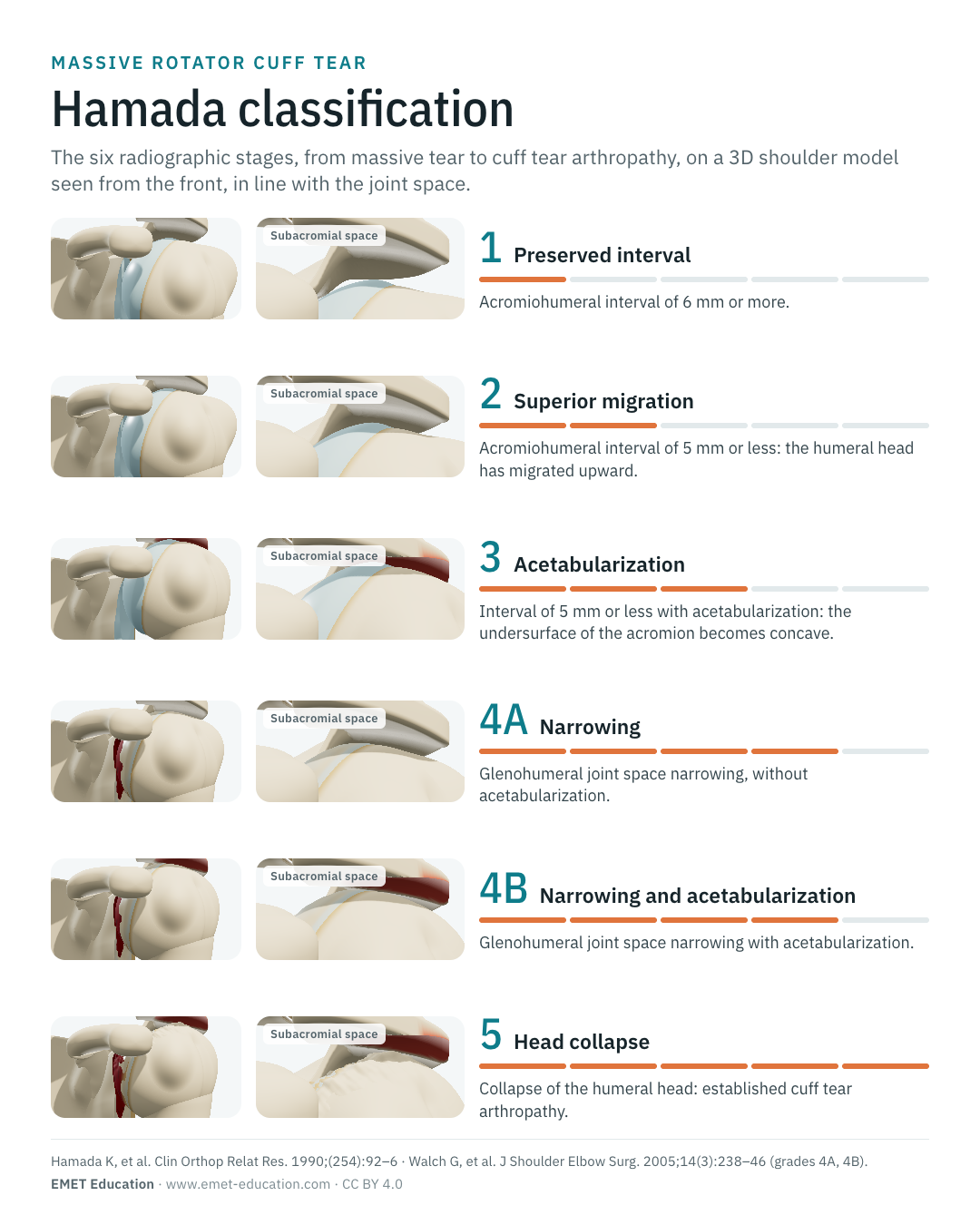
Cite this figure
Ready to paste. For a journal figure, add the original references (Hamada et al., 1990; Walch et al., 2005).
EMET Education. Hamada classification of massive rotator cuff tears: 3D visualization [Internet]. 2026 [cited]. Available from: https://www.emet-education.com/en/classifications/hamada/. CC BY 4.0.EMET Education. (2026). Hamada classification of massive rotator cuff tears: 3D visualization [Infographic]. https://www.emet-education.com/en/classifications/hamada/ (CC BY 4.0)<a href="https://www.emet-education.com/en/classifications/hamada/"><img src="https://www.emet-education.com/en/classifications/hamada/img/infographic.webp" alt="Hamada classification in 3D" width="960"></a><br>Figure: <a href="https://www.emet-education.com/en/classifications/hamada/">EMET Education</a>, CC BY 4.0Questions
Frequently asked questions
What is the difference between Hamada grades 4A and 4B?
Both combine narrowing of the glenohumeral joint space with an upward-migrated head. At grade 4B, the undersurface of the acromion is also hollowed into a cup (acetabularization); at grade 4A, it is not. This subdivision comes from Walch et al. (2005), who had observed glenohumeral arthritis without acetabularization, which the original grade 4 did not describe.
At what acromiohumeral interval is the head considered to have migrated upward?
The normal acromiohumeral interval measures 6 to 14 mm on an anteroposterior view. Hamada grade 1 corresponds to an interval of 6 mm or more, grade 2 to an interval of 5 mm or less. Narrowing below 6 to 7 mm suggests a tear extending into the infraspinatus.
What is acetabularization of the acromion?
Under repeated contact with the upward-migrated head, the undersurface of the acromion becomes hollowed and sclerotic, until it forms a cup that fits the head, like an acetabulum. It often comes with rounding of the greater tuberosity, called "femoralization". It is what distinguishes grade 3 from grade 2, and grade 4B from grade 4A.
How does it differ from the Seebauer classification?
Hamada describes the successive stages of a massive tear, from simple superior migration of the head to arthropathy. Seebauer classifies established arthropathy by the centering and stability of the head, up to anterosuperior escape, to guide the choice of prosthesis. Both have similar reliability; Hamada is often said to be more useful at early stages, Seebauer at late stages.
Does the Hamada grade decide the treatment?
No, it guides it. Grades 1 and 2 leave room for joint-preserving surgery, grades 4 and 5 point rather toward arthroplasty, often reverse. The decision also takes into account age, function, tendon and muscle quality on MRI, and the patient's wishes.
Can I reuse these images in a course or an article?
Yes. The images and infographics on this page are under the Creative Commons Attribution 4.0 license: free reuse, including commercial, with credit to EMET Education and a link to this page. The "Cite this figure" box gives ready-to-paste citations.
How were the 3D images made?
They are renderings of the EMET Education shoulder osteoarthritis model, in its eccentric form, set to each grade: the height of the head is computed to give the intended acromiohumeral interval, then the model hollows the acromion, rounds off the greater tuberosity, wears the upper glenoid and collapses the head according to the grade. It is a schematic model for teaching and patient explanation; it does not reproduce a radiograph.
Sources
Author and references
- Hamada K, Fukuda H, Mikasa M, Kobayashi Y. Roentgenographic findings in massive rotator cuff tears. A long-term observation. Clin Orthop Relat Res. 1990;(254):92–6. doi:10.1097/00003086-199005000-00014
- Walch G, Edwards TB, Boulahia A, Nové-Josserand L, Neyton L, Szabo I. Arthroscopic tenotomy of the long head of the biceps in the treatment of rotator cuff tears: clinical and radiographic results of 307 cases. J Shoulder Elbow Surg. 2005;14(3):238–46. doi:10.1016/j.jse.2004.07.008
- Hamada K, Yamanaka K, Uchiyama Y, Mikasa T, Mikasa M. A radiographic classification of massive rotator cuff tear arthritis. Clin Orthop Relat Res. 2011;469(9):2452–60. doi:10.1007/s11999-011-1896-9
- Brolin TJ, Updegrove GF, Horneff JG. Classifications in brief: Hamada classification of massive rotator cuff tears. Clin Orthop Relat Res. 2017;475(11):2819–23. doi:10.1007/s11999-017-5340-7
- Kappe T, Cakir B, Reichel H, Elsharkawi M. Reliability of radiologic classification for cuff tear arthropathy. J Shoulder Elbow Surg. 2011;20(4):543–7. doi:10.1016/j.jse.2011.01.012
- Schumaier AP, Bedeir YH, Dines JS, et al. Quantifying the impact of patient-specific factors and disease severity on clinical decision making in cuff tear arthropathy: a case-based survey. HSS J. 2019;15(3):276–85. doi:10.1007/s11420-019-09695-x
- Iannotti JP, McCarron J, Raymond CJ, et al. Agreement study of radiographic classification of rotator cuff tear arthropathy. J Shoulder Elbow Surg. 2010;19(8):1243–9. doi:10.1016/j.jse.2010.02.010
- Neer CS 2nd, Craig EV, Fukuda H. Cuff-tear arthropathy. J Bone Joint Surg Am. 1983;65(9):1232–44. doi:10.2106/00004623-198365090-00003
- Weiner DS, Macnab I. Superior migration of the humeral head. A radiological aid in the diagnosis of tears of the rotator cuff. J Bone Joint Surg Br. 1970;52(3):524–7. doi:10.1302/0301-620X.52B3.524
- Nové-Josserand L, Lévigne C, Noël E, Walch G. L'espace sous-acromio-huméral. Étude des facteurs influençant sa hauteur. Rev Chir Orthop Reparatrice Appar Mot. 1996;82(5):379–85.
- Visotsky JL, Basamania C, Seebauer L, Rockwood CA, Jensen KL. Cuff tear arthropathy: pathogenesis, classification, and algorithm for treatment. J Bone Joint Surg Am. 2004;86-A Suppl 2:35–40.
- Sirveaux F, Favard L, Oudet D, et al. Grammont inverted total shoulder arthroplasty in the treatment of glenohumeral osteoarthritis with massive rupture of the cuff. Results of a multicentre study of 80 shoulders. J Bone Joint Surg Br. 2004;86(3):388–95. doi:10.1302/0301-620X.86B3.14024
- Samilson RL, Prieto V. Dislocation arthropathy of the shoulder. J Bone Joint Surg Am. 1983;65(4):456–60.
- Kääb MJ, Kohut G, Irlenbusch U, Joudet T, Reuther F. Reverse total shoulder arthroplasty in massive rotator cuff tears: does the Hamada classification predict clinical outcomes? Arch Orthop Trauma Surg. 2022;142(7):1405–11. doi:10.1007/s00402-021-03755-w
3D models
Show the grade to your patient
The images on this page come from an interactive model. In clinic, set the grade from your patient's radiograph, then show the treatment discussed, up to reverse shoulder arthroplasty.
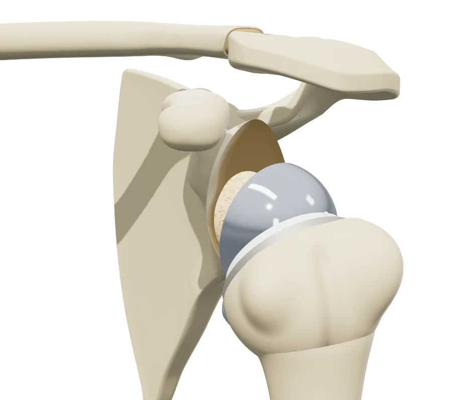
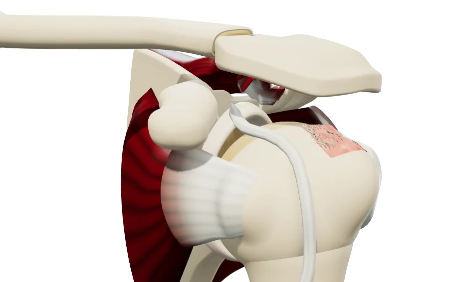
Explain the stage in 3D
The shoulder osteoarthritis model opens in your browser, at the grade you choose, with no sign-up.
Share this page with a colleague or a resident: