- EMET Education
- 3D classifications
- Samilson-Prieto classification
Classification · Shoulder osteoarthritis
Samilson-Prieto Classification: 3D Visualization
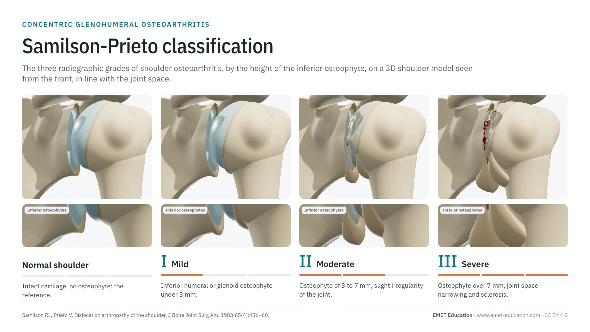
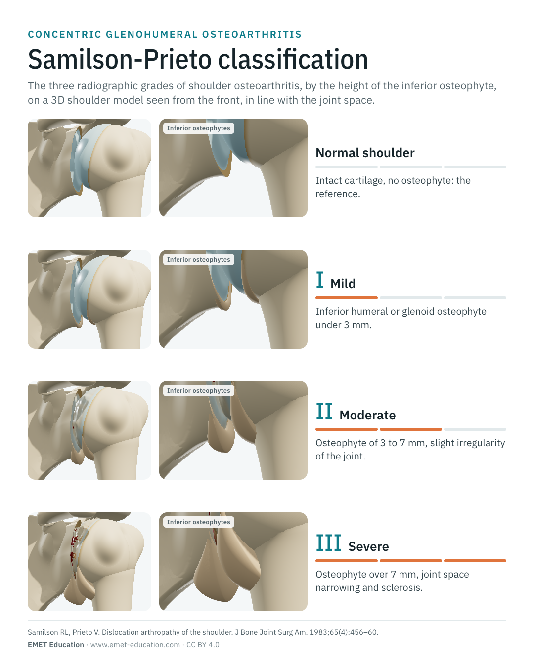
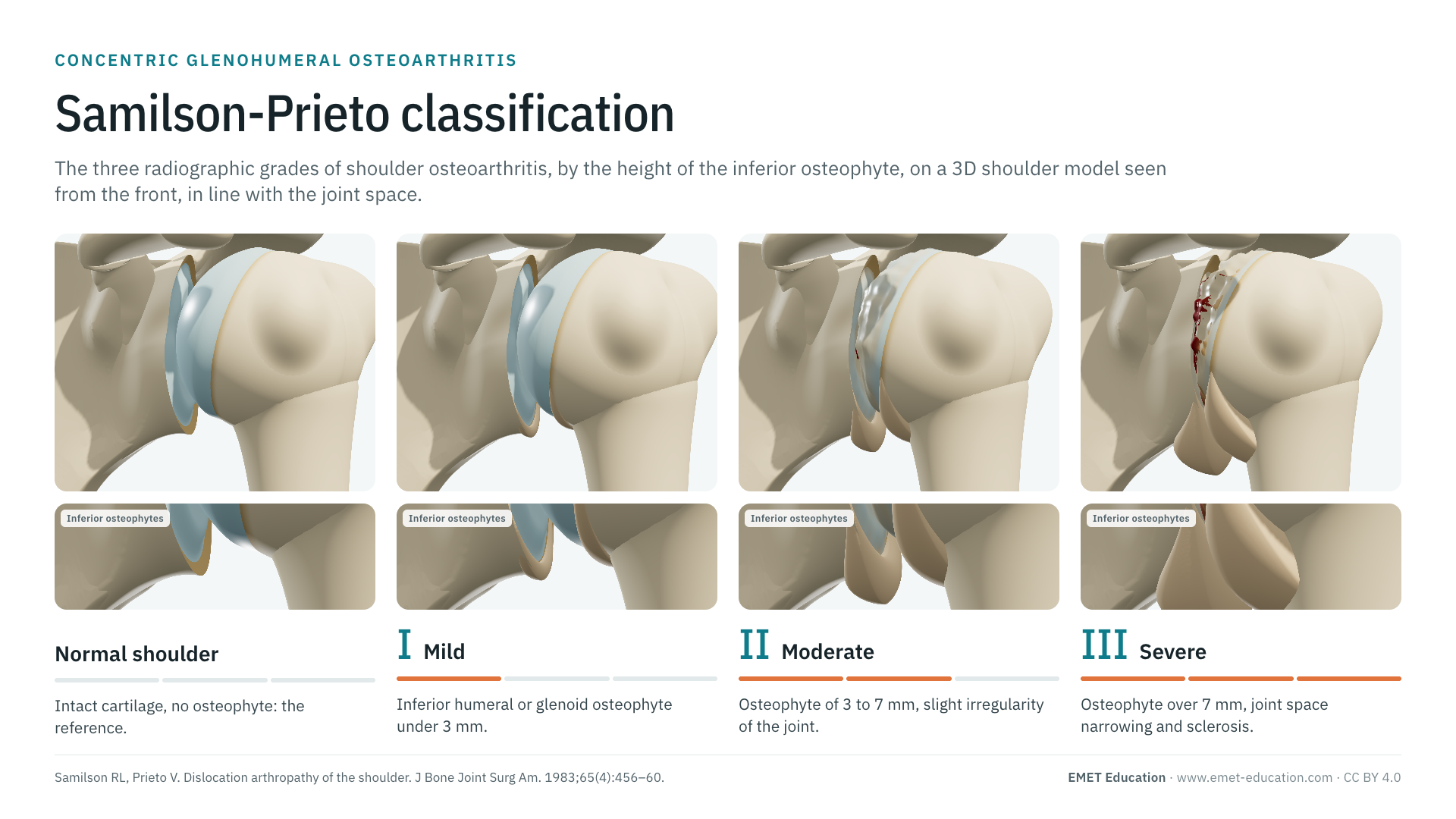
The three radiographic grades of glenohumeral osteoarthritis, by the height of the inferior osteophyte, on a 3D model of the shoulder seen as on an anteroposterior radiograph. With the original article, the variants, reliability data and free infographics for teaching and publication.
Designed by an orthopedic surgeon
The 3D model opens in your browser, at the grade shown, with no sign-up. Infographics are free to reuse under CC BY 4.0.
- 1983Published by Samilson and Prieto in the Journal of Bone and Joint Surgery
- I–IIIThree grades: mild, moderate, severe osteoarthritis
- 911Citations of the original article (OpenAlex, October 5, 2026)
- 3 · 7 mmThe two inferior osteophyte heights that separate the grades
The three grades
From grade I to grade III, in 3D
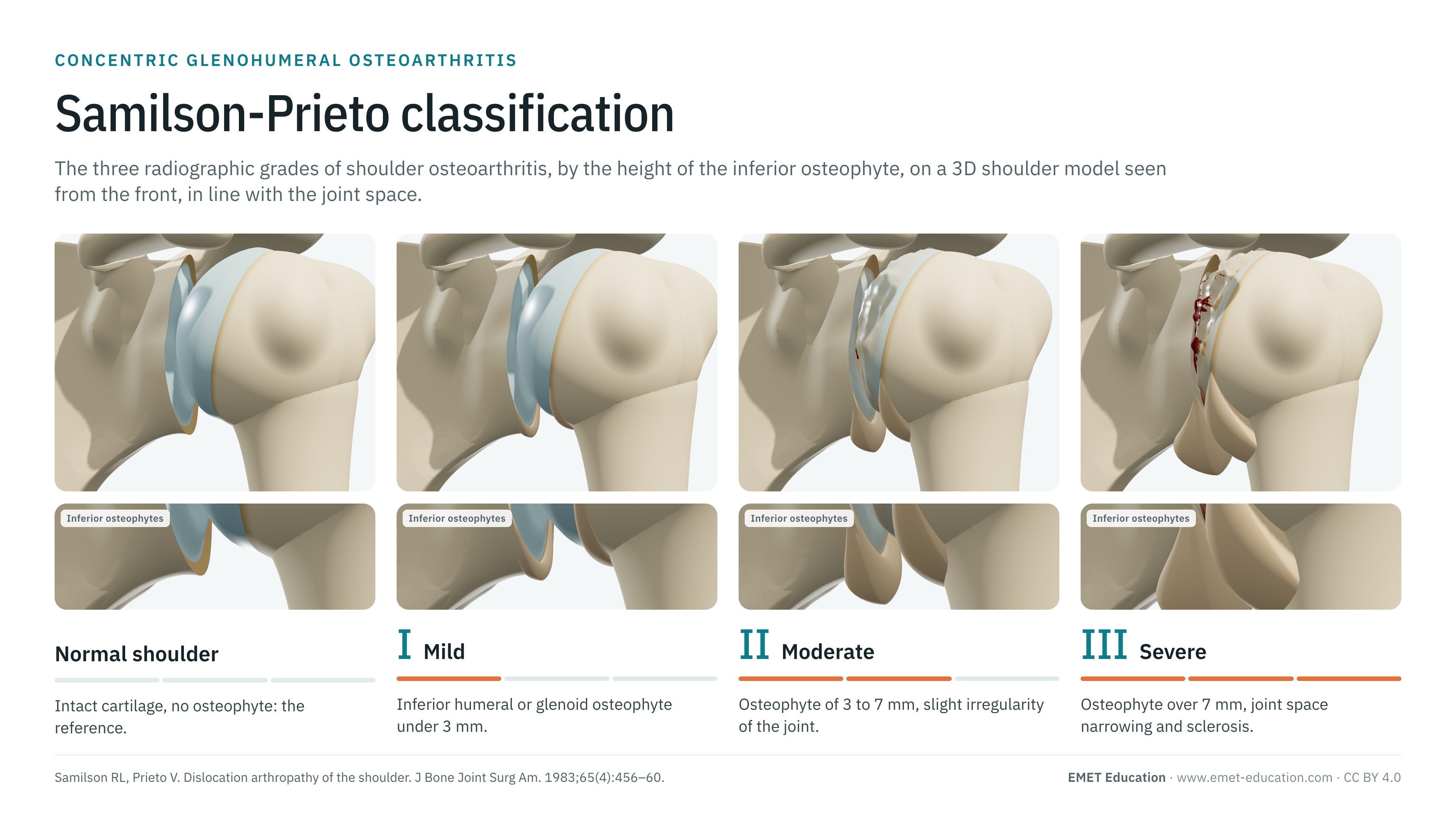
Each image is a rendering of the EMET Education shoulder osteoarthritis model in its concentric form, at that grade, seen as on the anteroposterior radiograph on which the osteophyte is measured: in line with the glenohumeral joint space, the coracoid process projecting over the upper glenoid. The normal shoulder serves as a reference. The model grows the humeral osteophyte under the head (the "goat's beard") and the glenoid osteophyte under the glenoid, wears the cartilage, narrows the joint space and deforms the head according to the grade.
-
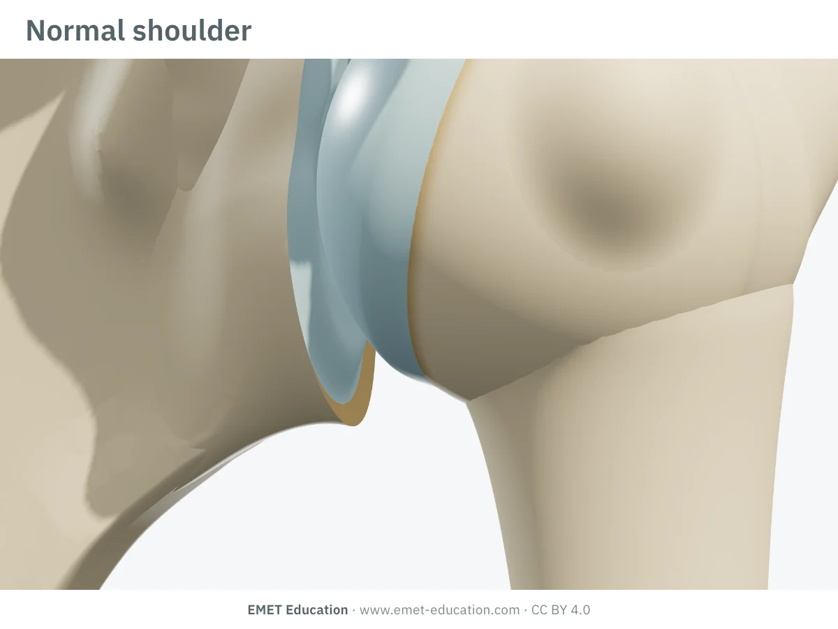
Normal shoulder (reference)
- In the 3D model
- Intact cartilage on the head and glenoid, no osteophyte, regular joint space. It is not a grade of the classification: it is there for comparison.
-
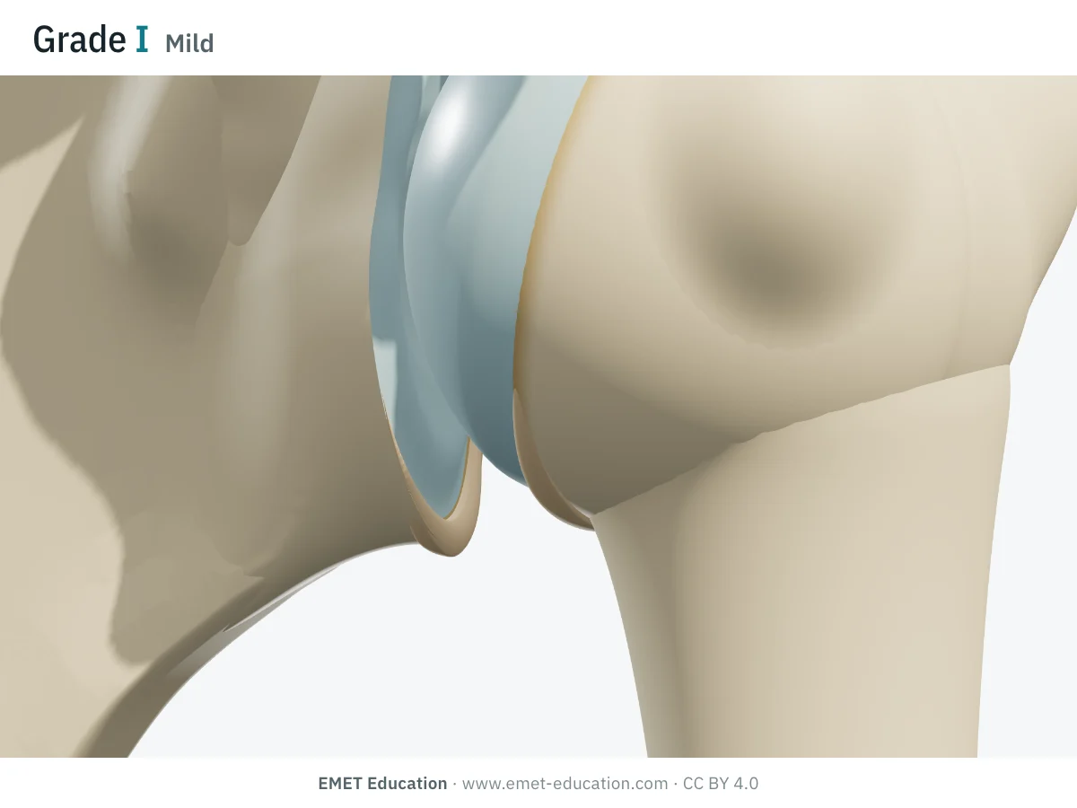
IMild
- Radiographic criteria
- Inferior humeral or glenoid osteophyte (or both) less than 3 mm high.
- In the 3D model
- Humeral osteophyte of 2.8 mm, glenoid osteophyte of 1.6 mm; early cartilage wear at the center of the contact zone.
-
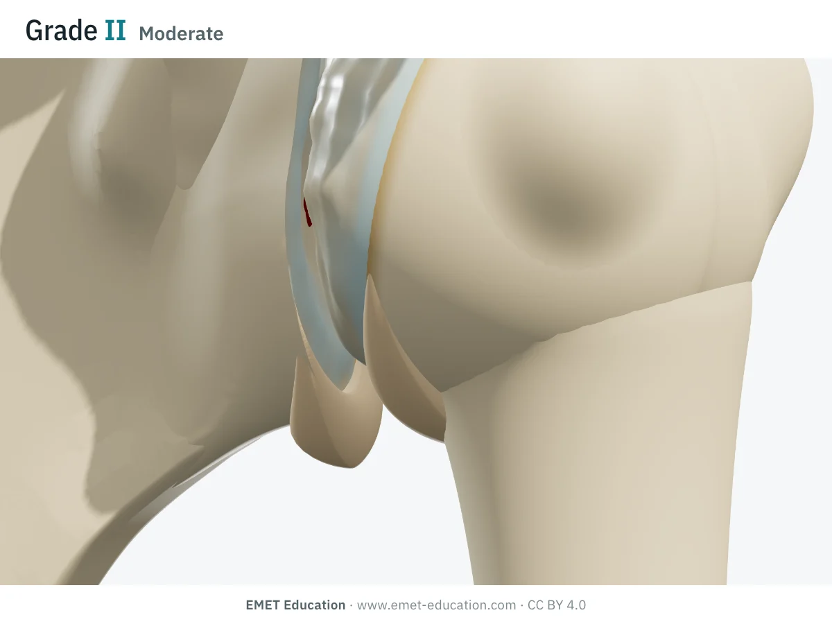
IIModerate
- Radiographic criteria
- Inferior osteophyte of 3 to 7 mm, slight irregularity of the glenohumeral joint.
- In the 3D model
- Humeral osteophyte of 7 mm, glenoid osteophyte of 4.5 mm; thinned, irregular cartilage with small areas of exposed bone; slight narrowing, head flattened in the contact zone (about 4 mm).
-
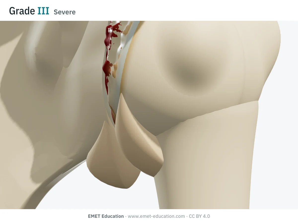
IIISevere
- Radiographic criteria
- Inferior osteophyte over 7 mm, glenohumeral joint space narrowing and sclerosis.
- In the 3D model
- Humeral osteophyte of 14 mm, glenoid osteophyte of 9 mm; cartilage almost gone centrally, bone exposed; narrowed joint space, markedly deformed head (flattened by about 6 mm and irregular).
Schematic rendering, for teaching: the model illustrates each grade on a single shoulder, it does not reproduce a radiograph. Osteophyte heights are taken at the top of each class so that they show clearly. Criteria reworded from the original description; the 1983 figures are not reproduced.
The original article
Samilson and Prieto, 1983
R. L. Samilson and V. Prieto published Dislocation arthropathy of the shoulder in the Journal of Bone and Joint Surgery in 1983 (65:456–60, doi:10.2106/00004623-198365040-00005). They described 74 shoulders in 70 patients with one or more dislocations and radiographic glenohumeral arthropathy, the opposite shoulder being normal: 62 anterior dislocations, 11 posterior and one multidirectional. They graded the arthropathy by the height of the inferior osteophyte of the humeral head or glenoid: mild, moderate or severe.
Their main observations:
- posterior dislocations were followed far more often by moderate or severe arthropathy, which they related to delayed diagnosis;
- neither the number of recurrences nor bony defects of the glenoid rim or humeral head were related to the severity of the arthropathy;
- operations in which fixation devices intruded on the joint cartilage were often followed by moderate to severe arthropathy;
- arthropathy also occurred without surgery: 10 of the 15 shoulders with severe arthropathy and 6 of the 14 with moderate arthropathy had never been operated on.
Designed for arthropathy after dislocation, the classification has become the standard grading of concentric shoulder osteoarthritis in general, primary osteoarthritis included. As of October 5, 2026, OpenAlex counts 911 citations of the original article.
Reliability
How reproducible is the grade?
Well, and better than the other radiographic classifications of the shoulder: a single measurement, the height of the osteophyte, with two simple cut-offs.
| Study | Measure | Value |
|---|---|---|
| Brox et al., 2003 | Interobserver kappa, 40 shoulders operated on for instability (Kellgren-Lawrence: 0.36) | 0.76 |
| Kappe et al., 2011 | Interobserver kappa, 52 shoulders with cuff tear arthropathy | 0.51 |
| Linke et al., 2022 | Weighted interobserver kappa, 352 primary osteoarthritic shoulders | 0.88 |
| Longo et al., 2024 | Weighted intraobserver kappa, two readers, 752 patients | 0.82 and 0.82 |
| Longo et al., 2024 | Weighted interobserver kappa (Kellgren-Lawrence: 0.70) | 0.87 |
In the two studies that compared them on the same radiographs, Samilson-Prieto did better than Kellgren-Lawrence (0.76 versus 0.36 in Brox et al.; 0.87 versus 0.70 in Longo et al.), and Brox et al. recommended it for its simplicity. Agreement drops when the joint is heavily remodeled: in cuff tear arthropathy, it was only 0.51 in Kappe et al.
Variants and related classifications
Three variants, and what the osteophyte does not tell
Several groups have adjusted the cut-offs or added a grade, mostly in series of operated instability; their reliability is comparable to the original's (Longo et al., 2024). Above all, osteophyte height describes neither glenoid shape nor posterior subluxation of the head, which CT or MRI show in the axial plane: in Linke et al., the Samilson-Prieto grade did not correlate with the Walch glenoid type.
When reporting a grade, say which version you used:
| Classification | What it grades | Typical use |
|---|---|---|
| Allain (1998) | Four grades: humeral osteophyte of 1 to 3 mm, 4 to 7 mm, over 7 mm, then narrowing and sclerosis. | Long-term follow-up after the Latarjet procedure; also separates concentric from eccentric osteoarthritis. |
| Gerber (1988) | Osteophyte under 3 mm; 3 to 5 mm with irregularity and sclerosis; beyond that. | Series of operated instability (Trillat procedure). |
| Buscayret (2004) | The three original grades and a grade 4: bone-on-bone contact, with or without osteophytes. | Osteoarthritis before and after instability surgery (570 patients). |
| Walch (1999) | Glenoid shape and head centering in the axial plane (types A, B, C, later D). | Arthroplasty planning; requires CT or MRI. |
| Hamada (1990) | Eccentric osteoarthritis with a massive cuff tear: superior migration of the head, acetabularization, collapse. | Cuff tear arthropathy: the other form in the 3D model. |
Use
In research and in clinical practice
In research
The classification remains the reference measure of osteoarthritis after instability. In 570 patients operated on for anterior instability, Buscayret et al. found 9.2% osteoarthritis before surgery and 19.7% new osteoarthritis after, at 6.5 years' follow-up; risk rose with age at first dislocation and at surgery, delay before surgery, bony glenoid rim lesions and the number of dislocations. After the Latarjet procedure followed for 14 years on average, Allain et al. found concentric osteoarthritis in 34 of 58 shoulders, mostly grade 1, and noted that grade 1, unlike the higher grades, did not affect function.
In clinical practice
The grade summarizes the severity of osteoarthritis on an anteroposterior view, simply and reproducibly. It measures neither pain nor disability: in Brox et al., no patient without osteoarthritis had pain, but only one in five patients with osteoarthritis did. Treatment decisions rest on the clinical picture.
Before arthroplasty, the grade is not enough: posterior glenoid wear and posterior subluxation of the head, which weigh on the choice and position of the implant, are only seen in the axial plane. Linke et al. concluded that CT or MRI remained essential to gauge the complexity of the osteoarthritis.
Grading pitfalls
Five pitfalls when grading a shoulder
- Grade on a true anteroposterior view. In line with the joint space, the inferior osteophyte stands clear; arm rotation changes its projection and can shorten it. Allain et al. took three anteroposterior views, in external, neutral and internal rotation.
- Measure the largest osteophyte. The criterion applies to the humeral or glenoid osteophyte, or both: the taller one sets the grade. Measure its height in millimeters, on a calibrated image.
- Say which version you use. Gerber's cut-offs (5 mm) and Allain's or Buscayret's grade 4 do not match the three original grades.
- Keep the classification for concentric osteoarthritis. A head migrated up under the acromion, with a massive cuff tear, falls under the Hamada classification.
- Add axial imaging before arthroplasty. The grade says nothing about posterior glenoid wear or head subluxation (Walch classification).
Self-test
Which grade?
The same shoulder seen from the front and below, normal or at one of the three grades, in random order. Find the grade of each one; the answer comes with the criteria.
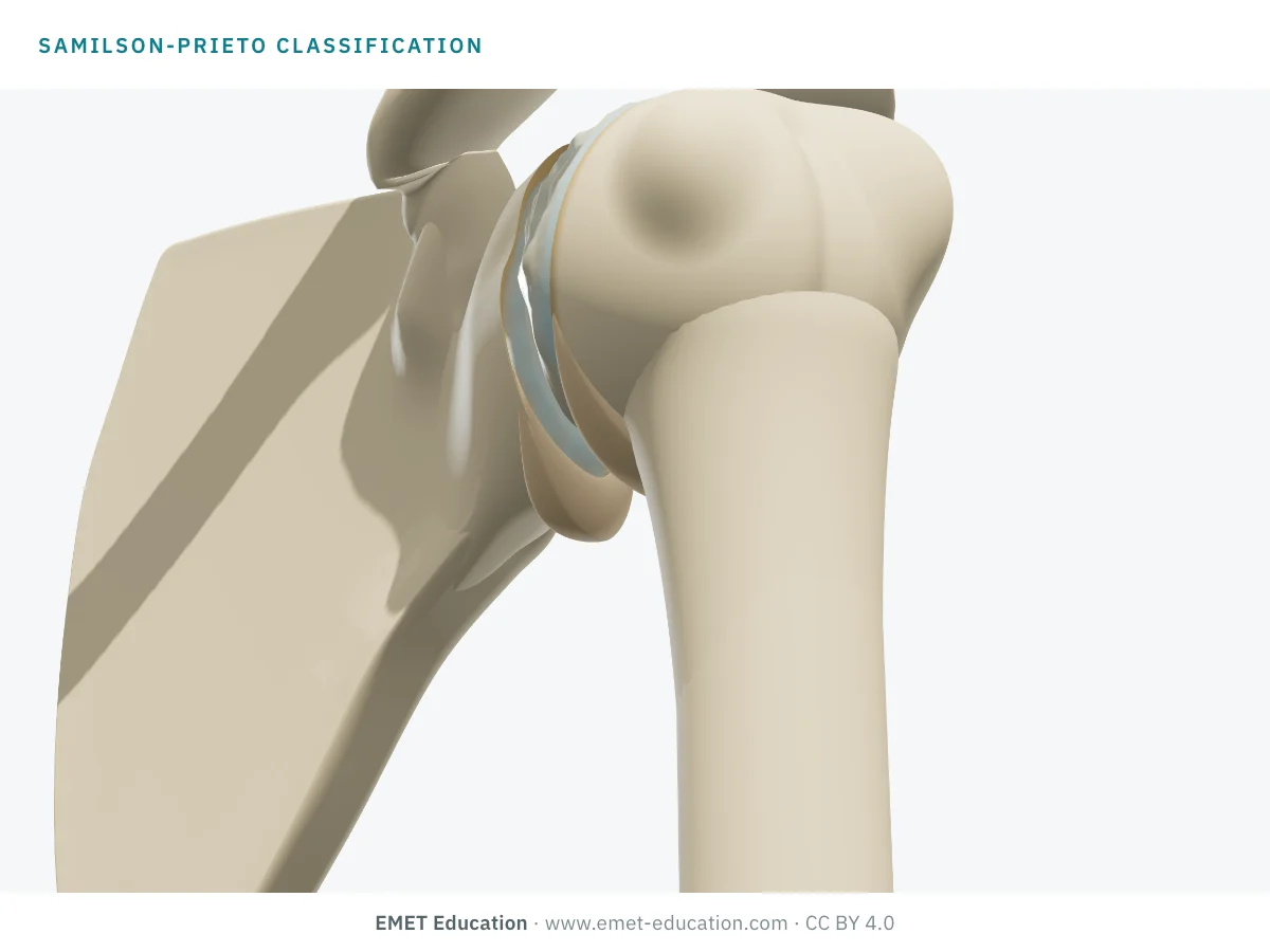
Which Samilson-Prieto grade is this?
Download and cite
Free infographics, for teaching and publication
Use them in a course, a presentation, a social media post or a scientific article, under the Creative Commons Attribution 4.0 license: free reuse, including commercial, provided EMET Education is credited with a link to this page.
Cite this figure
Ready to paste. For a journal figure, add the original reference (Samilson and Prieto, 1983).
EMET Education. Samilson-Prieto classification of shoulder osteoarthritis: 3D visualization [Internet]. 2026 [cited]. Available from: https://www.emet-education.com/en/classifications/samilson-prieto/. CC BY 4.0.EMET Education. (2026). Samilson-Prieto classification of shoulder osteoarthritis: 3D visualization [Infographic]. https://www.emet-education.com/en/classifications/samilson-prieto/ (CC BY 4.0)<a href="https://www.emet-education.com/en/classifications/samilson-prieto/"><img src="https://www.emet-education.com/en/classifications/samilson-prieto/img/infographic.webp" alt="Samilson-Prieto classification in 3D" width="960"></a><br>Figure: <a href="https://www.emet-education.com/en/classifications/samilson-prieto/">EMET Education</a>, CC BY 4.0Questions
Frequently asked questions
What is the difference between Samilson-Prieto grades II and III?
The height of the inferior osteophyte: 3 to 7 mm at grade II, over 7 mm at grade III. Grade III also comes with joint space narrowing and subchondral sclerosis, whereas grade II has only slight irregularity of the joint.
Should the humeral or the glenoid osteophyte be measured?
Both count: the criterion is the inferior osteophyte of the humeral head or the glenoid, or both. The taller one is used. The humeral osteophyte, extending the bottom of the head like a "goat's beard", is usually the larger.
Is the classification only for osteoarthritis after dislocation?
It was described in 1983 in osteoarthritis after dislocation, but it is now used to grade concentric shoulder osteoarthritis in general, primary osteoarthritis included. It does not suit cuff tear arthropathy, where the head rises under the acromion (Hamada classification).
How does it differ from the Walch classification?
Samilson-Prieto grades the severity of osteoarthritis on an anteroposterior view, by the osteophyte. Walch describes, on axial CT or MRI, glenoid shape and head centering (types A, B, C). The two do not correlate: before arthroplasty, the latter is essential.
Samilson-Prieto or Kellgren-Lawrence for the shoulder?
For the shoulder, Samilson-Prieto is more reproducible: interobserver kappa of 0.76 versus 0.36 in Brox et al., 0.87 versus 0.70 in Longo et al. It is also simpler, since it rests on a single measurement.
Can I reuse these images in a course or an article?
Yes. The images and infographics on this page are under the Creative Commons Attribution 4.0 license: free reuse, including commercial, with credit to EMET Education and a link to this page. The "Cite this figure" box gives ready-to-paste citations.
How were the 3D images made?
They are renderings of the EMET Education shoulder osteoarthritis model, in its concentric form, set to each grade: the model grows the humeral and glenoid osteophytes to the grade's height, wears the cartilage, narrows the joint space along its axis without migration of the head, and flattens the head in its contact zone. It is a schematic model for teaching and patient explanation; it does not reproduce a radiograph.
Sources
Author and references
- Samilson RL, Prieto V. Dislocation arthropathy of the shoulder. J Bone Joint Surg Am. 1983;65(4):456–60. doi:10.2106/00004623-198365040-00005
- Allain J, Goutallier D, Glorion C. Long-term results of the Latarjet procedure for the treatment of anterior instability of the shoulder. J Bone Joint Surg Am. 1998;80(6):841–52. doi:10.2106/00004623-199806000-00008
- Brox JI, Lereim P, Merckoll E, Finnanger AM. Radiographic classification of glenohumeral arthrosis. Acta Orthop Scand. 2003;74(2):186–9. doi:10.1080/00016470310013932
- Kappe T, Cakir B, Reichel H, Elsharkawi M. Reliability of radiologic classification for cuff tear arthropathy. J Shoulder Elbow Surg. 2011;20(4):543–7. doi:10.1016/j.jse.2011.01.012
- Linke PM, Zemke K, Ecker NU, Neumann J, Werner AW. Standard radiological classification of glenohumeral osteoarthritis does not correlate with the complexity of the arthritic glenoid deformity. Arch Orthop Trauma Surg. 2022;142(7):1413–20. doi:10.1007/s00402-021-03758-7
- Longo UG, de Sire A, De Salvatore S, et al. Imaging of glenohumeral osteoarthritis: reliability and reproducibility of radiological classifications. J Back Musculoskelet Rehabil. 2024;37(6):1729–39. doi:10.3233/BMR-240187
- Buscayret F, Edwards TB, Szabo I, Adeleine P, Coudane H, Walch G. Glenohumeral arthrosis in anterior instability before and after surgical treatment: incidence and contributing factors. Am J Sports Med. 2004;32(5):1165–72. doi:10.1177/0363546503262686
- Gerber C, Terrier F, Ganz R. The Trillat procedure for recurrent anterior instability of the shoulder. J Bone Joint Surg Br. 1988;70(1):130–4. doi:10.1302/0301-620X.70B1.3339045
- Walch G, Badet R, Boulahia A, Khoury A. Morphologic study of the glenoid in primary glenohumeral osteoarthritis. J Arthroplasty. 1999;14(6):756–60. doi:10.1016/S0883-5403(99)90232-2
- Hamada K, Fukuda H, Mikasa M, Kobayashi Y. Roentgenographic findings in massive rotator cuff tears. A long-term observation. Clin Orthop Relat Res. 1990;(254):92–6. doi:10.1097/00003086-199005000-00014
- Kellgren JH, Lawrence JS. Radiological assessment of osteo-arthrosis. Ann Rheum Dis. 1957;16(4):494–502. doi:10.1136/ard.16.4.494
3D models
Show the grade to your patient
The images on this page come from an interactive model. In clinic, set the grade from your patient's radiograph, then show the treatment discussed, up to shoulder arthroplasty.
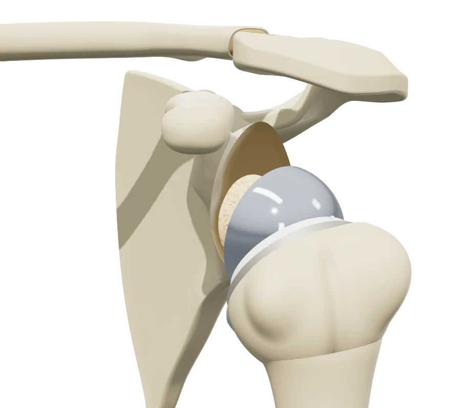 ShoulderShoulder osteoarthritisSamilson-Prieto and Hamada grades, humeral head osteonecrosis, anatomic and reverse shoulder arthroplasty.
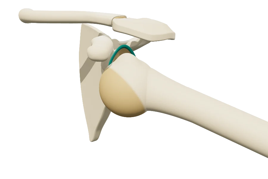 ShoulderShoulder instabilityDislocation, Bankart lesion, Hill-Sachs lesion, glenoid bone loss.
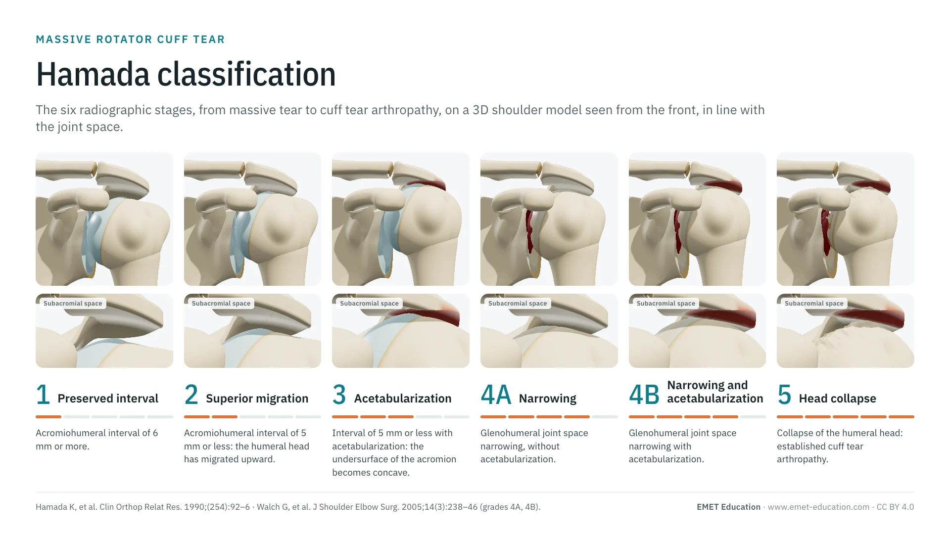 ClassificationsHamada classificationEccentric osteoarthritis with a massive cuff tear, grade by grade.
Explain the grade in 3D
The shoulder osteoarthritis model opens in your browser, at the grade you choose, with no sign-up.
Share this page with a colleague or a resident: