- EMET Education
- 3D classifications
- Mayo classification (olecranon)
Classification · Olecranon fracture
Mayo Classification of Olecranon Fractures: 3D Visualization
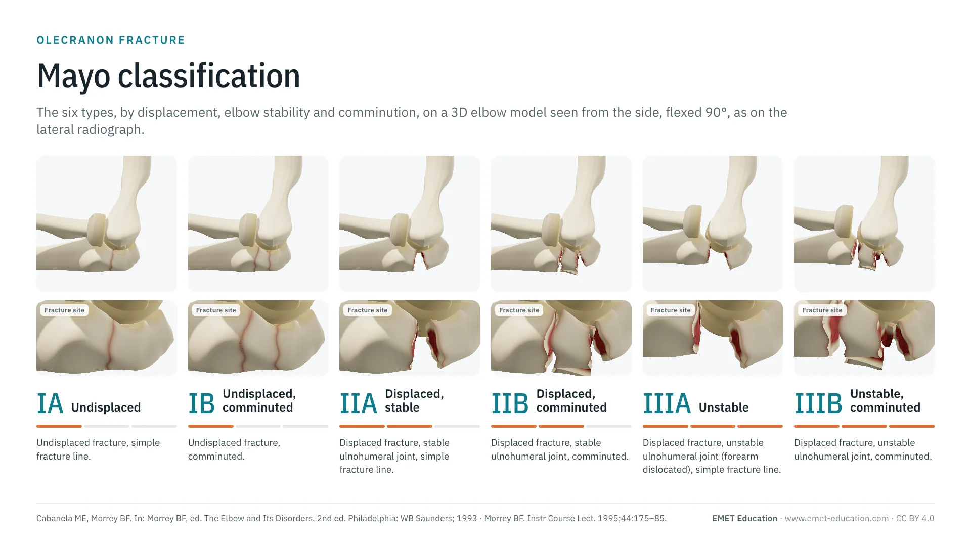
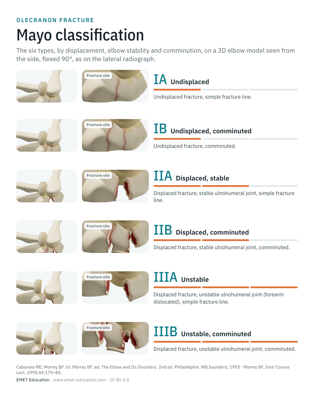
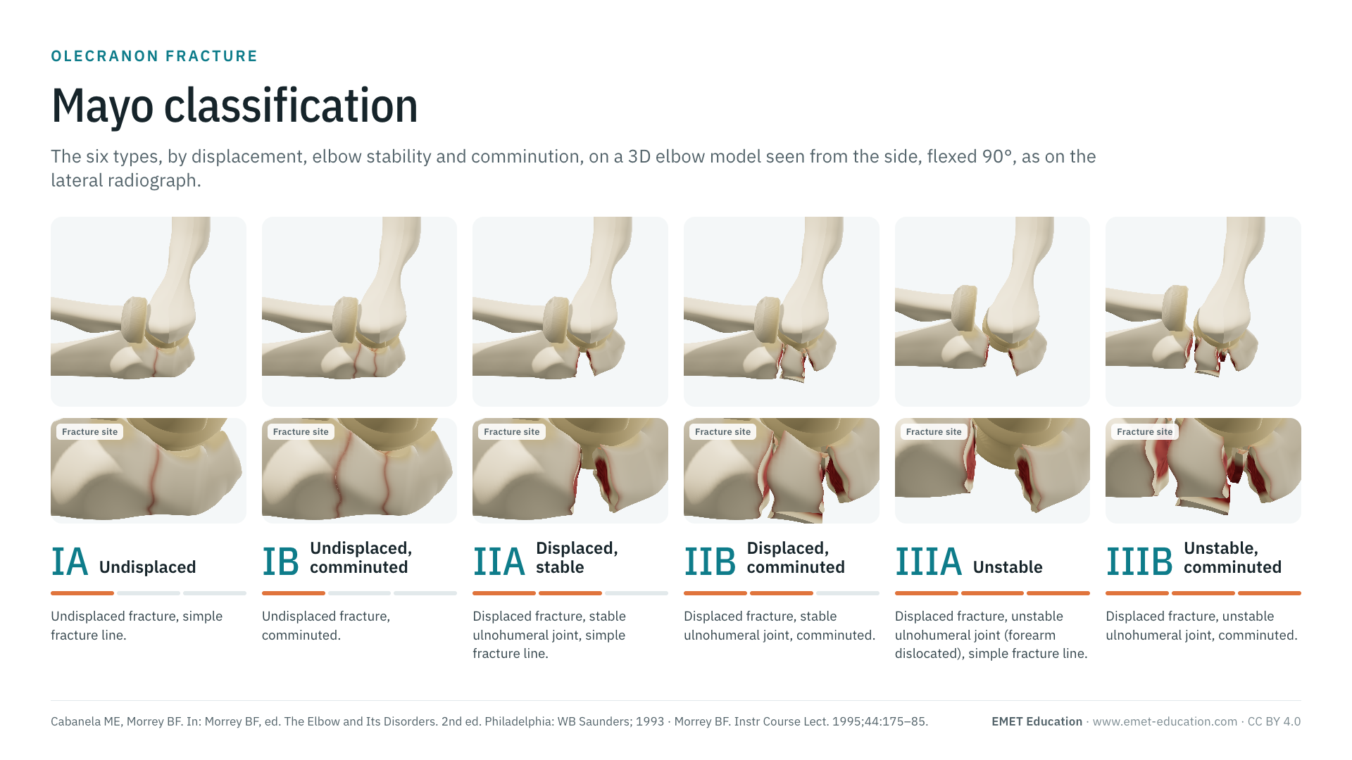
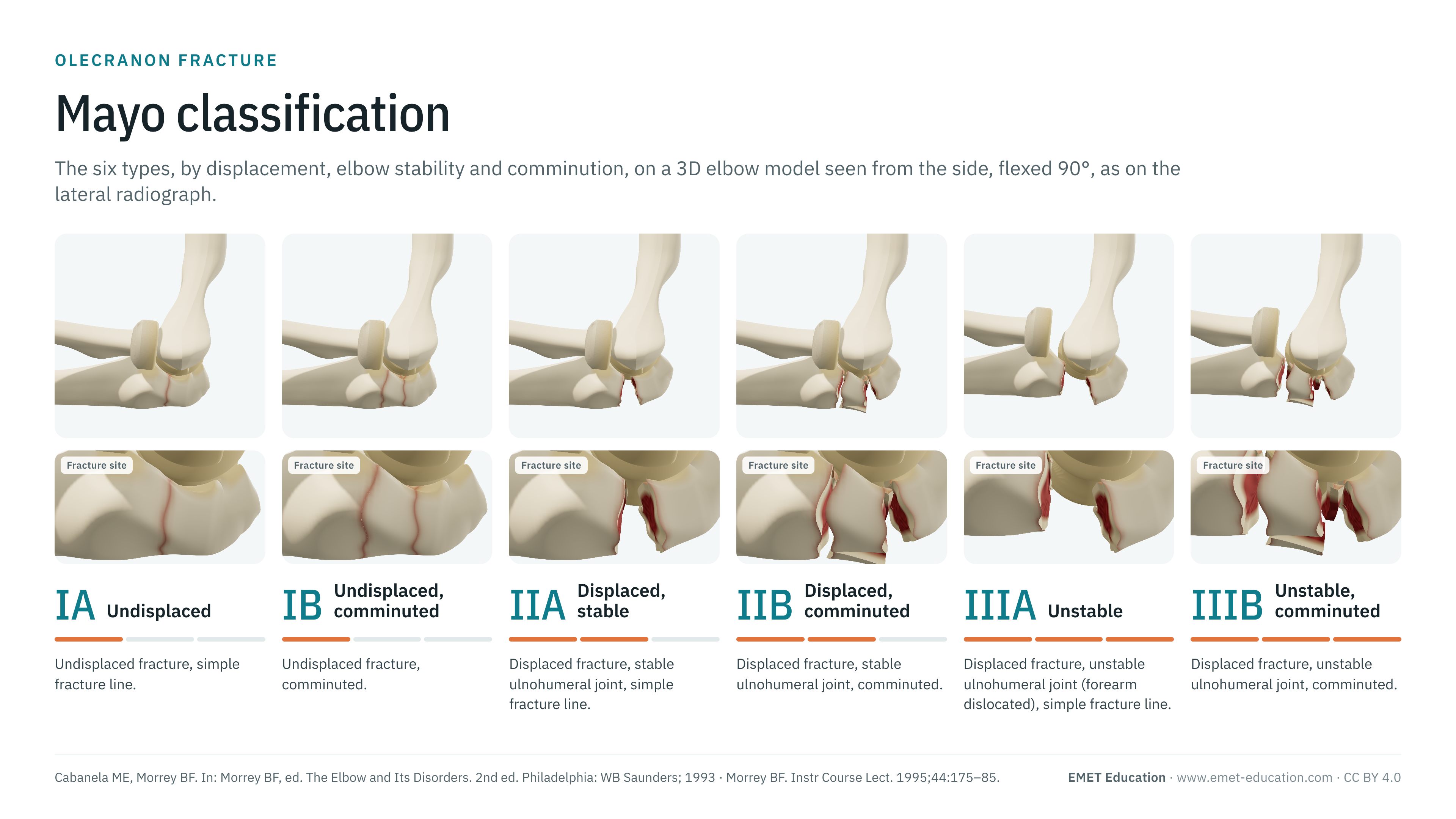
The six types of the Mayo Clinic classification, by displacement, elbow stability and comminution, shown on a 3D elbow model seen as on the lateral radiograph. With the origin of the classification, reliability data, related classifications and free infographics for teaching and publication.
Designed by an orthopedic surgeon
The 3D model opens in your browser, at the type shown, with no sign-up. Infographics are free to reuse under CC BY 4.0.
- 1993Described by Cabanela and Morrey in The Elbow and Its Disorders
- I–III · A/BThree types by displacement and stability, each simple (A) or comminuted (B)
- 80–85 %Of olecranon fractures are type II: displaced, stable elbow
- κ 0.32Interobserver agreement on radiographs, in two studies
The six types
From type IA to type IIIB, in 3D
Each image is a rendering of the EMET Education olecranon fracture model at that type, seen as on the lateral radiograph on which the classification is read: from the lateral side, elbow flexed 90°, forearm in neutral rotation; the radial head projects over the coronoid, as on the radiograph. The fracture line starts in the middle of the trochlear notch and exits the posterior cortex; in types II and III, the gap opens by 10 mm posteriorly.
-
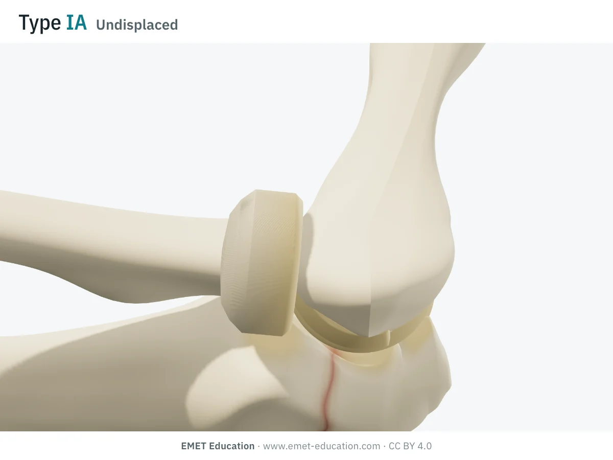
IAUndisplaced
- Criteria
- Undisplaced fracture, simple (noncomminuted) fracture line.
- In the 3D model
- Transverse, slightly oblique line from the middle of the trochlear notch to the posterior cortex; the fragments stay in place, a red rim outlines the line.
-
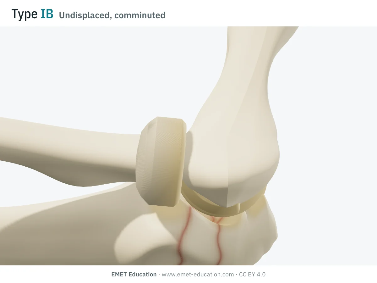
IBUndisplaced, comminuted
- Criteria
- Undisplaced fracture, comminuted.
- In the 3D model
- A zone of about 9 mm along the posterior cortex, between two lines; five fragments (two articular, three cortical) left in place.
-
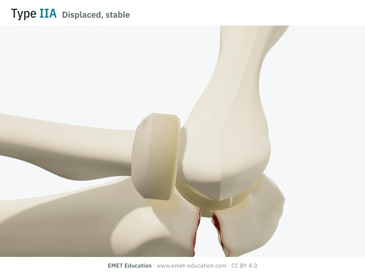
IIADisplaced, stable
- Criteria
- Displaced fracture, stable ulnohumeral joint (collateral ligaments intact, particularly the anterior bundle of the medial collateral ligament), simple fracture line.
- In the 3D model
- The triceps pulls the proximal fragment: it tilts around the tip, still hooked on the trochlea, and rides up; the gap opens by 10 mm posteriorly. The distal ulna and radius stay in place.
-
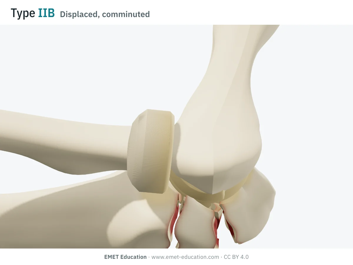
IIBDisplaced, comminuted
- Criteria
- Displaced fracture, stable ulnohumeral joint, comminuted.
- In the 3D model
- Same displacement of the proximal fragment; the fragments follow, the articular ones impacted, the cortical ones spreading apart.
-
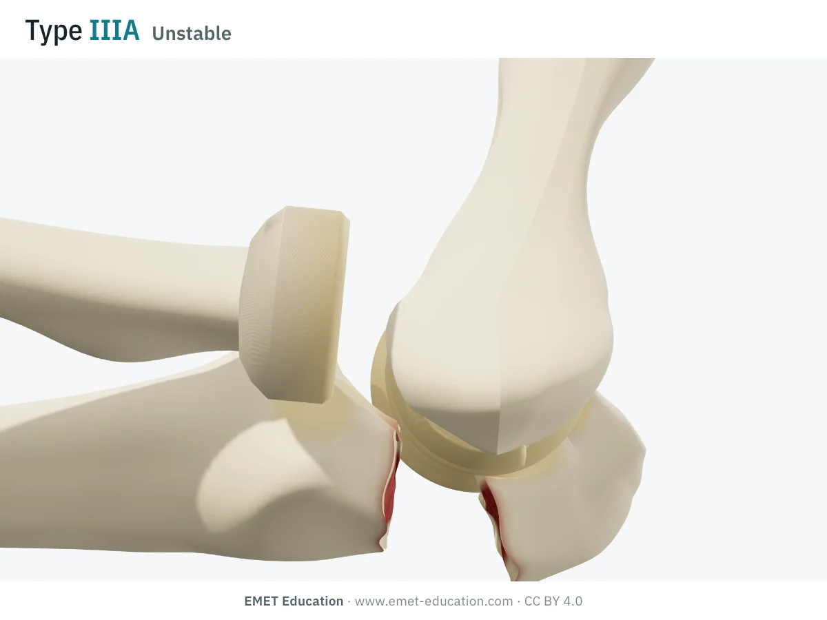
IIIAUnstable
- Criteria
- Displaced fracture, unstable ulnohumeral joint (collateral ligaments torn), simple fracture line.
- In the 3D model
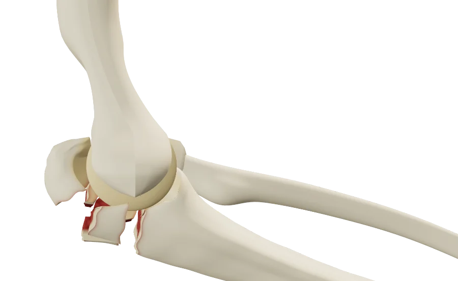
- The distal ulna and radius, held together by the annular ligament, slide anteriorly over the trochlea in front of the distal humerus; the olecranon stays behind with the triceps: a transolecranon fracture-dislocation.
-
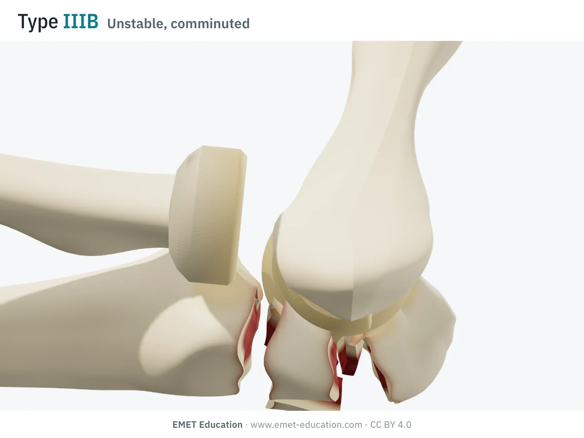
IIIBUnstable, comminuted
- Criteria
- Displaced fracture, unstable ulnohumeral joint, comminuted.
- In the 3D model
- Anterior dislocation of the forearm, with an olecranon broken into fragments.
Schematic rendering, for teaching: the model illustrates each type on a single elbow, it does not reproduce a radiograph. Criteria reworded from Cabanela and Morrey (1993) and Morrey (1995); their figures are not reproduced.
The origin
Cabanela and Morrey, 1993
M. E. Cabanela and B. F. Morrey, of the Mayo Clinic, proposed this classification in 1993, in the second edition of The Elbow and Its Disorders, and Morrey restated it in the 1995 Instructional Course Lectures (44:175–85). It aimed to simplify the existing classifications — Colton's (1973), based on fracture pattern and mechanism, and the AO's (1987) — by keeping only what guides treatment.
It rests on three questions, read on the lateral radiograph:
- is the fracture displaced? Type I if not; articular displacement of more than 2 mm is usually taken as displacement;
- is the elbow stable? Type II if the ulnohumeral joint is stable, the collateral ligaments intact (particularly the anterior bundle of the medial collateral ligament); type III if it is unstable;
- is the fracture comminuted? A if not, B if so;
- prognosis follows the type: excellent for type I (about 5% of fractures), good for type II (80% to 85%), guarded for type III.
It is now the most widely used classification in olecranon studies. In a prospective Edinburgh database, type IIA accounted for 47 of the 78 proximal ulna fractures seen in one year (Duckworth et al., 2012); in a French multicenter series, types IIA and IIB made up 66% of 109 olecranon fractures (Niéto et al., 2015).
Reliability
How reproducible is the type?
Poorly, between observers. A reader repeats themselves fairly well, but two readers agree poorly, and CT changes little: Sullivan and Desai judged this low reliability enough to question its use.
| Study | Measure | Value |
|---|---|---|
| Tamaoki et al., 2014 | Mean intraobserver kappa, 4 readers, 59 fractures, three readings | 0.64 |
| Tamaoki et al., 2014 | Interobserver kappa (AO 0.35; Schatzker 0.29; Colton 0.12) | 0.32 |
| Benetton et al., 2015 | Interobserver kappa, 8 surgeons (Colton 0.67; as reported by Sullivan and Desai) | 0.19 |
| Harbrecht et al., 2024 | Intraobserver kappa, 4 surgeons, 20 fractures: radiograph, then CT | 0.76 · 0.82 |
| Harbrecht et al., 2024 | Interobserver kappa: radiograph, then CT | 0.32 · 0.44 |
None of the olecranon classifications really does better: in Tamaoki et al., all four classifications compared had low reproducibility, and Colton's ranking flips from one study to the next (0.12 in Tamaoki, 0.67 in Benetton). Mayo's weak point is type III: elbow stability is hard to judge on a static radiograph.
Related classifications
What the other classifications add
Mayo sorts fractures by what guides treatment; other classifications describe the fracture pattern, the fragments or the associated injuries in more detail. In fracture-dislocations, the coronoid is the key to stability, and is classified separately.
The most used:
| Classification | What it grades | Typical use |
|---|---|---|
| Colton (1973) | Fracture pattern and mechanism: undisplaced fractures, then avulsion, oblique or transverse, comminuted, fracture-dislocation. | Historical classification; reliability varies widely between studies. |
| Schatzker | Fracture pattern, from transverse to fracture-dislocation, through impacted, oblique and comminuted. | Fixation planning; correlated with arthritis in Rommens et al. |
| AO/OTA | An alphanumeric code for the proximal forearm, by extra- or intra-articular location and comminution. | Research and registries. |
| Regan-Morrey (1989) | The associated coronoid fracture: tip (I), under 50% (II), over 50% of the process (III). | Elbow fracture-dislocations; it is also shown in the 3D model. |
| Fragment-specific classification (2024) | The fragments of complex fractures on CT (posterior, intermediate, tricipital, supinator crest, coronoid…). | Surgical planning of comminuted fractures (interobserver ICC 0.71). |
Use
In research and in clinical practice
In research
The Mayo type is used mostly to describe series and compare techniques. In Rommens et al. (58 patients reviewed), unstable elbows (type III) lost more function, and the authors recommended Mayo or Schatzker. In Tarallo et al. (78 type IIA and IIB fractures), tension band wiring and plating gave comparable functional results, but tension band wiring had more complications (48% versus 17% for type IIA, 40% versus 23% for type IIB) and its hardware was removed more often. Studies, however, generally find no difference in outcome between types, which questions its prognostic value.
In clinical practice
The type guides treatment: for type I, brief immobilization then active motion; for type IIA, tension band wiring; for type IIB, a plate; for type III, a plate, with treatment of the associated injuries (coronoid, radial head, ligaments). It takes neither age nor bone quality into account.
Yet age matters: in older low-demand patients (43 patients, mean age 76), nonoperative treatment of displaced fractures gave 72% good or excellent short-term results, a mean DASH score of 2.9 at six years, and 91% satisfaction, with no surgery for nonunion (Duckworth et al., 2014).
Grading pitfalls
Five pitfalls when classifying an olecranon fracture
- Grade on a true lateral view. Elbow flexed 90°, epicondyles superimposed: a rotated elbow hides the gap or the fragments. If comminution is in doubt, CT defines the fragments.
- Do not judge stability on a single radiograph. Type III implies an unstable ulnohumeral joint; a static radiograph may not show it. Look for subluxation of the forearm relative to the humerus, and for radiocapitellar congruence.
- In type III, look for associated injuries. Coronoid fracture (Regan-Morrey), radial head fracture, lateral collateral ligament tear: they change the operation.
- Measure displacement at the articular surface. The articular gap or step is what moves a fracture from type I to type II; 2 mm is the usual cut-off.
- Do not treat the type alone. A displaced fracture in an older, low-demand patient can be treated without surgery; the type says nothing about the patient.
Self-test
Which type?
The same elbow seen from the medial side, at the six types, in random order. Find the type of each one; the answer comes with the criteria.

Which Mayo type is this?
Download and cite
Free infographics, for teaching and publication
Use them in a course, a presentation, a social media post or a scientific article, under the Creative Commons Attribution 4.0 license: free reuse, including commercial, provided EMET Education is credited with a link to this page.
Cite this figure
Ready to paste. For a journal figure, add the original reference (Morrey, 1995).
EMET Education. Mayo classification of olecranon fractures: 3D visualization [Internet]. 2026 [cited]. Available from: https://www.emet-education.com/en/classifications/mayo-olecranon/. CC BY 4.0.EMET Education. (2026). Mayo classification of olecranon fractures: 3D visualization [Infographic]. https://www.emet-education.com/en/classifications/mayo-olecranon/ (CC BY 4.0)<a href="https://www.emet-education.com/en/classifications/mayo-olecranon/"><img src="https://www.emet-education.com/en/classifications/mayo-olecranon/img/infographic.webp" alt="Mayo classification of olecranon fractures in 3D" width="960"></a><br>Figure: <a href="https://www.emet-education.com/en/classifications/mayo-olecranon/">EMET Education</a>, CC BY 4.0Questions
Frequently asked questions
What is the difference between Mayo types IIA and IIB?
Both are displaced fractures in a stable elbow. Type IIA has a simple fracture line, type IIB is comminuted. The difference guides fixation: tension band wiring rather for type IIA, a plate for type IIB.
How is a type III recognized?
By instability of the ulnohumeral joint: the forearm (distal ulna and radius) dislocates or subluxates, most often anteriorly, while the olecranon stays behind with the triceps. A static radiograph can underestimate it; associated injuries of the coronoid, radial head and ligaments should then be sought.
Is a type I fracture treated surgically?
Usually not: brief immobilization, then gradually increasing active motion. Follow-up radiographs check that the fracture does not displace secondarily.
Mayo, Colton or Schatzker?
Mayo sorts fractures by what guides treatment (displacement, stability, comminution); Colton and Schatzker mainly describe the fracture pattern. None is highly reproducible; Mayo is the most used in recent studies.
Should a displaced fracture be operated on in an older person?
Not always. In older low-demand patients, nonoperative treatment of displaced fractures gives good functional results and high satisfaction, even when the fracture does not unite: the decision takes the patient into account, not just the type.
Can I reuse these images in a course or an article?
Yes. The images and infographics on this page are under the Creative Commons Attribution 4.0 license: free reuse, including commercial, with credit to EMET Education and a link to this page. The "Cite this figure" box gives ready-to-paste citations.
How were the 3D images made?
They are renderings of the EMET Education olecranon fracture model, set to each type: the fragments are cut from the model's ulna, the proximal fragment tilts under the pull of the triceps, and in type III the forearm dislocates in front of the humerus. It is a schematic model for teaching and patient explanation; it does not reproduce a radiograph.
Sources
Author and references
- Cabanela ME, Morrey BF. Fractures of the proximal ulna and olecranon. In: Morrey BF, editor. The Elbow and Its Disorders. 2nd ed. Philadelphia: WB Saunders; 1993.
- Morrey BF. Current concepts in the treatment of fractures of the radial head, the olecranon, and the coronoid. Instr Course Lect. 1995;44:175–85.
- Sullivan CW, Desai K. Classifications in brief: Mayo classification of olecranon fractures. Clin Orthop Relat Res. 2019;477(4):908–10. doi:10.1097/CORR.0000000000000614
- Tamaoki MJ, Matsunaga FT, Silveira JD, Balbachevsky D, Matsumoto MH, Belloti JC. Reproducibility of classifications for olecranon fractures. Injury. 2014;45 Suppl 5:S18–20. doi:10.1016/S0020-1383(14)70015-4
- Benetton CA, Cesa G, El-Kouba Junior G, et al. Agreement of olecranon fractures before and after the exposure to four classification systems. J Shoulder Elbow Surg. 2015;24(3):358–63. doi:10.1016/j.jse.2014.10.025
- Harbrecht A, Hackl M, Ott N, et al. Mayo classification of olecranon fractures revisited: assessment of intra- and interobserver reliability based on CT scans. J Orthop. 2024;52:102–6. doi:10.1016/j.jor.2024.02.026
- Colton CL. Fractures of the olecranon in adults: classification and management. Injury. 1973;5(2):121–9. doi:10.1016/S0020-1383(73)80088-9
- Regan W, Morrey B. Fractures of the coronoid process of the ulna. J Bone Joint Surg Am. 1989;71(9):1348–54.
- Rommens PM, Küchle R, Schneider RU, Reuter M. Olecranon fractures in adults: factors influencing outcome. Injury. 2004;35(11):1149–57. doi:10.1016/j.injury.2003.12.002
- Tarallo L, Mugnai R, Adani R, et al. Simple and comminuted displaced olecranon fractures: a clinical comparison between tension band wiring and plate fixation techniques. Arch Orthop Trauma Surg. 2014;134(8):1107–14. doi:10.1007/s00402-014-2021-9
- Duckworth AD, Bugler KE, Clement ND, Court-Brown CM, McQueen MM. Nonoperative management of displaced olecranon fractures in low-demand elderly patients. J Bone Joint Surg Am. 2014;96(1):67–72. doi:10.2106/JBJS.L.01137
- Duckworth AD, Clement ND, Aitken SA, Court-Brown CM, McQueen MM. The epidemiology of fractures of the proximal ulna. Injury. 2012;43(3):343–6. doi:10.1016/j.injury.2011.10.017
- Chapleau J, Joly-Chevrier M, Tohmé P, et al. A novel fragment specific classification of complex olecranon fractures: 3-dimensional model design, radiological validation, and proposed surgical algorithm. J Shoulder Elbow Surg. 2024;33(5):1084–91. doi:10.1016/j.jse.2023.12.021
3D models
Show the fracture to your patient
The images on this page come from an interactive model. In clinic, set the type, comminution and gap from your patient's radiograph, then show the proposed fixation.
Explain the fracture in 3D
The olecranon fracture model opens in your browser, at the type you choose, with no sign-up.
Share this page with a colleague or a resident: