
- EMET Education
- 3D classifications
- Lanz classification
Classification · Carpal tunnel
Lanz Classification of Median Nerve Variations: 3D Visualization
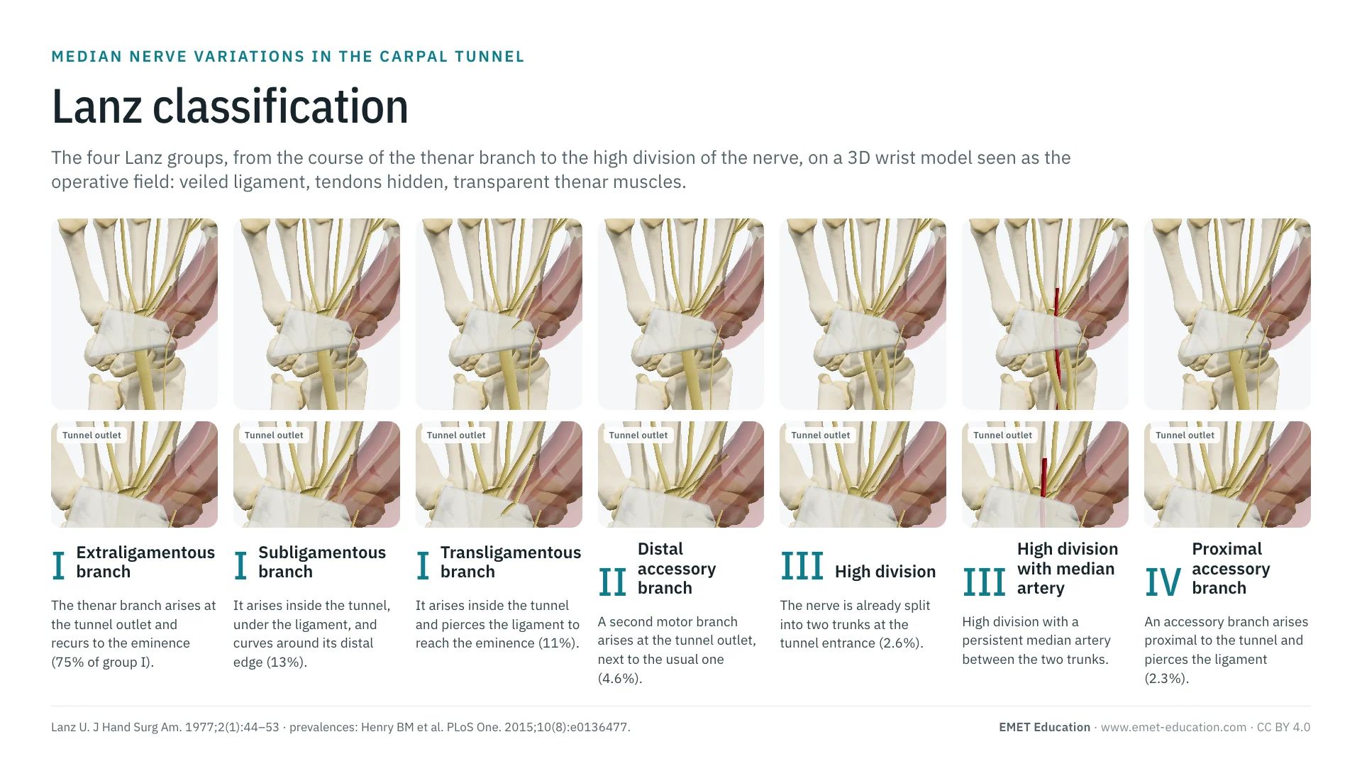
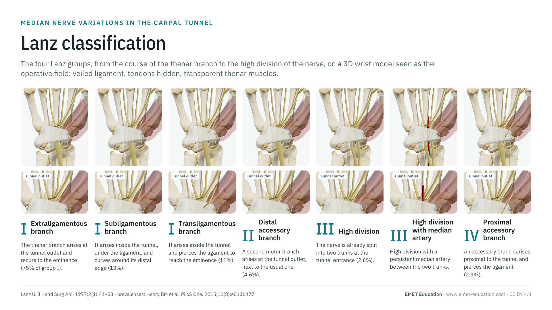
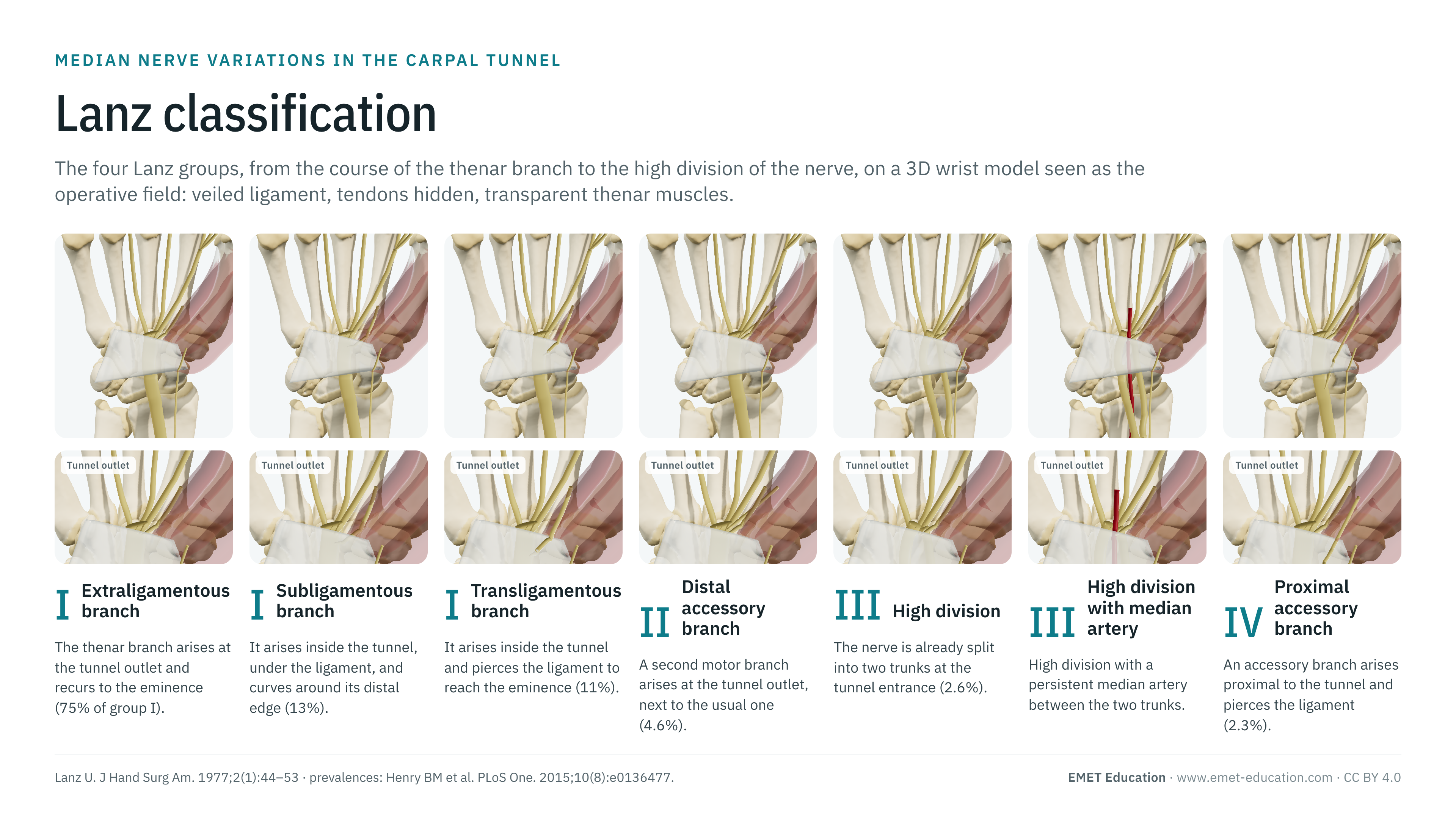
The four Lanz groups, which sort the variations of the median nerve in the carpal tunnel, shown on a 3D wrist model seen as the operative field: veiled transverse carpal ligament, tendons hidden, transparent thenar muscles. With the original article, the prevalence of each type, the variants the classification leaves out, what it changes for surgery, and free infographics for teaching and publication.
Designed by an orthopedic surgeon
The 3D model opens in your browser, at the form shown, with no sign-up. Infographics are free to reuse under CC BY 4.0.
- 1977Published by Lanz, from 246 operated carpal tunnels
- I–IVGroups, including three courses of the thenar branch in group I
- 489Citations of the 1977 article (OpenAlex, October 2026)
- 75%Extraligamentous course, the most frequent (meta-analysis, 3,918 hands)
The groups
From group I to group IV, in 3D
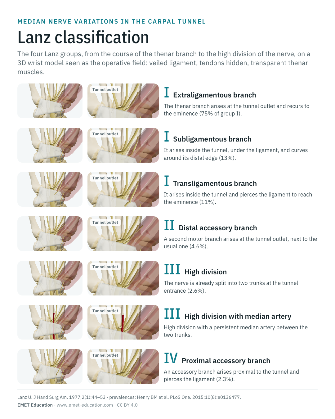
Each image is a rendering of the EMET Education carpal tunnel model at that form, seen as the operative field: wrist from the front, palm forward, veiled transverse carpal ligament, flexor tendons hidden and thenar muscles transparent, so that the median nerve (yellow) and its motor branches can be followed to the thenar eminence. Group I is shown in its three courses, group III without and with a persistent median artery (red).
-
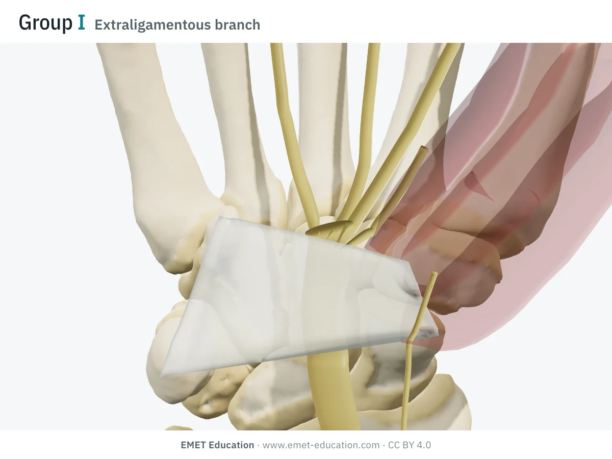
IExtraligamentous branch
- Definition
- Group I, usual course: the thenar branch arises from the radial side of the nerve at the tunnel outlet, then curves back (recurrent course) to the thenar muscles, without crossing the ligament.
- Prevalence
- 75.2% of thenar branch courses.
- In the 3D model
- The branch leaves the nerve's fan at the distal edge of the ligament and reaches the base of the eminence.
-
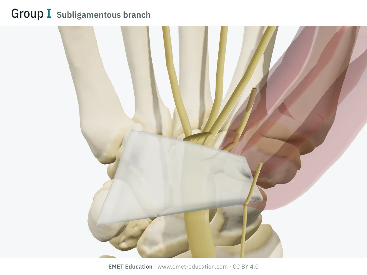
ISubligamentous branch
- Definition
- Group I: the branch arises inside the tunnel, runs under the ligament and curves around its distal edge before reaching the eminence.
- Prevalence
- 13.5%.
- In the 3D model
- The branch leaves the trunk under the ligament and exits at its distal edge, radially.
-
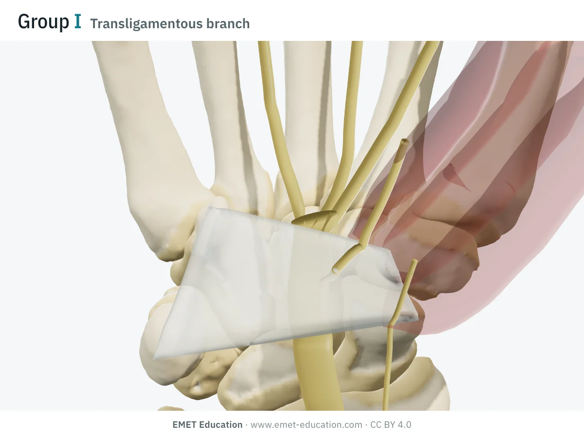
ITransligamentous branch
- Definition
- Group I: the branch arises inside the tunnel and pierces the ligament, from its deep to its superficial surface, to reach the eminence over it.
- Prevalence
- 11.3%.
- In the 3D model
- The branch passes through the radial half of the ligament, two-thirds of the way along it.
-
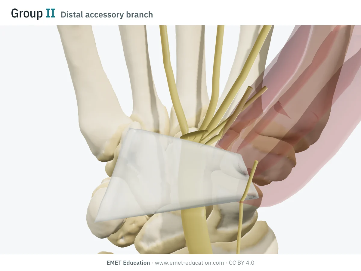
IIDistal accessory branch
- Definition
- Group II: one or more accessory motor branches arise at the distal part of the tunnel, in addition to the thenar branch.
- Prevalence
- 4.6%.
- In the 3D model
- A second branch arises at the end of the trunk and reaches the middle of the eminence; it runs under the thenar muscles.
-
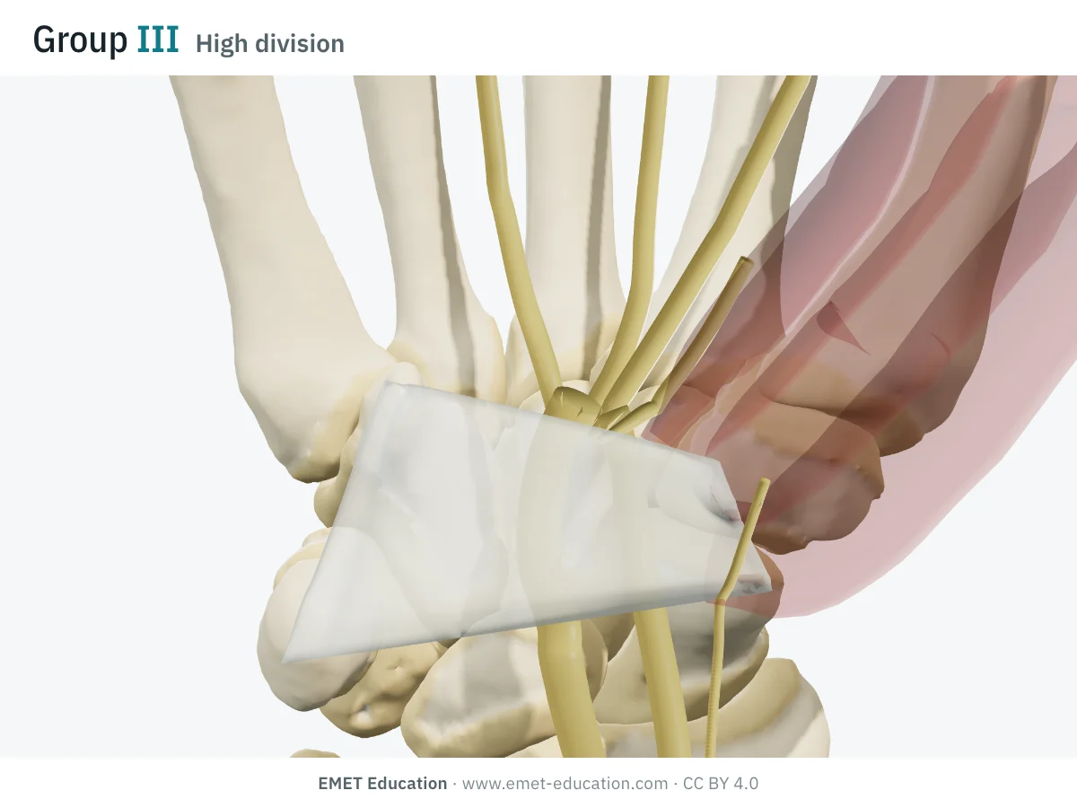
IIIHigh division
- Definition
- Group III: the median nerve divides into two trunks before or within the tunnel (bifid nerve), instead of at its outlet.
- Prevalence
- 2.6%.
- In the 3D model
- A radial division arising in the distal forearm runs under the ligament radial to the trunk, which has become the ulnar division.
-
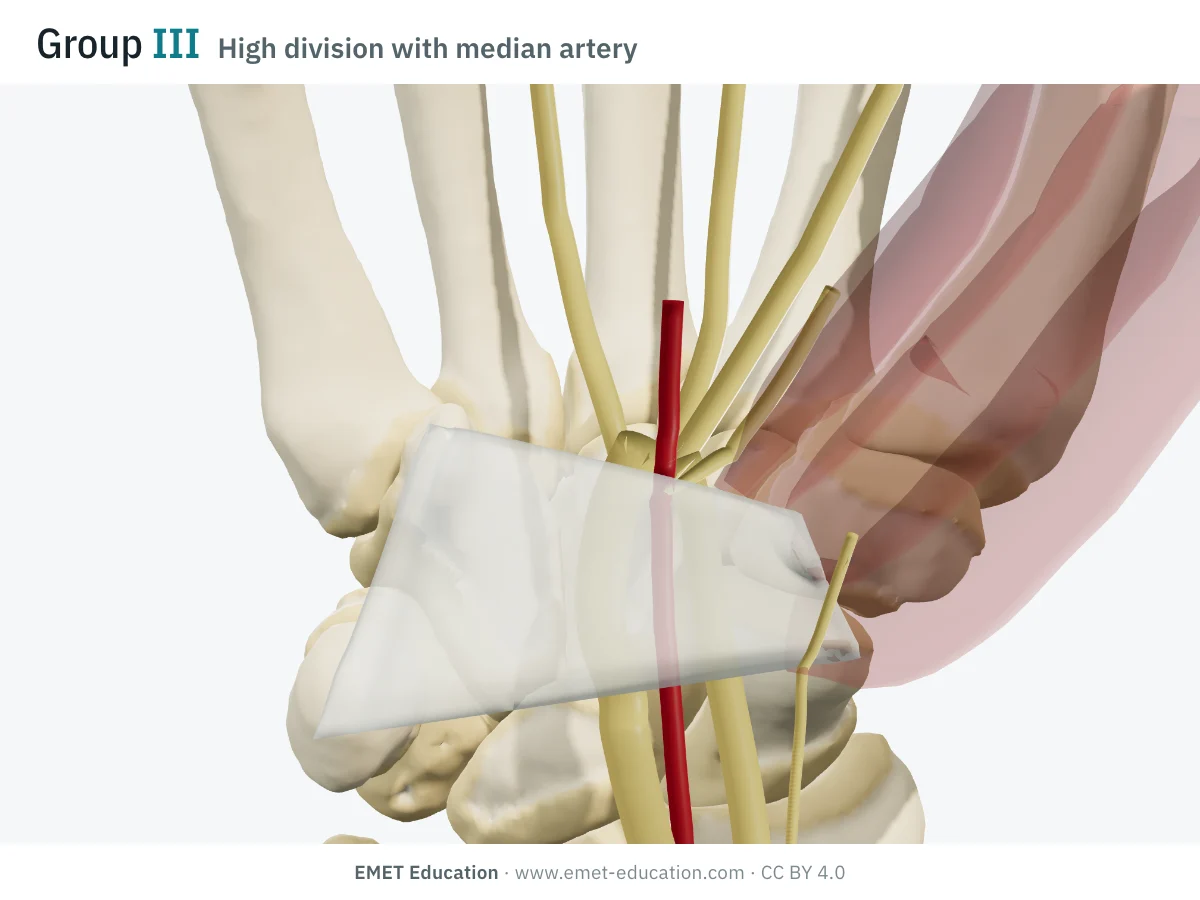
IIIHigh division with median artery
- Definition
- Group III with a persistent median artery: the small embryonic artery, retained in the adult, runs between the two trunks. Lanz distinguishes the forms with and without the artery.
- Prevalence
- Artery present in over 60% of bifid or trifid nerves (Honis 2023).
- In the 3D model
- The artery, in red, runs between the radial and ulnar divisions, under the ligament.
-
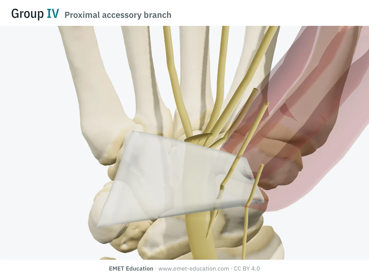
IVProximal accessory branch
- Definition
- Group IV: an accessory branch arises proximal to the tunnel; it may accompany the nerve through the tunnel or, as here, pierce the ligament.
- Prevalence
- 2.3%.
- In the 3D model
- The branch leaves the trunk just before the tunnel, runs under the ligament, then pierces it at its proximal third.
Prevalences: meta-analysis by Henry et al. (2015), 31 studies, 3,918 hands. These are not severity grades: one hand may combine several variations.
The original article
Lanz, 1977: 246 carpal tunnels opened
In 1977, in the first volume of the Journal of Hand Surgery, Ulrich Lanz reported the median nerve variations found when opening 246 carpal tunnels: 29 variations, including 18 accessory branches at the distal part of the tunnel, 7 high divisions and 4 accessory branches proximal to the tunnel.
He added published series, including Poisel's cadaveric series (100 hands, 1974), which described the three courses of the thenar branch, and sorted everything into four groups:
- I — variations in the course of the thenar branch (extra-, sub-, transligamentous);
- II — accessory branches at the distal part of the tunnel;
- III — high divisions of the median nerve (with or without a persistent median artery, with or without a lumbrical);
- IV — accessory branches proximal to the tunnel.
His practical conclusion still stands: approach the median nerve from its ulnar side when opening the tunnel.
Prevalences
How many hands for each type?
An anatomical classification is not measured by interobserver agreement: it is observed at surgical exploration, or on ultrasound. What matters is the prevalence of each form. It varies widely between series, especially for group I courses.
| Form | Lanz, 1977 | Al-Qattan, 2010 | Henry et al., 2015 |
|---|---|---|---|
| I — extraligamentous | 46%¹ | 56% | 75.2% |
| I — subligamentous | 31%¹ | 34% | 13.5% |
| I — transligamentous | 23%¹ | 9% | 11.3% |
| II — distal accessory branches | 18 of 246 | — | 4.6% |
| III — high division | 7 of 246 | — | 2.6% |
| IV — proximal accessory branches | 4 of 246 | — | 2.3% |
¹ Poisel's cadaveric series (100 hands), cited by Lanz. Al-Qattan: 100 consecutive open releases, with one preligamentous course (1%). Henry et al.: meta-analysis of 31 studies; the thenar branch arises from the ulnar side of the nerve in 2.1% of hands, and the course is identical on both sides in 72% of patients. In one surgeon's practice (Lindley and Kleinert, 526 releases), only 1% of hands showed an anomaly of the median nerve or its branches clear enough to be recorded: most variations are seen only when looked for.
What Lanz leaves out
Variants described since, and related classifications
The Lanz classification remains the reference for describing the median nerve at the tunnel: the meta-analysis by Henry et al. sorts all its data into it. Several forms described since fit poorly; they are described alongside it.
| Form or classification | What it describes | Relation to Lanz |
|---|---|---|
| Poisel (1974) | The three courses of a single thenar branch: extra-, sub-, transligamentous. | Taken over as group I. |
| Preligamentous course | The branch arises proximal to the tunnel and runs over the ligament, without entering the tunnel (1 in 100 in Al-Qattan). | Close to group IV, but without perforation. |
| Supraligamentous course | The branch arises in the tunnel then runs on the palmar surface of the ligament to the eminence (Honis 2023). | Outside the three group I courses. |
| Ulnar origin | The thenar branch arises from the ulnar side of the nerve and crosses it to reach the eminence (2.1%). | Recorded separately: it is the form exposed to an ulnar-side release. |
| Persistent median artery | Embryonic artery retained in the adult; palmar form in 4–20% of wrists, mostly on one side. | A group III subtype when the nerve is bifid; also possible with a single nerve. |
Use
What the classification changes for surgery
In the operating room
The median nerve branches at the tunnel lie almost all on the radial side: the "danger zone". The ligament is therefore always divided on the ulnar side of the nerve, layer by layer, as recommended by Lanz and later by Henry et al. The transligamentous course and group IV branches are the most exposed, since they pass through the ligament being cut.
A fleshy muscle lying over the ligament should suggest a transligamentous course: in Al-Qattan's series, every trans- and preligamentous course came with such a hypertrophic muscle. On the ulnar side, however, the Berrettini branch (sensory median-ulnar anastomosis) sometimes crosses the distal edge of the ligament.
On ultrasound
With an 18–22 MHz probe, the thenar branch was seen in every subject of a prospective study, including its course relative to the ligament (Riegler et al., 2017). Ultrasound-guided releases therefore begin by identifying the Lanz form, the median artery and any branches crossing the working space.
In research
Anatomical and surgical studies report their variations in Lanz groups, which made the meta-analysis by Henry et al. (31 studies, 3,918 hands) and comparisons between populations possible.
Pitfalls
Five pitfalls with median nerve variations
- One hand may combine several forms. A bifid nerve (III) often carries accessory branches, and a transligamentous course may coexist with a distal branch: describe each element rather than a single group.
- One side does not fully predict the other. The course is identical on both sides in only about 7 patients out of 10.
- Do not mistake an accessory branch for the thenar branch. A branch crossing over the trunk, between two trunks of a bifid nerve, can mislead; the true branch is often more distal.
- Think of the artery when the nerve is bifid. More than six bifid nerves out of ten come with a persistent median artery, sometimes eccentric on the ulnar side, where the cut is made.
- An ulnar-side release does not protect from everything. A branch arising from the ulnar side (2%) and the Berrettini branch lie on the cutting side: identify them before cutting.
Self-test
Which form?
The same tunnel seen close up, slightly from below, at the model's seven forms, in random order. Find the form of each one; the answer comes with its definition.
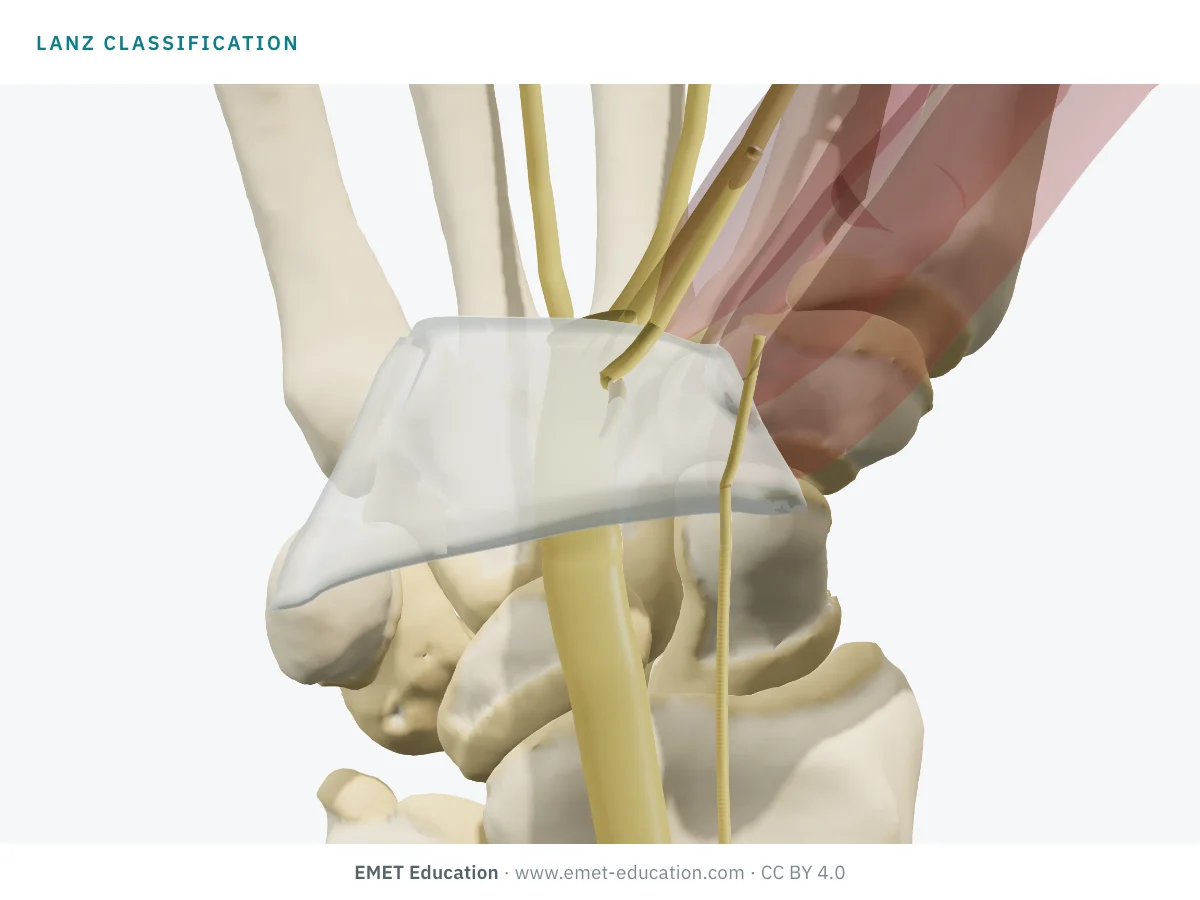
Which Lanz form is this?
Download and cite
Free infographics, for teaching and publication
Use them in a course, a presentation, a social media post or a scientific article, under the Creative Commons Attribution 4.0 license: free reuse, including commercial, provided EMET Education is credited with a link to this page.
Cite this figure
Ready to paste. For a journal figure, add the original reference (Lanz, 1977).
EMET Education. Lanz classification of median nerve variations: 3D visualization [Internet]. 2026 [cited]. Available from: https://www.emet-education.com/en/classifications/lanz/. CC BY 4.0.EMET Education. (2026). Lanz classification of median nerve variations: 3D visualization [Infographic]. https://www.emet-education.com/en/classifications/lanz/ (CC BY 4.0)<a href="https://www.emet-education.com/en/classifications/lanz/"><img src="https://www.emet-education.com/en/classifications/lanz/img/infographic.webp" alt="Lanz classification of median nerve variations in 3D" width="960"></a><br>Figure: <a href="https://www.emet-education.com/en/classifications/lanz/">EMET Education</a>, CC BY 4.0Questions
Frequently asked questions
What is the Lanz classification?
It is the classification of median nerve variations in the carpal tunnel, published by Ulrich Lanz in 1977: group I, course of the thenar branch (extra-, sub-, transligamentous); II, accessory branches at the distal part of the tunnel; III, high division of the nerve; IV, accessory branches proximal to the tunnel.
What is the most frequent course of the thenar branch?
The extraligamentous course: the branch arises at the tunnel outlet and curves back to the thenar eminence, in about three hands out of four according to the meta-analysis by Henry et al. The sub- and transligamentous courses share the rest, with wide differences between series.
Why divide the ligament on the ulnar side?
Because the thenar branch and almost all its variants lie on the radial side of the nerve. A release along the ulnar edge of the nerve avoids them, whatever the Lanz form. On the ulnar side remain the rare branch arising from the ulnar edge and the Berrettini branch.
Does a bifid median nerve favor carpal tunnel syndrome?
It is associated with it in several studies, often together with a persistent median artery. On ultrasound, the area of both trunks must be measured together so as not to miss swelling.
Can the thenar branch be seen on ultrasound?
Yes, with a high-frequency probe: it was seen in every subject of a prospective study, with its course relative to the ligament (Riegler et al., 2017). It is the first step of ultrasound-guided releases.
Can I reuse these images in a course or an article?
Yes. The images and infographics on this page are under the Creative Commons Attribution 4.0 license: free reuse, including commercial, with credit to EMET Education and a link to this page. The "Cite this figure" box gives ready-to-paste citations.
How were the 3D images made?
They are renderings of the EMET Education carpal tunnel model, built on bones taken from CT scans, set to each Lanz form: the branches are drawn from the model's ligament and thenar eminence landmarks. It is a model for teaching and patient explanation; it replaces neither exploration nor ultrasound.
Sources
Author and references
- Lanz U. Anatomical variations of the median nerve in the carpal tunnel. J Hand Surg Am. 1977;2(1):44–53. doi:10.1016/s0363-5023(77)80009-9
- Poisel S. Cadaveric series of 100 hands (1974), cited by Lanz U, ref. 1; original publication in German.
- Henry BM, Zwinczewska H, Roy J, Vikse J, Ramakrishnan PK, Walocha JA, Tomaszewski KA. The prevalence of anatomical variations of the median nerve in the carpal tunnel: a systematic review and meta-analysis. PLoS One. 2015;10(8):e0136477. doi:10.1371/journal.pone.0136477
- Al-Qattan MM. Variations in the course of the thenar motor branch of the median nerve and their relationship to the hypertrophic muscle overlying the transverse carpal ligament. J Hand Surg Am. 2010;35(11):1820–4. doi:10.1016/j.jhsa.2010.08.011
- Honis HR, Gruber H, Honold S, Konschake M, Moriggl B, Brenner E, Skalla-Oberherber E, Loizides A. Anatomical considerations of US-guided carpal tunnel release in daily clinical practice. J Ultrason. 2023;23(94):e131–43. doi:10.15557/JoU.2023.0022
- Lindley SG, Kleinert JM. Prevalence of anatomic variations encountered in elective carpal tunnel release. J Hand Surg Am. 2003;28(5):849–55. doi:10.1016/s0363-5023(03)00365-4
- Riegler G, Pivec C, Platzgummer H, Lieba-Samal D, Brugger P, Jengojan S, Vierhapper M, Bodner G. High-resolution ultrasound visualization of the recurrent motor branch of the median nerve: normal and first pathological findings. Eur Radiol. 2017;27(7):2941–9. doi:10.1007/s00330-016-4671-1
- Mizia E, Tomaszewski KA, Goncerz G, Kurzydło W, Walocha J. Median nerve thenar motor branch anatomical variations. Folia Morphol (Warsz). 2012;71(3):183–6.
- Agarwal P, Gupta S, Yadav P, Sharma D. Cadaveric study of anatomical variations of the median nerve and persistent median artery at wrist. Indian J Plast Surg. 2014;47(1):95–101. doi:10.4103/0970-0358.129632
- Natsis K, Iordache G, Gigis I, et al. Persistent median artery in the carpal tunnel: anatomy, embryology, clinical significance, and review of the literature. Folia Morphol (Warsz). 2009;68(4):193–200.
3D models
Show the carpal tunnel to your patient
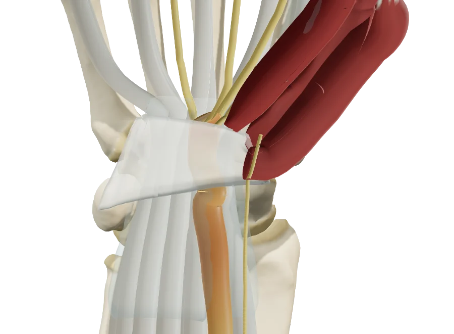
The images on this page come from an interactive model. In clinic, show the nerve compression, severity, cause, Lanz form and danger zone, then the proposed treatment: splint, injection, open or endoscopic release.
Explain the carpal tunnel in 3D
The carpal tunnel model opens in your browser, at the form you choose, with no sign-up.
Share this page with a colleague or a resident: