- EMET Education
- 3D classifications
- Neer classification (clavicle)
Classification · Clavicle fracture
Neer Classification of Distal Clavicle Fractures: 3D Visualization
The types of the modified Neer classification, by the position of the fracture line relative to the coracoclavicular ligaments, shown on a 3D shoulder model seen as on the cephalic-tilt AP radiograph. With the origin of the classification, reliability data, related classifications and free infographics for teaching and publication.
Designed by an orthopedic surgeon
The 3D model opens in your browser, at the type shown, with no sign-up. Infographics are free to reuse under CC BY 4.0.
- 1963First described by Neer, in the Journal of Trauma
- I–VTypes in the modified version, including IIA and IIB (Craig) and IV, in children
- 558Citations of the 1968 article (OpenAlex, October 2026)
- κ 0.28–0.57Interobserver agreement on radiographs, in four studies
The types
From type I to type V, in 3D
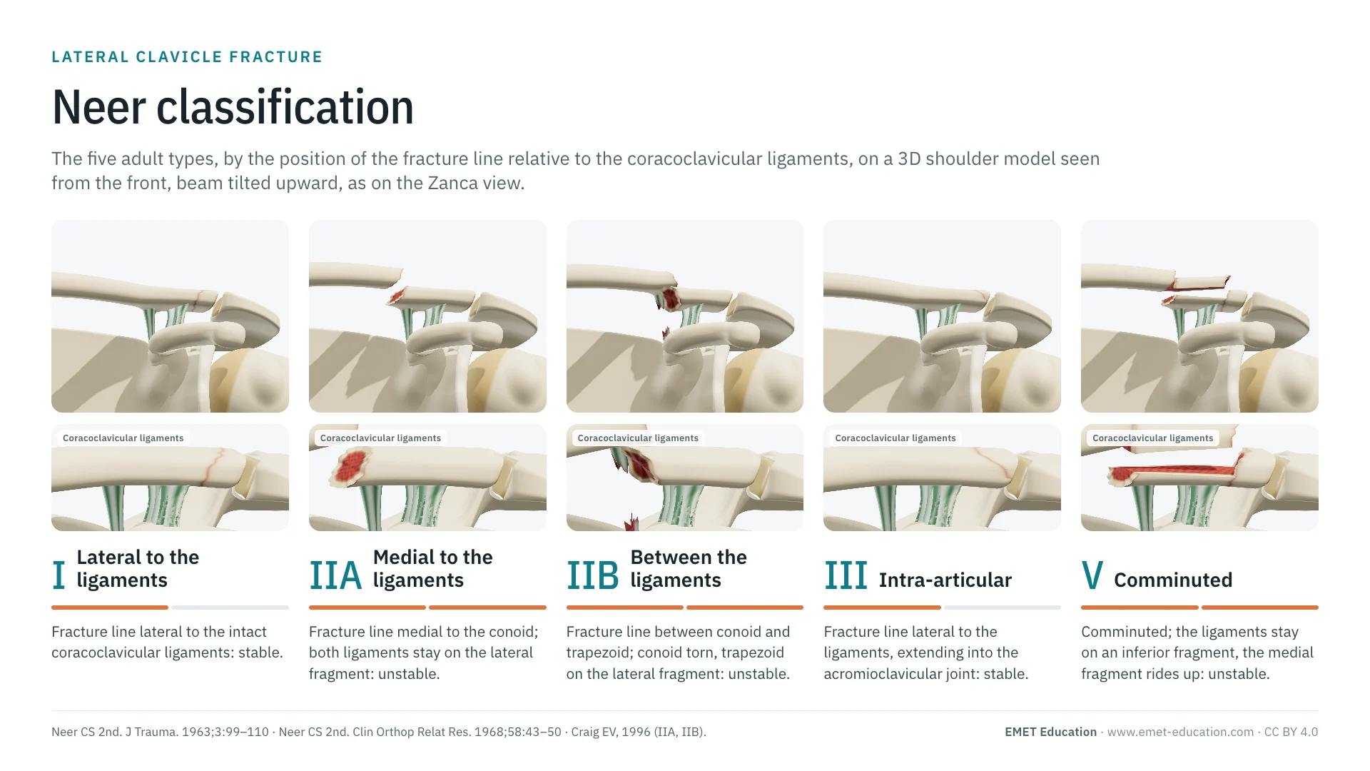
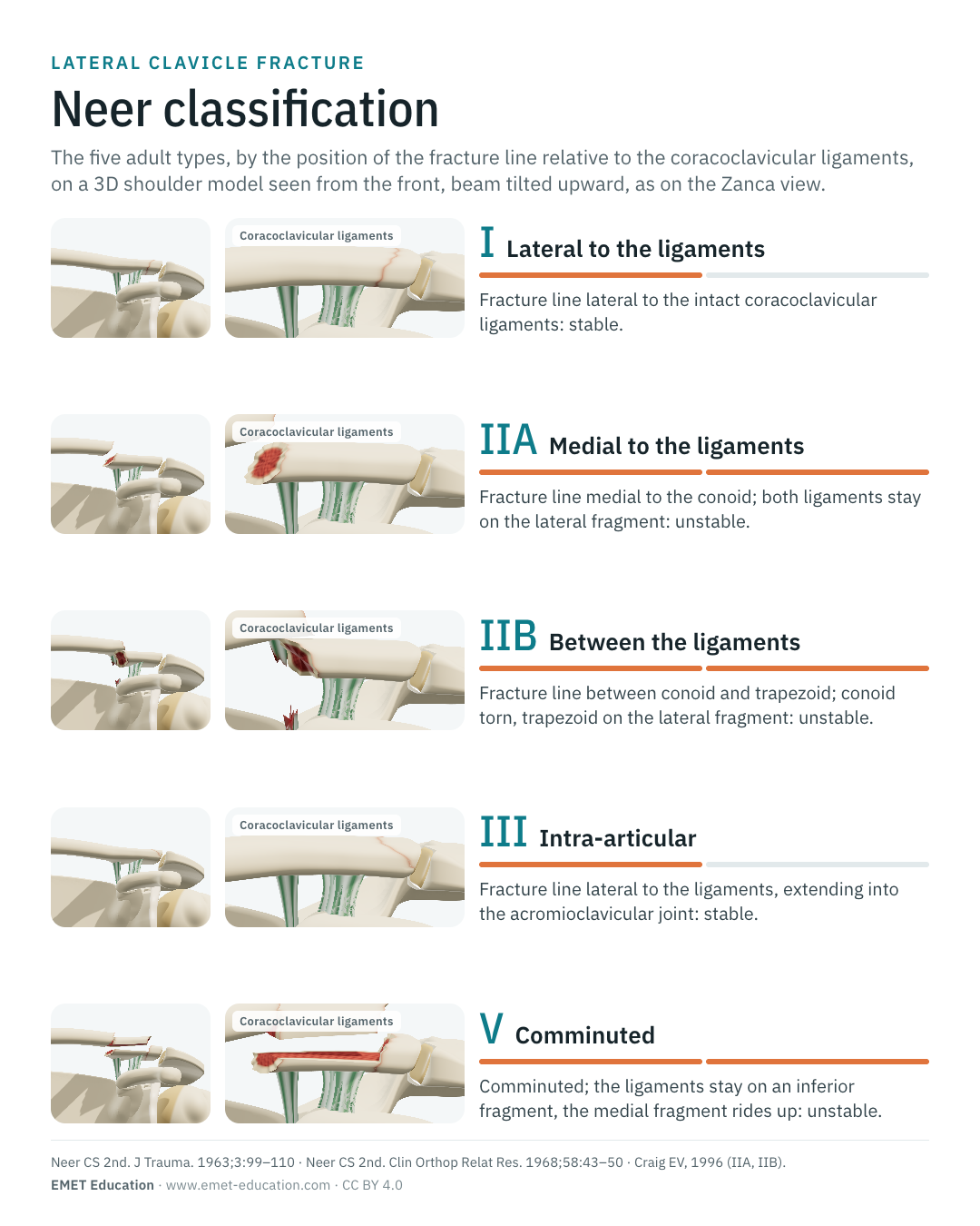
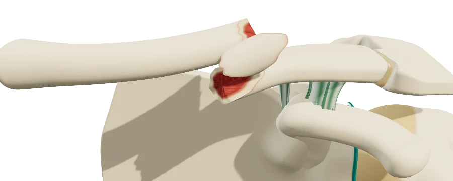
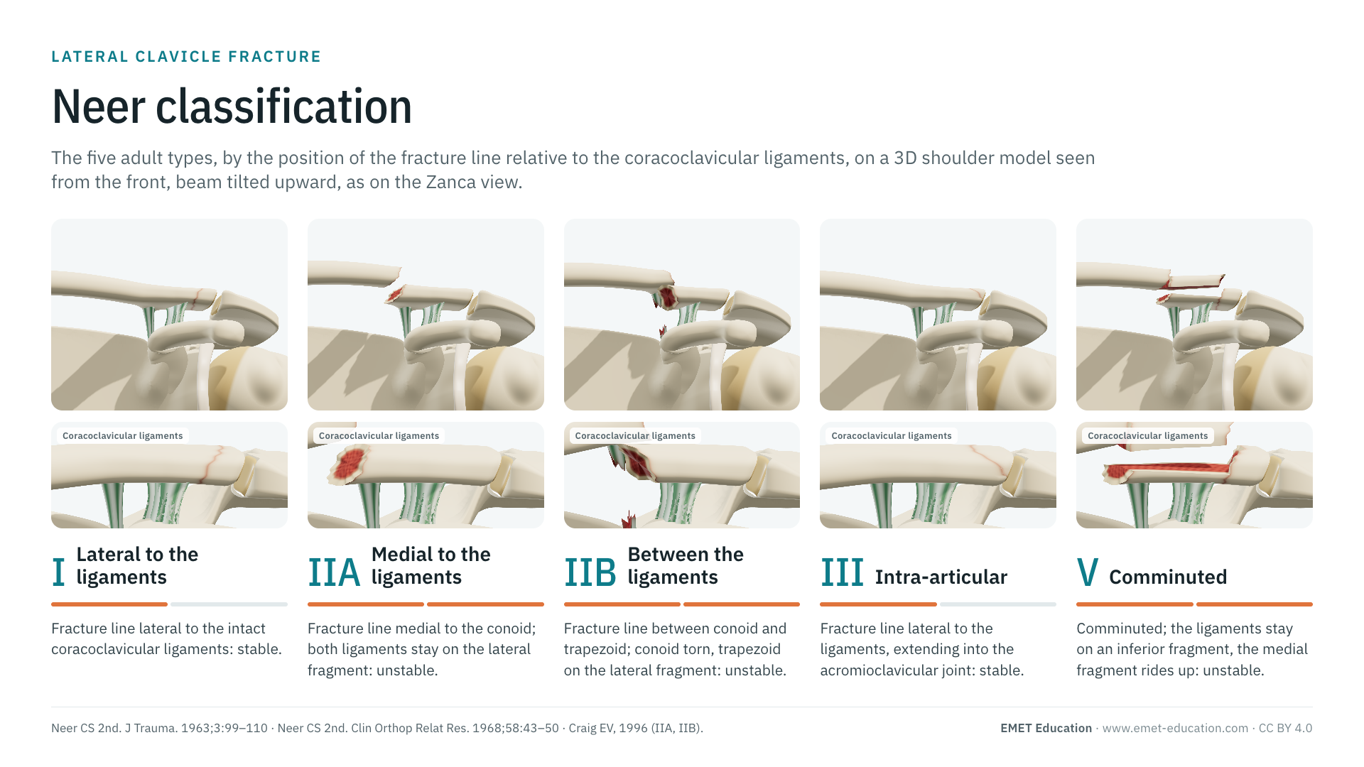
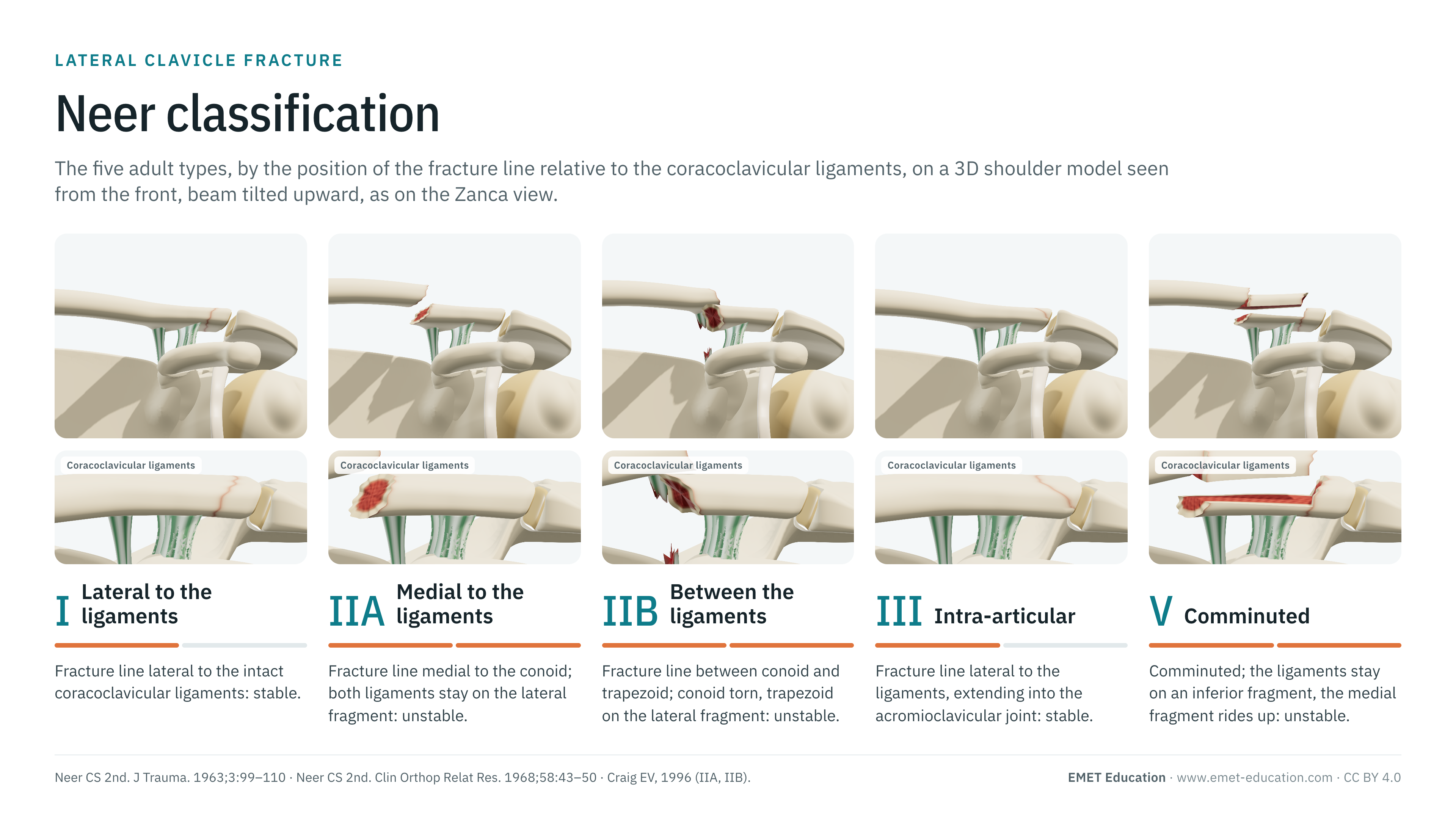
Each image is a rendering of the EMET Education clavicle fracture model at that type, seen as on the radiograph on which the classification is read: from the front, beam tilted 12° upward (Zanca view), the coracoid projecting below the clavicle. Everything depends on the two coracoclavicular ligaments, in green: the conoid medially, the trapezoid laterally. In types IIA, IIB and V, the medial fragment is shown displaced, raised by one bone thickness.
-
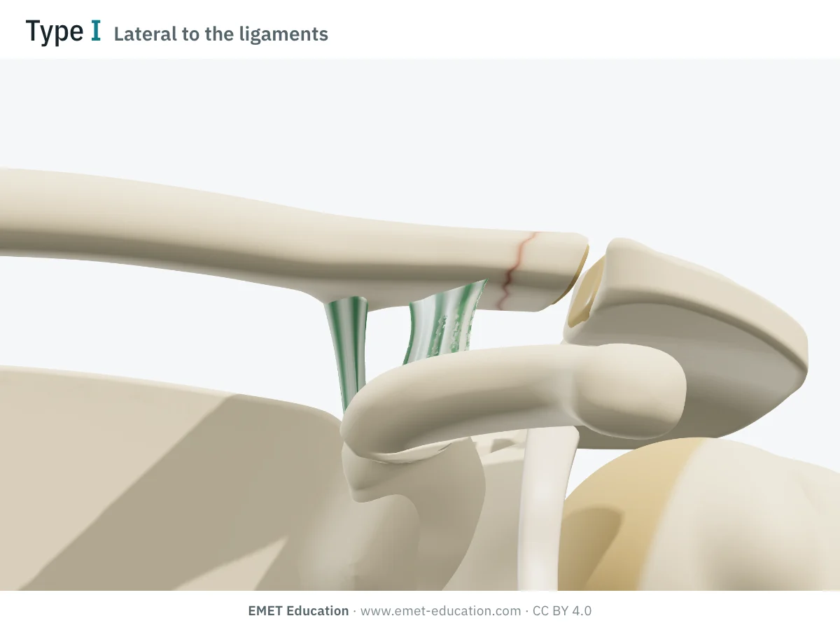
ILateral to the ligaments
- Criteria
- Fracture line lateral to the intact coracoclavicular ligaments, sparing the acromioclavicular joint: a stable, minimally displaced fracture.
- In the 3D model
- Fracture line about 4 mm lateral to the trapezoid; both ligaments stay on the medial fragment, which does not move. A red rim outlines the line.
-
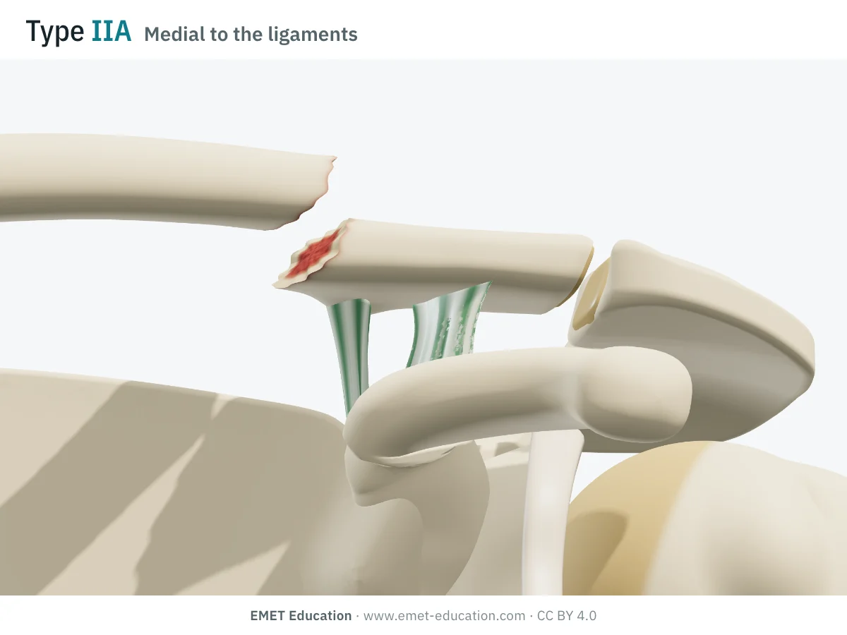
IIAMedial to the ligaments
- Criteria
- Fracture line medial to the conoid; both ligaments, intact, stay attached to the lateral fragment. The medial fragment is no longer held down: an unstable fracture.
- In the 3D model
- Fracture line about 5 mm medial to the conoid; the medial fragment rises by one bone thickness (about 9 mm) and moves back, the shoulder stays in place.
-
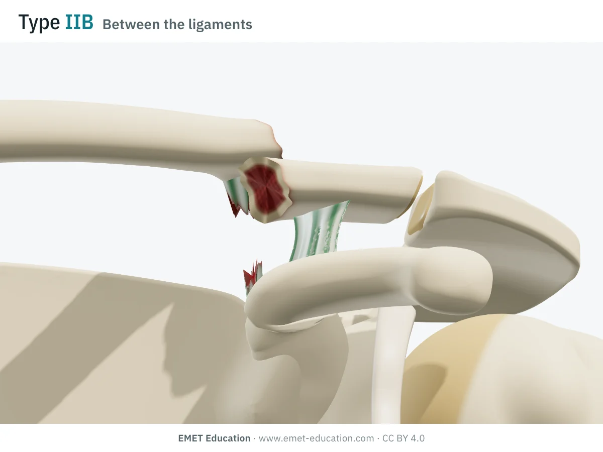
IIBBetween the ligaments
- Criteria
- Fracture line between the conoid and the trapezoid; the conoid is torn, the trapezoid stays attached to the lateral fragment: an unstable fracture.
- In the 3D model
- Fracture line between the two ligament footprints; the torn conoid leaves a stump under the medial fragment, which rises and moves back.
-
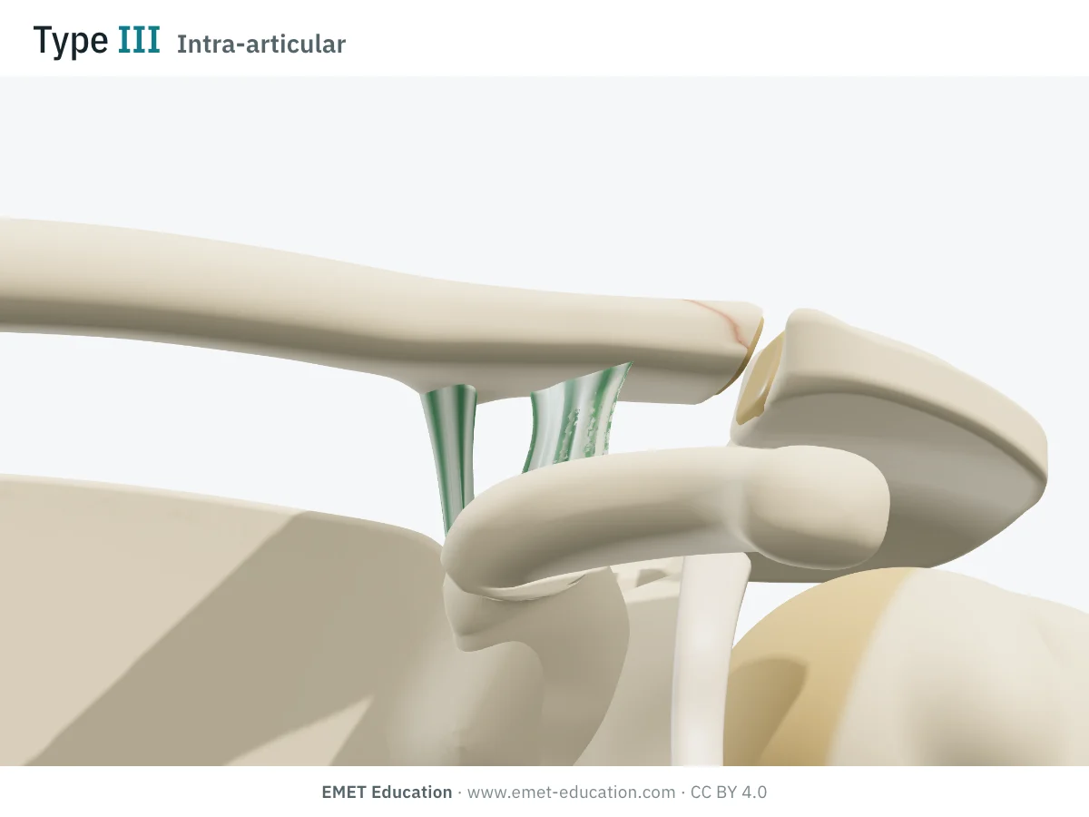
IIIIntra-articular
- Criteria
- Fracture line lateral to the intact coracoclavicular ligaments, extending into the acromioclavicular joint: a stable fracture, at risk of acromioclavicular arthritis.
- In the 3D model
- The upper half of the lateral end breaks off, from the articular facet to about 8 mm medially; no displacement. From the front, the line is barely visible, as on a standard radiograph.
-
IVPediatric
- Criteria
- In children and adolescents: the lateral end of the clavicle slips out of its periosteal sleeve (physeal separation), the coracoclavicular ligaments staying attached to the periosteum. It can mimic an acromioclavicular dislocation.
- In the 3D model
- Not shown: the model is an adult shoulder. Remodeling from the periosteal sleeve explains why these fractures are usually treated without surgery.
-
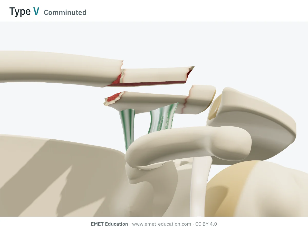
VComminuted
- Criteria
- Comminuted fracture; the coracoclavicular ligaments stay attached to an inferior fragment, neither to the medial nor to the lateral fragment: an unstable fracture.
- In the 3D model
- Two fracture lines, lateral to the trapezoid and medial to the conoid, and a horizontal split: the inferior fragment keeps the ligaments, the medial and superior fragments rise.
Schematic rendering, for teaching: the model illustrates each type on a single shoulder, it does not reproduce a radiograph. Criteria reworded from Neer (1963, 1968), Craig (1996) and Stenson and Baker (2021); their figures are not reproduced. The bar shows stability: one segment, stable; two, unstable.
The origin
Neer, 1963 and 1968; Craig, 1996
Charles S. Neer II, in New York, became interested in fractures of the lateral third of the clavicle while studying nonunions (1960). In 1963, in the Journal of Trauma, he described the fracture of the lateral end with "detachment" of the coracoclavicular ligaments from the medial fragment: freed, it is pulled upward, and the fracture heals poorly. In 1968, in Clinical Orthopaedics and Related Research, he sorted these fractures into types by the position of the fracture line relative to the ligaments.
The version used today, called "modified", comes down to one idea: is the medial fragment still held by the ligaments?
- yes: the line runs lateral to the ligaments, without (type I) or with (type III) involvement of the acromioclavicular joint; the fracture is stable;
- no: the line runs medial to the conoid (IIA) or between a torn conoid and the trapezoid (IIB) — the subdivision of type II is Craig's (1996) — or the ligaments stay on an inferior fragment of a comminuted fracture (V); the fracture is unstable;
- in children, type IV: the lateral end slips out of its periosteal sleeve, to which the ligaments stay attached;
- prognosis follows stability: type II fractures heal poorly without surgery, with 21% to 33% nonunion depending on the series.
Fractures of the lateral end account for 10% to 28% of clavicle fractures. In the Edinburgh series of 1000 adult clavicle fractures, those of the lateral fifth occurred mostly in older people after a simple fall, and displaced fractures more often led to delayed union and nonunion (Robinson, 1998). The modified Neer classification remains the most widely used for these fractures.
Reliability
How reproducible is the type?
Rather poorly. On radiographs, two readers agree only fairly, and 3D CT does not help; Stenson and Baker judged these values too low to base the operative decision on alone. Readers agree better on fracture stability and on the indication than on the type.
| Study | Measure | Value |
|---|---|---|
| Bishop et al., 2015 | Interobserver kappa, 14 shoulder surgeons, 30 fractures (as reported by Stenson and Baker); intraobserver 0.42 | 0.11–0.35 |
| Cho et al., 2015 | Inter- then intraobserver kappa, 18 readers, 52 fractures, radiographs | 0.34 · 0.50 |
| Cho et al., 2015 | The same, radiographs and 3D CT | 0.32 · 0.51 |
| Rauer et al., 2020 | Mean interobserver kappa, 5 radiologists and 5 trauma surgeons, 20 fractures (OTA 0.34) | 0.28 |
| Lin et al., 2023 | Mean interobserver kappa, 16 surgeons, 20 fractures (OTA 0.42) | 0.37 |
| Akgün et al., 2025 | Inter- then intraobserver kappa, 4 shoulder surgeons, 100 fractures | 0.57 · 0.69 |
Agreement is lowest for types IIB and III (Bishop et al.): those that require placing the line relative to an invisible ligament, or seeing a subtle articular line. In Akgün et al., 28 of the 100 fractures could not be classified by at least one reader, and none of the 30 surgeons surveyed could describe the Neer types.
Related classifications
What the other classifications add
Neer sorts fractures by the ligaments, and thus by presumed stability; other classifications take measured displacement, joint involvement or the size of the lateral fragment into account, or seek better reliability.
The most used:
| Classification | What it grades | Typical use |
|---|---|---|
| Robinson (1998) | The lateral fifth (type 3): undisplaced (A) or displaced (B), extra-articular (1) or intra-articular (2). | Epidemiology and prognosis; it is also shown in the 3D model. |
| AO/OTA (15.3) | A code for the lateral end of the clavicle: extra-articular (A), intra-articular (B), comminuted (C). | Research and registries; reliability close to Neer's. |
| Jäger and Breitner | The position of the fracture line relative to the coracoclavicular ligaments, in four types. | German-speaking countries; somewhat more reliable than Neer in Akgün et al. |
| Cho et al. (2018) | A classification designed for implant choice. | Interobserver kappa 0.43, intraobserver 0.64. |
| Akgün et al. (2025) | Three zones relative to the ligaments (lateral, at their insertion, medial), undisplaced (a) or displaced (b). | Recently proposed; interobserver kappa 0.80; 99% of fractures classified. |
Use
In research and in clinical practice
In research
Neer type II defines the population of most surgical studies. In the review of 425 fractures by Oh et al., nonoperative treatment left 33% nonunion, surgery 1.6%, but with 22% other complications, up to 41% with the hook plate. The meta-analysis by Stegeman et al. (350 operated patients) found 98% union and similar functional results across techniques; the hook plate increased the risk of a major complication 11-fold compared with intramedullary fixation.
In clinical practice
The type points toward treatment: for stable types I and III, treatment is almost always nonoperative; for unstable type II, fixation is discussed (locking plate, coracoclavicular fixation, or both), as for type V. But in Bishop et al., it was stability judged on the radiograph, more than the type, that predicted the decision to operate (89% correct predictions).
And a nonunion is not always troublesome: of 127 displaced fractures initially treated without surgery, 14% of patients later had surgery, 21% kept a nonunion, and the mean Constant score of patients not operated on was 93, with no clear difference between those whose fracture had united and those whose had not (Robinson and Cairns, 2004).
Grading pitfalls
Five pitfalls when classifying a distal clavicle fracture
- Ask for a Zanca view. On a standard AP shoulder view, the lateral end overlaps the acromion and the scapular spine: an intra-articular line (type III) is missed. A beam tilted 10° to 15° upward clears the acromioclavicular joint.
- The ligaments are not visible. Their status is inferred from the line and the displacement: an increased coracoclavicular distance, compared with the healthy side, suggests an unstable fracture. In a series studied with MRI, 3 of 59 fractures (5%) graded Neer I on radiographs were actually unstable (Fleischhacker et al., 2022).
- Measure the lateral fragment. The type says nothing about its size, yet a plate needs about 1 cm of lateral bone for its screws. A small fragment points toward coracoclavicular fixation.
- IIA or IIB: locate the conoid. Its tubercle lies about a quarter of the clavicle's length from its lateral end; a line running at the level of the conoid footprint, with a raised medial fragment, suggests IIB.
- In children, think of type IV. Before physeal closure, a displaced lateral end is often a physeal separation within its periosteal sleeve, not an acromioclavicular dislocation.
Self-test
Which type?
The same shoulder seen from the front, slightly laterally, at the five adult types, in random order. Find the type of each one; the answer comes with the criteria.
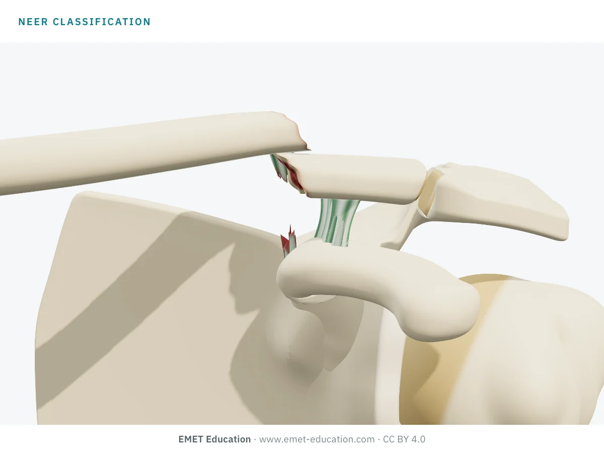
Which Neer type is this?
Download and cite
Free infographics, for teaching and publication
Use them in a course, a presentation, a social media post or a scientific article, under the Creative Commons Attribution 4.0 license: free reuse, including commercial, provided EMET Education is credited with a link to this page.
Cite this figure
Ready to paste. For a journal figure, add the original reference (Neer, 1968).
EMET Education. Neer classification of distal clavicle fractures: 3D visualization [Internet]. 2026 [cited]. Available from: https://www.emet-education.com/en/classifications/neer-clavicle/. CC BY 4.0.EMET Education. (2026). Neer classification of distal clavicle fractures: 3D visualization [Infographic]. https://www.emet-education.com/en/classifications/neer-clavicle/ (CC BY 4.0)<a href="https://www.emet-education.com/en/classifications/neer-clavicle/"><img src="https://www.emet-education.com/en/classifications/neer-clavicle/img/infographic.webp" alt="Neer classification of distal clavicle fractures in 3D" width="960"></a><br>Figure: <a href="https://www.emet-education.com/en/classifications/neer-clavicle/">EMET Education</a>, CC BY 4.0Questions
Frequently asked questions
What is the difference between Neer types IIA and IIB?
Both are unstable: the medial fragment is no longer held by the coracoclavicular ligaments. In type IIA, the line runs medial to the conoid and both ligaments stay on the lateral fragment; in type IIB, it runs between the torn conoid and the trapezoid, which stays on the lateral fragment. The subdivision is Craig's.
Why does type II displace?
Because the medial fragment, freed from the ligaments, is pulled upward and backward (sternocleidomastoid, trapezius), while the lateral fragment, still tied to the scapula by the ligaments, drops with the weight of the arm. The resulting gap explains the high nonunion rate without surgery.
Are type I or III fractures treated surgically?
Usually not: these fractures are stable, and are treated with a sling and gradual rehabilitation. In type III, acromioclavicular arthritis may develop later.
Is the clavicle "Neer classification" the same as the humeral one?
No. Neer also described the four-part classification of proximal humerus fractures (1970) and the stages of subacromial impingement. This page covers fractures of the lateral end of the clavicle.
Should every type II fracture be operated on?
Not always. Surgery clearly lowers the nonunion risk, but at the cost of its own complications; and a nonunion of the lateral end is often well tolerated, especially in an older, low-demand person. The decision takes into account displacement, the size of the lateral fragment and the patient, not just the type.
Can I reuse these images in a course or an article?
Yes. The images and infographics on this page are under the Creative Commons Attribution 4.0 license: free reuse, including commercial, with credit to EMET Education and a link to this page. The "Cite this figure" box gives ready-to-paste citations.
How were the 3D images made?
They are renderings of the EMET Education clavicle fracture model, set to each type: the fracture line is placed from the insertions of the model's coracoclavicular ligaments, and the medial fragment rises when it is no longer held. It is a schematic model for teaching and patient explanation; it does not reproduce a radiograph.
Sources
Author and references
- Neer CS 2nd. Fracture of the distal clavicle with detachment of the coracoclavicular ligaments in adults. J Trauma. 1963;3:99–110. doi:10.1097/00005373-196303000-00001
- Neer CS 2nd. Fractures of the distal third of the clavicle. Clin Orthop Relat Res. 1968;58:43–50.
- Stenson J, Baker W. Classifications in brief: the modified Neer classification for distal-third clavicle fractures. Clin Orthop Relat Res. 2021;479(1):205–9. doi:10.1097/CORR.0000000000001456
- Robinson CM. Fractures of the clavicle in the adult. Epidemiology and classification. J Bone Joint Surg Br. 1998;80(3):476–84. doi:10.1302/0301-620x.80b3.8079
- Craig EV. Fractures of the clavicle. In: Rockwood CA Jr, Green DP, Bucholz RW, Heckman JD, editors. Rockwood and Green's Fractures in Adults. 4th ed. Philadelphia: Lippincott-Raven; 1996. p. 1109–93.
- Bishop JY, Jones GL, Lewis B, Pedroza A; MOON Shoulder Group. Intra- and interobserver agreement in the classification and treatment of distal third clavicle fractures. Am J Sports Med. 2015;43(4):979–84. doi:10.1177/0363546514563281
- Cho CH, Oh JH, Jung GH, et al. The interrater and intrarater agreement of a modified Neer classification system and associated treatment choice for lateral clavicle fractures. Am J Sports Med. 2015;43(10):2431–6. doi:10.1177/0363546515593949
- Rauer T, Boos M, Neuhaus V, et al. Inter- and intraobserver agreement of three classification systems for lateral clavicle fractures – reliability comparison between two specialist groups. Patient Saf Surg. 2020;14:4. doi:10.1186/s13037-019-0228-y
- Lin J, Sun WJ, Chen JH, et al. Comparison of interobserver agreement of four classification systems for lateral clavicle fractures between two groups of surgeons: a multicenter study. Orthop Surg. 2023;15(8):2138–43. doi:10.1111/os.13659
- Akgün D, Moroder P, Lacheta L, et al. Differentiating and treating lateral clavicle fractures: a new simple classification system. J Shoulder Elbow Surg. 2026;35(1):296–302 (epub 2025). doi:10.1016/j.jse.2025.04.021
- Cho CH, Kim BS, Kim DH, Choi CH, Dan J, Lee H. Distal clavicle fractures: a new classification system. Orthop Traumatol Surg Res. 2018;104(8):1231–5. doi:10.1016/j.otsr.2018.05.015
- Oh JH, Kim SH, Lee JH, Shin SH, Gong HS. Treatment of distal clavicle fracture: a systematic review of treatment modalities in 425 fractures. Arch Orthop Trauma Surg. 2011;131(4):525–33. doi:10.1007/s00402-010-1196-y
- Stegeman SA, Nacak H, Huvenaars KH, Stijnen T, Krijnen P, Schipper IB. Surgical treatment of Neer type-II fractures of the distal clavicle: a meta-analysis. Acta Orthop. 2013;84(2):184–90. doi:10.3109/17453674.2013.786637
- Robinson CM, Cairns DA. Primary nonoperative treatment of displaced lateral fractures of the clavicle. J Bone Joint Surg Am. 2004;86(4):778–82. doi:10.2106/00004623-200404000-00016
- Fleischhacker E, Siebenbürger G, Gleich J, Böcker W, Gilbert F, Helfen T. The accuracy of distal clavicle fracture classifications — do we need an amendment to imaging modalities or fracture typing? J Clin Med. 2022;11(19):5638. doi:10.3390/jcm11195638
3D models
Show the fracture to your patient
The images on this page come from an interactive model. In clinic, choose the type and displacement from your patient's radiograph, then show the proposed treatment: nonoperative treatment, locking plate or coracoclavicular fixation.
Explain the fracture in 3D
The clavicle fracture model opens in your browser, at the type you choose, with no sign-up.
Share this page with a colleague or a resident: