- EMET Education
- 3D classifications
- AO/OTA classification (radial head)
Classification · Radial head fracture
AO/OTA Classification of Radial Head Fractures in 3D
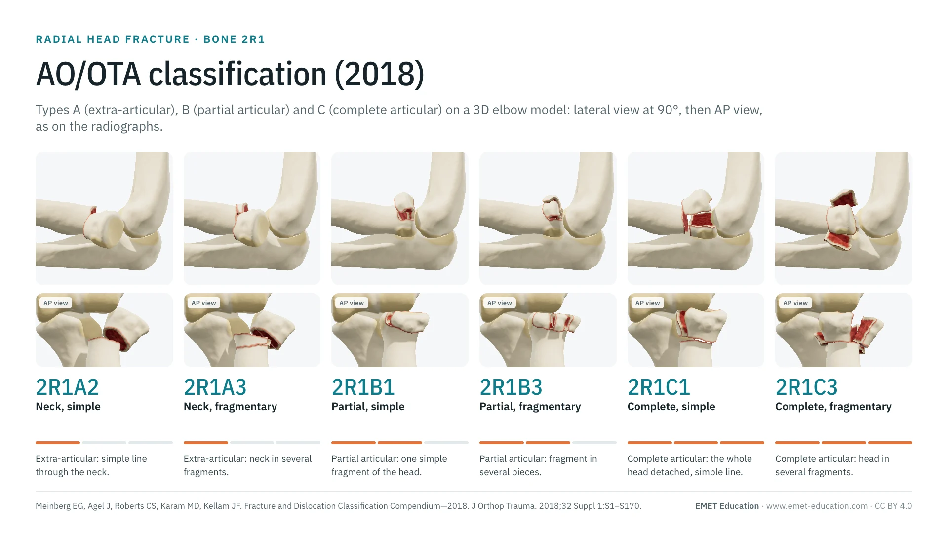
The seven codes of the AO/OTA 2018 classification of the proximal end of the radius (bone 2R1: A extra-articular, B partial articular, C complete articular), on the radial head fracture 3D model, seen in lateral view at 90° and AP view as on elbow radiographs. With how to read the code, its link with the Mason classification, reliability data, grading pitfalls and free infographics for teaching and publication.
Designed by an orthopedic surgeon
The 3D model opens in your browser, at the code shown, with no sign-up. Infographics are free to reuse under CC BY 4.0.
- 2018Revision of the AO/OTA compendium, in the Journal of Orthopaedic Trauma (Meinberg et al.)
- 7Codes of the proximal end of the radius, from 2R1A1 to 2R1C3: the code stops at the group, with no subgroup
- 2,771Citations of the 2018 compendium, all bones (OpenAlex, November 4, 2026)
- κ 0.26Interobserver agreement of the full AO code, 5 observers, 43 patients; Hotchkiss: 0.59 (Sheps et al.)
The codes
Types A, B and C, in 3D
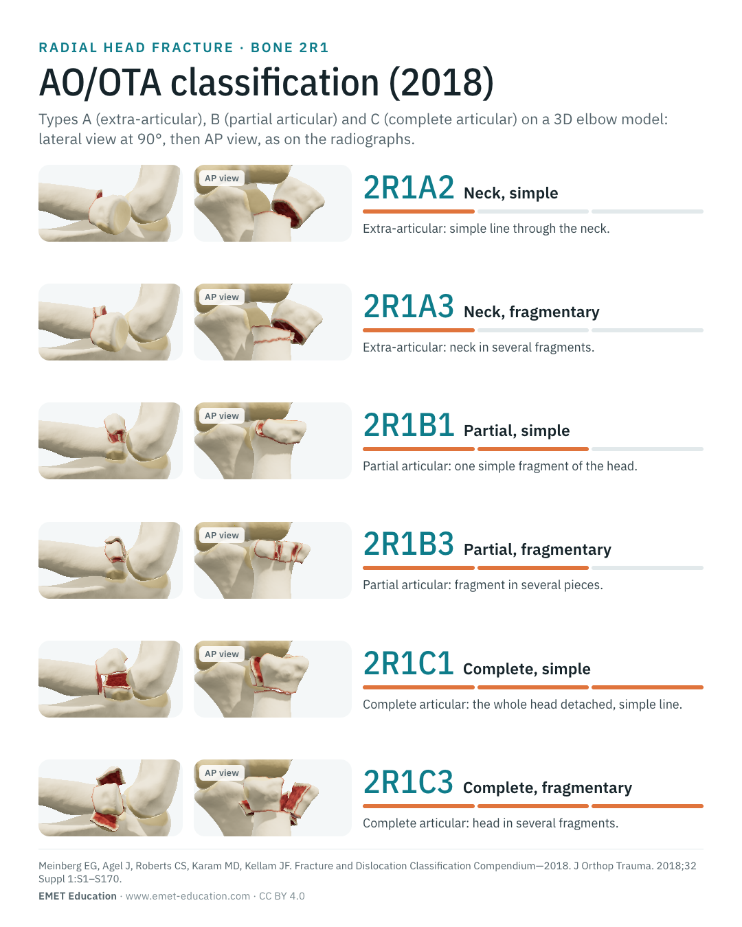
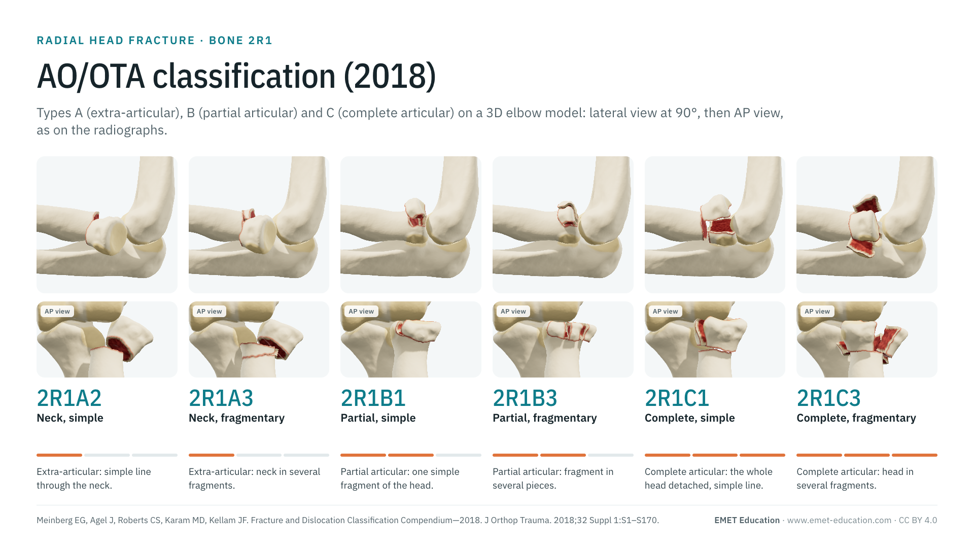
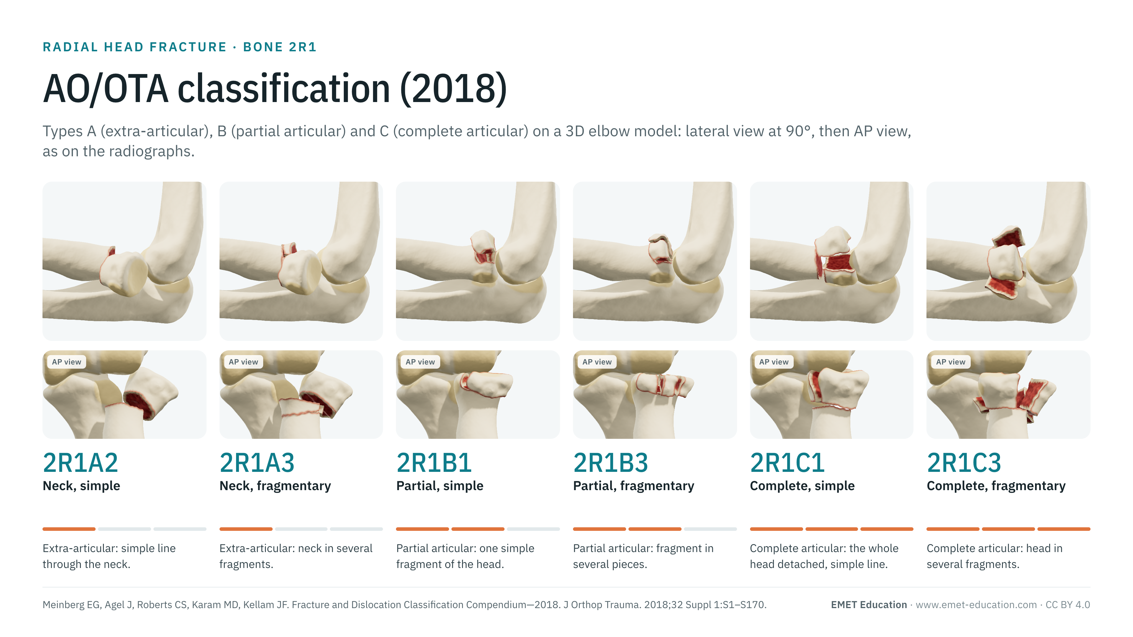
Each image is a rendering of the EMET Education radial head fracture model at that code, left elbow seen in lateral view at 90° of flexion, the head beneath the capitellum and the anterior fragment above it, with a narrow field to approach the projection of a lateral radiograph; the infographic adds the AP view. The model draws six of the seven codes: 2R1A1 (avulsion of the bicipital tuberosity) is described without an image. The displacements shown are typical cases, adjustable in the model: the code itself does not say whether the fracture is displaced, that is the role of the modifiers. Under each code, the closest type of the Mason classification according to the model.
-
2R1A1Bicipital tuberosity avulsion
- Definition
- Type A, group 1: avulsion of the bicipital tuberosity of the radius, where the biceps tendon inserts. Biceps tendon avulsions usually occur without a fracture; weakness of supination suggests one (AO Surgery Reference).
- Mason, indicative
- Outside Mason, which describes only the head.
- In the 3D model
- Not drawn: the model shows neither the bicipital tuberosity nor the distal biceps tendon.
-
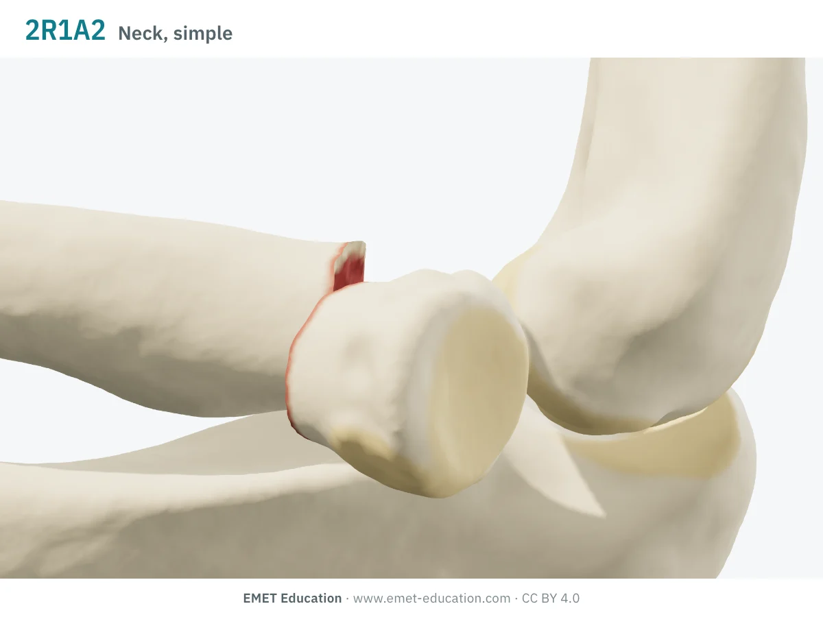
2R1A2Neck, simple
- Definition
- Type A, group 2: simple extra-articular fracture of the radial neck, one single line (AO Surgery Reference). When displaced and unstable, the AO Surgery Reference advises looking for injury to the medial collateral ligament (valgus mechanism), to both collateral ligaments (elbow dislocation) or to the interosseous ligament of the forearm (Essex-Lopresti injury).
- Mason, indicative
- Type II, by convention: Mason described only the head, reviews place a displaced or angulated neck in type II (van der Windt et al.).
- In the 3D model
- The head tilts 35° outward, translated 2 mm: one simple line through the neck, with no other fragment.
-
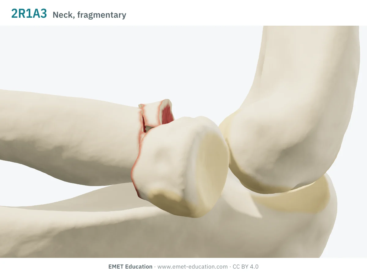
2R1A3Neck, multifragmentary
- Definition
- Type A, group 3: extra-articular multifragmentary fracture of the neck. It is uncommon and difficult to repair (AO Surgery Reference).
- Mason, indicative
- Type II, by convention, like the simple neck (EMET Education's reading: Mason's definition applies only to the head).
- In the 3D model
- The ring of the neck in three fragments, the head angulated 25° with a 2 mm gap.
-
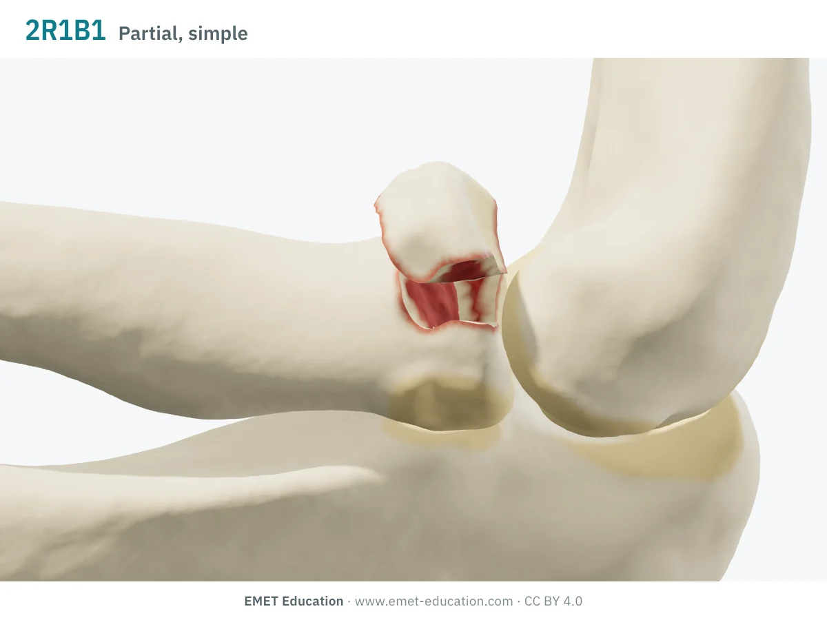
2R1B1Partial articular, simple
- Definition
- Type B, group 1: simple partial articular fracture: one fragment of the articular surface of the head breaks off, the rest of the head stays attached to the neck (AO Surgery Reference). The fracture is called stable when no ligament injury of the elbow or forearm goes with it; it is unstable with an elbow dislocation or in an Essex-Lopresti injury, and a wide gap between fragments suggests an associated ligament or bone injury.
- Mason, indicative
- Type I with modifier [1] (undisplaced); type II with [2] (displaced: at least 2 mm and 30% of the head according to the threshold attributed to Broberg and Morrey).
- In the 3D model
- Anterolateral fragment of 135° (about 34% of the head), displaced 3 mm, tilted 20° and depressed 1 mm.
-
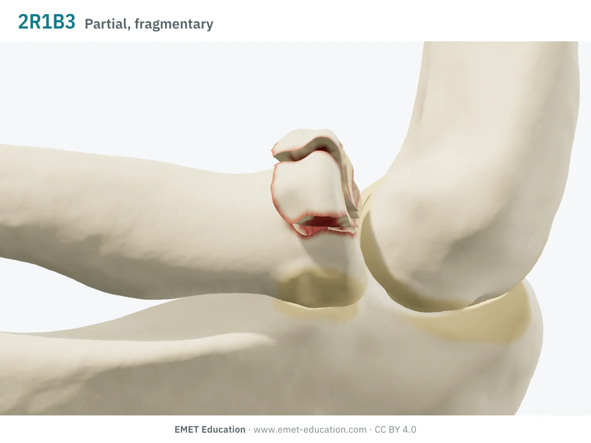
2R1B3Partial articular, fragmentary
- Definition
- Type B, group 3: fragmentary partial articular fracture: the detached portion of the articular surface is itself in several pieces, the rest of the head staying attached to the neck (AO Surgery Reference). The same stability conditions as for 2R1B1 apply.
- Mason, indicative
- Type II (Hotchkiss's reading: reconstructable); Mason reserved type III for the whole head being comminuted (van der Windt et al.). Modifier [1] or [2] says whether it is displaced.
- In the 3D model
- A 135° sector in 3 fragments, displaced 3 mm, tilted 15° and depressed 1.5 mm.
-
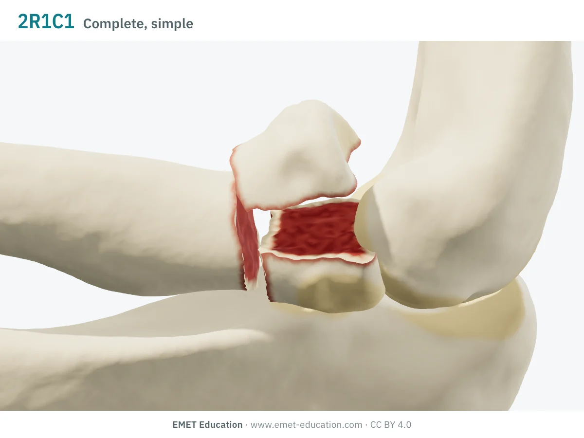
2R1C1Complete articular, simple
- Definition
- Type C, group 1: simple complete articular fracture: the whole articular surface of the head is detached from the neck, the fracture remaining simple, without fragmentation (AO Surgery Reference). It is stable without an associated ligament injury, unstable with an elbow dislocation or in an Essex-Lopresti injury; a gap between fragments suggests an associated ligament or bone injury.
- Mason, indicative
- Type III, the model's reading: the whole head is involved, but Mason does not describe this case separately (Hotchkiss: depending on whether the head is reconstructable).
- In the 3D model
- Fractured neck (3 mm gap, 10° angulation, 1 mm translation) and head split in two: the model's reading of "simple" (two lines).
-
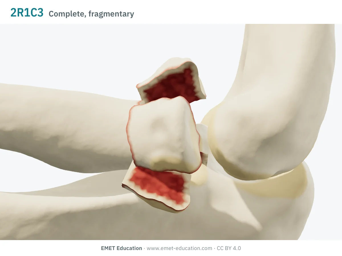
2R1C3Complete articular, multifragmentary
- Definition
- Type C, group 3: multifragmentary complete articular fracture: the whole head is detached from the neck and fragmented (AO Surgery Reference). Beyond 3 articular fragments, fixation becomes unreliable (13 of 14 unsatisfactory results: Ring et al.).
- Mason, indicative
- Type III: comminuted fracture of the whole head. Modifier [1] or [2] says whether it is displaced.
- In the 3D model
- The neck and four fragments of the head (3 to 5 if chosen), displaced 4 mm, tilted 20° and depressed 2 mm.
Schematic rendering, for teaching: the model illustrates each code on a single elbow, it does not reproduce a radiograph. Definitions reworded from the 2018 compendium (Meinberg et al.) and the AO Surgery Reference; their figures and pictograms are not reproduced. The bar shows the type: one segment A, two B, three C. The correspondences with Mason are the model's, for guidance.
Reading the code
From Müller (1990) to the 2018 compendium
The classification comes from Müller et al.'s classification of long-bone fractures, adopted by the Orthopaedic Trauma Association, whose 2018 compendium is the reference version. For the proximal end of the radius, it comes down to seven codes that stop at the group: there is no subgroup, and the numbers 2 of types B and C do not exist. The two reliability studies of the radial head (2009) predate this version: their results do not transfer as such to the 2018 code.
The code reads from left to right:
- 2R1: bone 2, the forearm; the letter R, the radius (U for the ulna: an injury to both bones is coded twice); its segment 1, the proximal end. End segments are delimited by a square, the two forearm bones being taken as a unit;
- the type: A, extra-articular fracture (the bicipital tuberosity or the neck); B, partial articular (part of the surface of the head stays attached to the neck); C, complete articular (the whole surface of the head is detached from the neck);
- the group: A1 avulsion of the bicipital tuberosity, A2 simple neck, A3 multifragmentary neck; B1 simple partial articular, B3 fragmentary partial articular; C1 simple complete articular, C3 multifragmentary complete articular;
- universal modifiers, as numbers in square brackets, shared by all bones: [1] undisplaced, [2] displaced, [3] impaction (articular or metaphyseal), [4] no impaction, [5] dislocation, with its direction ([5a] to [5f]) (Meinberg et al.). Thus 2R1B1[2]: simple partial articular fracture, displaced; 2R1B1[2,5b] with a posterior dislocation. See also the proximal humerus page, which goes into how to read the code.
The code describes the line and the number of fragments, not how far it is displaced or the neighbouring injuries: a simple fracture of the head may go with a coronoid fracture or a dislocation, which do not appear in the letters and numbers of the code (see the Mason classification and the Mayo suffixes).
Mason and AO/OTA
From the Mason type to the AO/OTA code
The two systems do not overlap: Mason mostly describes displacement and fragment size, the AO/OTA code the line and the number of fragments, with displacement as a modifier. A single code therefore covers several Mason types (2R1B1 is type I or II depending on displacement), and a single type several codes. The table gives the model's correspondence, on the Mason classification page.
| Mason type | AO/OTA 2018 code | Note |
|---|---|---|
| I, undisplaced | 2R1B1[1] | Fissure or marginal fracture with no displacement: simple partial articular fracture, with modifier [1] (Meinberg et al.). A displaced fragment of less than 2 mm or less than 30% of the head remains type I since Broberg and Morrey (attributed threshold). |
| II, displaced fragment | 2R1B1[2] | The same code, with modifier [2]: displacement makes the type, not the line. |
| II, several fragments | 2R1B3 | Fragmented sector, head continuous with the neck: read as type II as in Hotchkiss; Mason reserved III for the whole head being comminuted. |
| II, neck | 2R1A2 (2R1A3 if multifragmentary) | Type II by convention (van der Windt et al.): the neck is extra-articular in the AO/OTA, therefore type A. |
| III, head detached from the neck and split | 2R1C1 | The model's reading: the whole head is involved, Mason does not describe this case separately. |
| III, comminuted | 2R1C3 | The whole head in several fragments: this is Mason's definition. |
| IV, with elbow dislocation | the head's code, with [5] | Dislocation becomes a modifier, with its direction ([5a] to [5f]); Johnston's type IV (1962) has no code of its own. The head's code remains that of its fracture. |
Reliability
How reproducible is the code?
Poorly, on radiographs, in the two studies we found: both date from 2009 and concern a version earlier than the 2018 one; we found no reliability study of the 2018 code for the radial head. In both, the Mason (or Hotchkiss) classification agrees better than the AO; in Sheps et al., dropping the last digit of the code improves agreement.
| Study | Measure | Value |
|---|---|---|
| Matsunaga et al., 2009 | AO/ASIF, 4 observers, 3 sessions: inter- then intraobserver kappa, rated poor by the authors (Mason: 0.43–0.56 · 0.58) | 0.25–0.48 · 0.48 |
| Sheps et al., 2009 | Full AO code, 5 observers, 43 patients: interobserver kappa, "fair" (Hotchkiss: 0.59, "moderate"), mean raw agreement 37.7% | 0.26 |
| Sheps et al., 2009 | AO code without its last digit: interobserver kappa, "moderate" (Hotchkiss with types II and III combined: 0.76, "substantial"), mean raw agreement 67.4% | 0.46 |
In these series, the full AO code therefore agrees less well than Mason or Hotchkiss (Matsunaga et al., Sheps et al.), and the simplification, which amounts to keeping only the type and the group, only reaches the low end of the "moderate" range. In practice: the type to communicate, the full code for research, with written thresholds, and the Mason classification or Hotchkiss's to decide.
Neighbouring classifications
Mason, Broberg and Morrey, Hotchkiss, Mayo
For the radial head, the AO/OTA is mostly the code of registries and studies; the neighbouring classifications, older, steer decisions or describe the associated injuries. Their principles, from the reviews and the original papers:
| Classification | Principle | Relation to the AO/OTA |
|---|---|---|
| Mason, 1954 and Johnston, 1962 | I: undisplaced fissure or marginal fracture; II: displaced marginal sector; III: whole head comminuted; IV (Johnston): head fracture with elbow dislocation | Describes displacement and dislocation, which the AO/OTA leaves to modifiers. See the Mason page. |
| Broberg and Morrey, 1987 | Type II: at least 2 mm of displacement and at least 30% of the articular surface; takes up Johnston's type IV | Threshold attributed to these authors by the reviews: it gives a numerical meaning to modifier [2] for the head. |
| Hotchkiss, 1997 | I: undisplaced or minimally displaced (under 2 mm), no block; II: displaced (often over 2 mm) or angulated neck, reconstructable; III: comminuted, not reconstructable, excision or replacement | Reasons in terms of management. Interobserver agreement "moderate" (κ 0.59) against "fair" for the AO (0.26) in Sheps et al. |
| Mayo suffixes (van Riet and Morrey, 2008) | The Mason type, followed by letters for associated injuries: c coronoid, o olecranon, l LCL, m MCL, d distal radioulnar joint | Associated injuries are not in the head's AO/OTA code: the model adds them to its reading. |
Use
In research and in clinical practice
In research
The AO/OTA code is the language of trauma registries and databases: it fits on one line, sorts and groups. In a prospective series of 201 radial head or neck fractures, increasing complexity according to the AO-OTA (version not specified in the abstract), radiographic comminution and age, and the choice to operate, were independent predictors of a poorer outcome (Duckworth et al.). The 2018 code is also used to label radiographs for artificial intelligence: across 5,367 elbow examinations and 54 fracture types, the weighted mean area under the curve was 0.80 for the proximal radius (Pettersson et al.).
In clinical practice
The type, A, B or C, sums up the essentials: neck, partial articular fracture (a fragment stays attached to the neck) or fracture of the whole head. The code alone tells neither whether the fracture is displaced nor whether it is stable. The AO Surgery Reference points out that a displaced, unstable neck fracture calls for a search for collateral or interosseous ligament injury, and that an articular fracture is unstable with an elbow dislocation or in an Essex-Lopresti injury (AO Surgery Reference).
It is the Mason classification (or Hotchkiss's), more than the AO/OTA code, that steers treatment: fragment displaced at least 2 mm and 30% of the head, number of articular fragments (beyond three, fixation is unreliable: Ring et al.), mechanical block, associated injuries. The AO/OTA code is recorded alongside, with displacement modifier [1] or [2], for the report and for research.
Grading pitfalls
Five pitfalls when coding a radial head fracture
- Displacement is not in the code. A simple articular fragment is a 2R1B1, displaced or not: record [1] or [2], and state the threshold used (2 mm and 30% of the head, attributed to Broberg and Morrey). Without a modifier, the code does not say whether the fracture is operated.
- The neck is an A, even if the head is tilted. A line through the neck alone is extra-articular: 2R1A2 if simple, 2R1A3 if fragmentary. The head is B or C only if its articular surface is broken.
- B or C? The question is whether any part of the articular surface stays attached to the neck. On radiographs, agreement between readers is poor (Sheps et al.): write down the criterion used and, when in doubt about the number of fragments, review the cross-sectional imaging.
- Simple or fragmentary? An articular fragment in several pieces is a B3, even if the head stays continuous with the neck: it is a Mason type II in Hotchkiss's scheme. Do not write 2R1B2 or 2R1C2: these numbers do not exist for the radial head.
- What is not in the code. The coronoid, the lateral collateral ligament, the distal radioulnar joint and the dislocation: the dislocation goes in modifier [5], the rest in the description (Mayo suffixes). A displaced, unstable neck fracture should prompt a search for an Essex-Lopresti injury (AO Surgery Reference).
Self-test
Which code?
The same elbow seen from three-quarters, at the six codes the model draws, in random order. Find the code of each one; the answer comes with the definition.
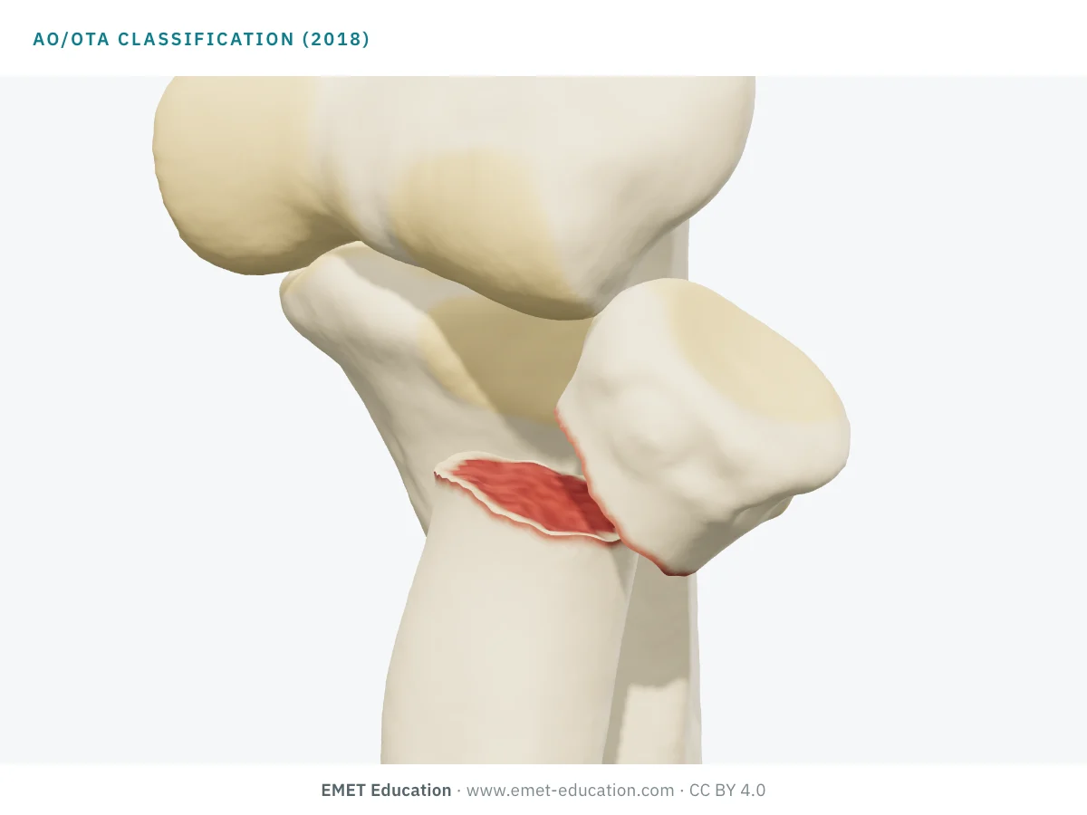
Which AO/OTA code is this?
Download and cite
Free infographics, for teaching and publication
Use them in a course, a presentation, a social media post or a scientific article, under the Creative Commons Attribution 4.0 license: free reuse, including commercial, provided EMET Education is credited with a link to this page. The license covers EMET Education's images, not the classification itself.
Cite this figure
Ready to paste. For a journal figure, add the original reference (Meinberg et al., 2018).
EMET Education. AO/OTA classification of radial head fractures: 3D visualization [Internet]. 2026 [cited]. Available from: https://www.emet-education.com/en/classifications/ao-ota-radial-head/. CC BY 4.0.EMET Education. (2026). AO/OTA classification of radial head fractures: 3D visualization [Infographic]. https://www.emet-education.com/en/classifications/ao-ota-radial-head/ (CC BY 4.0)<a href="https://www.emet-education.com/en/classifications/ao-ota-radial-head/"><img src="https://www.emet-education.com/en/classifications/ao-ota-radial-head/img/infographic.webp" alt="AO/OTA classification of radial head fractures in 3D" width="960"></a><br>Figure: <a href="https://www.emet-education.com/en/classifications/ao-ota-radial-head/">EMET Education</a>, CC BY 4.0Questions
Frequently asked questions
What does "2R1B1" mean in the code?
The 2 is the forearm, the letter R the radius (U: the ulna), the 1 the proximal segment. B is the type (partial articular fracture: a fragment of the head stays attached to the neck) and 1 the group (simple). There is no subgroup for the radial head. Modifiers, in square brackets ([1] undisplaced, [2] displaced, [5] dislocation), come after.
Which AO/OTA codes for Mason I, II, III and IV?
There is no official table; the page's correspondence is the model's, for guidance. Type I: 2R1B1[1]. Type II: 2R1B1[2] for one fragment, 2R1B3 for several fragments, 2R1A2 or 2R1A3 for the neck (by convention). Type III: 2R1C3 for the comminuted head, 2R1C1 for the detached, split head (the model's reading). Type IV: the head's code, with the dislocation modifier [5].
Why is a neck fracture "extra-articular"?
Because the compendium places it in type A: the AO Surgery Reference calls simple neck fractures 2R1A2 and multifragmentary neck fractures 2R1A3, under the heading "radius, extraarticular". Type B or C supposes that the articular surface of the head is itself broken.
AO/OTA or Mason: which one to use for the radial head?
To decide, Mason (or Hotchkiss): in the two 2009 studies we found, the AO code agrees less well (κ 0.26 against 0.59 for Hotchkiss in Sheps; 0.25–0.48 against 0.43–0.56 for Mason in Matsunaga). For a registry or a study, the AO/OTA code, with its displacement modifier. The two are recorded together.
Does the AO/OTA code say whether the fracture is displaced, stable or needs surgery?
No. Displacement is a modifier ([1] or [2]); stability is judged on association with an elbow dislocation, an Essex-Lopresti injury or a ligament injury, which the AO Surgery Reference mentions without including them in the code. The indication for surgery rests on displacement (2 mm and 30% of the head, threshold attributed to Broberg and Morrey), the number of fragments, mechanical block and associated injuries.
Can I reuse these images in a course or an article?
Yes. The images and infographics on this page are under the Creative Commons Attribution 4.0 license: free reuse, including commercial, with credit to EMET Education and a link to this page. The "Cite this figure" box gives ready-to-paste citations.
Do the images reproduce the AO drawings? How were they made?
No: no figure or pictogram from the compendium is reproduced, and the definitions are reworded. They are renderings of the EMET Education radial head fracture model, set to each code: the radius, humerus and ulna, taken from CT, are cut along the lines of the code and displaced so that they never cross each other. It is a schematic model for teaching; it does not reproduce a radiograph. EMET Education is not affiliated with the AO Foundation or the Orthopaedic Trauma Association.
Sources
Author and references
- Meinberg EG, Agel J, Roberts CS, Karam MD, Kellam JF. Fracture and Dislocation Classification Compendium—2018. J Orthop Trauma. 2018;32 Suppl 1:S1–S170. doi:10.1097/BOT.0000000000001063
- AO Foundation. AO Surgery Reference, orthopedic trauma, adult trauma, proximal forearm: radius, extraarticular (avulsion of bicipital tuberosity 2R1A1, simple 2R1A2, multifragmentary 2R1A3), partial articular (2R1B1, 2R1B3) and complete articular (2R1C1, 2R1C3), definitions. surgeryreference.aofoundation.org. Consulté le 4 novembre 2026.
- Müller ME, Nazarian S, Koch P, Schatzker J. The Comprehensive Classification of Fractures of Long Bones. Berlin: Springer; 1990.
- Matsunaga FT, Tamaoki MJ, Cordeiro EF, et al. Are classifications of proximal radius fractures reproducible? BMC Musculoskelet Disord. 2009;10:120. doi:10.1186/1471-2474-10-120
- Sheps DM, Kiefer KR, Boorman RS, et al. The interobserver reliability of classification systems for radial head fractures: the Hotchkiss modification of the Mason classification and the AO classification systems. Can J Surg. 2009;52(4):277–82. PMID 19680511
- Pettersson A, Axenhus M, Stukan T, Ljungberg O, Nåsell H, Razavian AS, Gordon M. Use of artificial intelligence for classification of fractures around the elbow in adults according to the 2018 AO/OTA classification system. BMC Musculoskelet Disord. 2025;26(1):848. doi:10.1186/s12891-025-09161-2
- Duckworth AD, Watson BS, Will EM, et al. Radial head and neck fractures: functional results and predictors of outcome. J Trauma. 2011;71(3):643–8. doi:10.1097/TA.0b013e3181f8fa5f
- Mason ML. Some observations on fractures of the head of the radius with a review of one hundred cases. Br J Surg. 1954;42(172):123–32. doi:10.1002/bjs.18004217203
- Johnston GW. A follow-up of one hundred cases of fracture of the head of the radius with a review of the literature. Ulster Med J. 1962;31(1):51–6. PMID 14452145
- Broberg MA, Morrey BF. Results of treatment of fracture-dislocations of the elbow. Clin Orthop Relat Res. 1987;(216):109–19. doi:10.1097/00003086-198703000-00017
- Hotchkiss RN. Displaced fractures of the radial head: internal fixation or excision? J Am Acad Orthop Surg. 1997;5(1):1–10. doi:10.5435/00124635-199701000-00001
- van Riet RP, Morrey BF. Documentation of associated injuries occurring with radial head fracture. Clin Orthop Relat Res. 2008;466(1):130–4. doi:10.1007/s11999-007-0064-8
- van der Windt AE, Langenberg LC, Colaris JW, Eygendaal D. Which radial head fractures are best treated surgically? EFORT Open Rev. 2024;9(5):413–21. doi:10.1530/EOR-24-0035
- Ring D, Quintero J, Jupiter JB. Open reduction and internal fixation of fractures of the radial head. J Bone Joint Surg Am. 2002;84(10):1811–5. doi:10.2106/00004623-200210000-00011
3D model
The 3D model and related classifications
The images on this page come from an interactive model, the radial head fracture 3D model. Choose the AO/OTA 2R1 code (or the Mason type), set the fragment size, displacement, tilt and number of fragments from the imaging, add the coronoid, the dislocation, the Osborne-Cotterill lesion or the LCL tear, then play the fixation (headless screws, plate) or the radial head replacement.
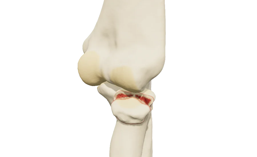 ElbowRadial head fractureMason and AO/OTA, coronoid, dislocation, Osborne-Cotterill lesion, LCL, headless screws, plate and replacement.
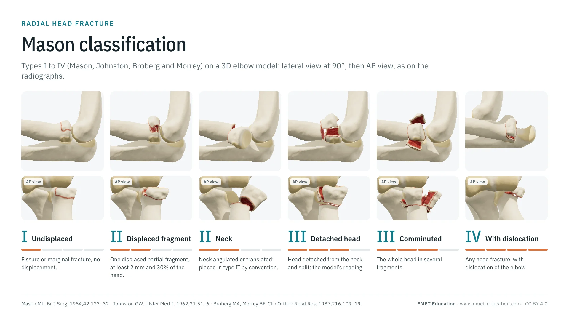 ClassificationsMason classification of the radial headTypes I to IV, the classification that steers treatment, in 3D.
 ClassificationsAll 3D classificationsArranged by joint.
Explain the fracture in 3D
The radial head fracture model opens in your browser, at the code you choose, with no sign-up.
Share this page with a colleague or a resident: