- EMET Education
- 3D classifications
- AO/OTA classification (proximal humerus)
Classification · Proximal humerus fracture
AO/OTA Classification of Proximal Humerus Fractures in 3D
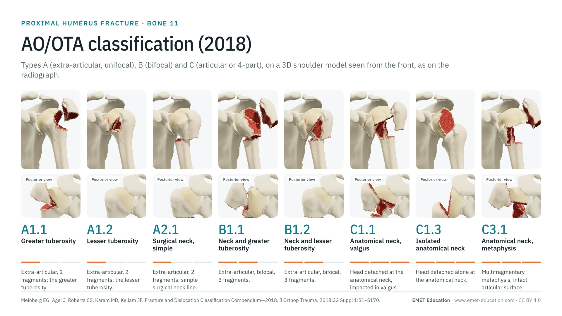
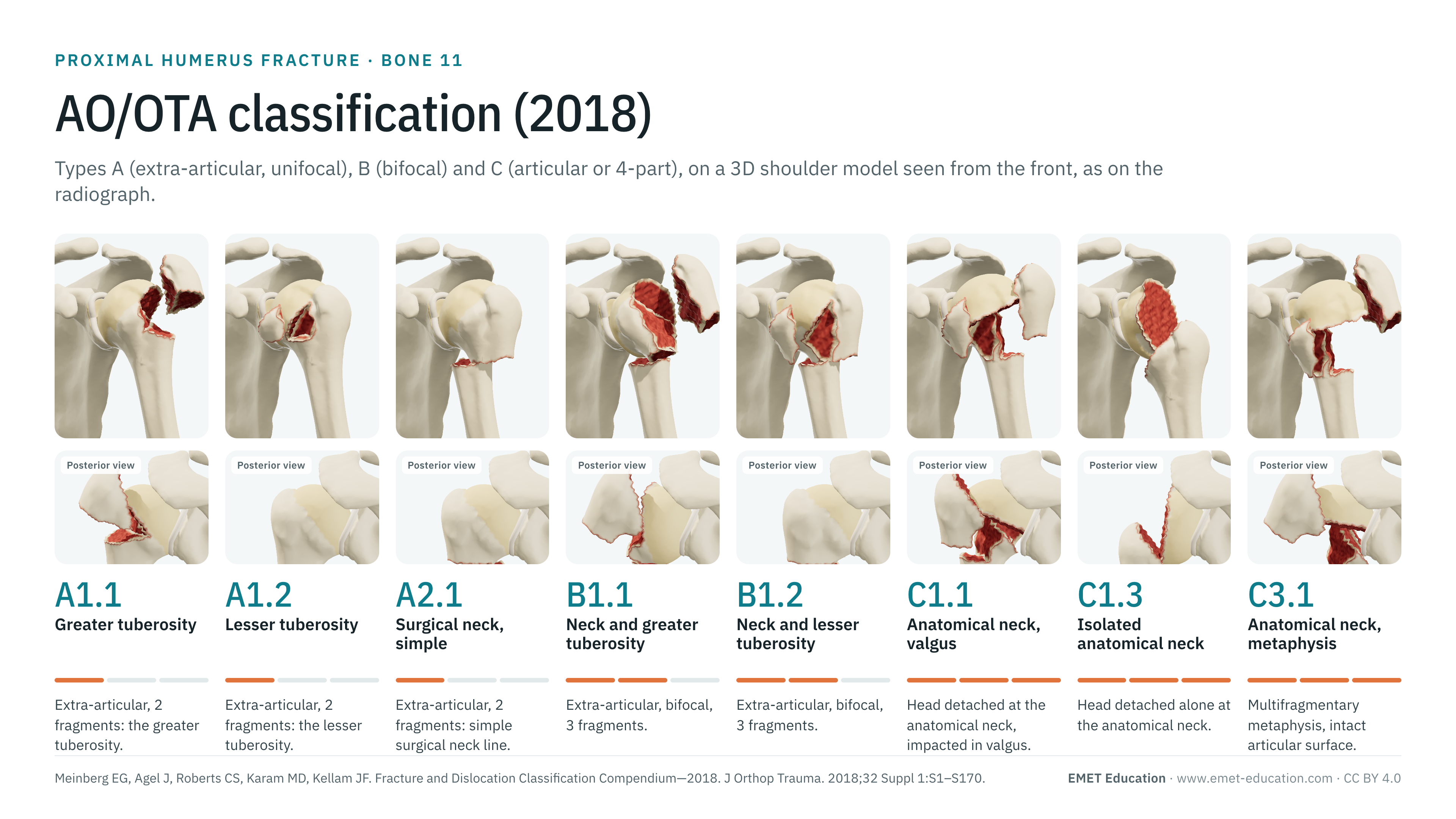
The types, groups and subgroups of the AO/OTA 2018 classification of the proximal humerus (bone 11), on the proximal humerus fracture 3D model, seen from the front as on the radiograph. With how to read the code, its qualifications and modifiers, the link with Neer, reliability data, grading pitfalls and free infographics for teaching and publication.
Designed by an orthopedic surgeon
The 3D model opens in your browser, at the code shown, with no sign-up. Infographics are free to reuse under CC BY 4.0.
- 2018Revision of the AO/OTA compendium, in the Journal of Orthopaedic Trauma (Meinberg et al.)
- 13Subgroups for the proximal humerus, against 27 in 2007
- 2,747Citations of the 2018 compendium, all bones (OpenAlex, October 2026)
- κ 0.11–0.78Interobserver agreement depending on study, version and code length
The codes
Types A, B and C, in 3D
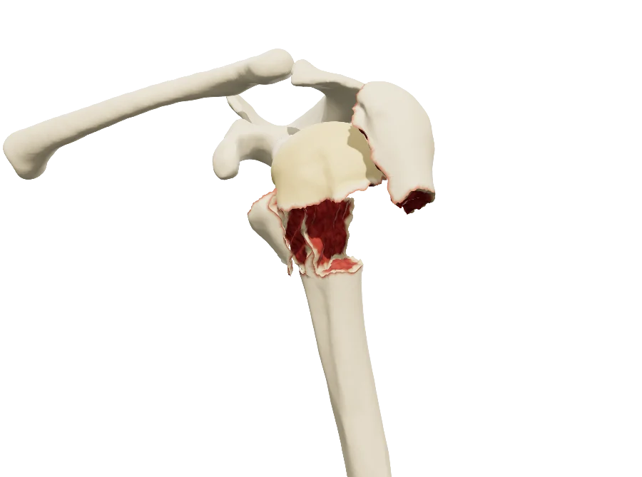
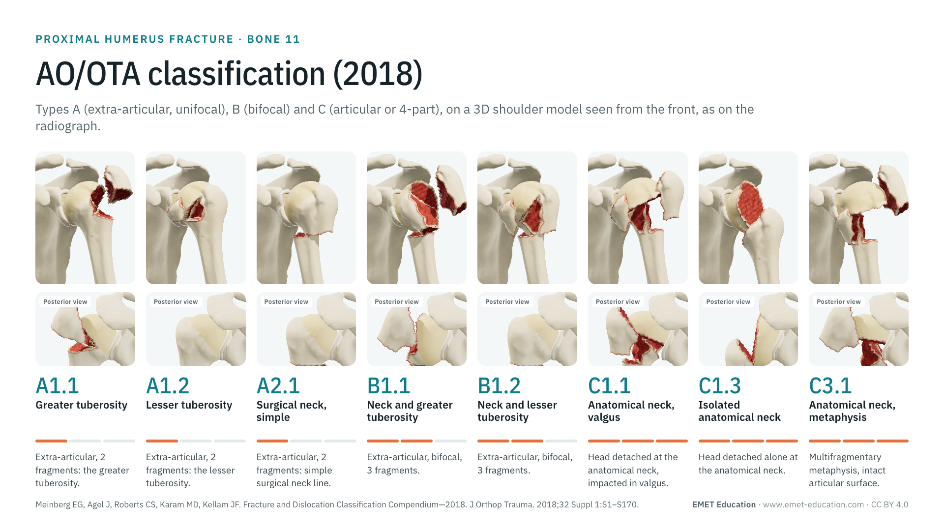
Each image is a rendering of the EMET Education proximal humerus fracture model at that code, left shoulder seen from the front, with a narrow field to approach the projection of an AP radiograph. The model draws eight of the thirteen subgroups; the other five are described without an image. The displacements shown are typical cases: the code itself does not say whether the fracture is displaced, which is the role of the modifiers. Under each code, its Neer category.
-
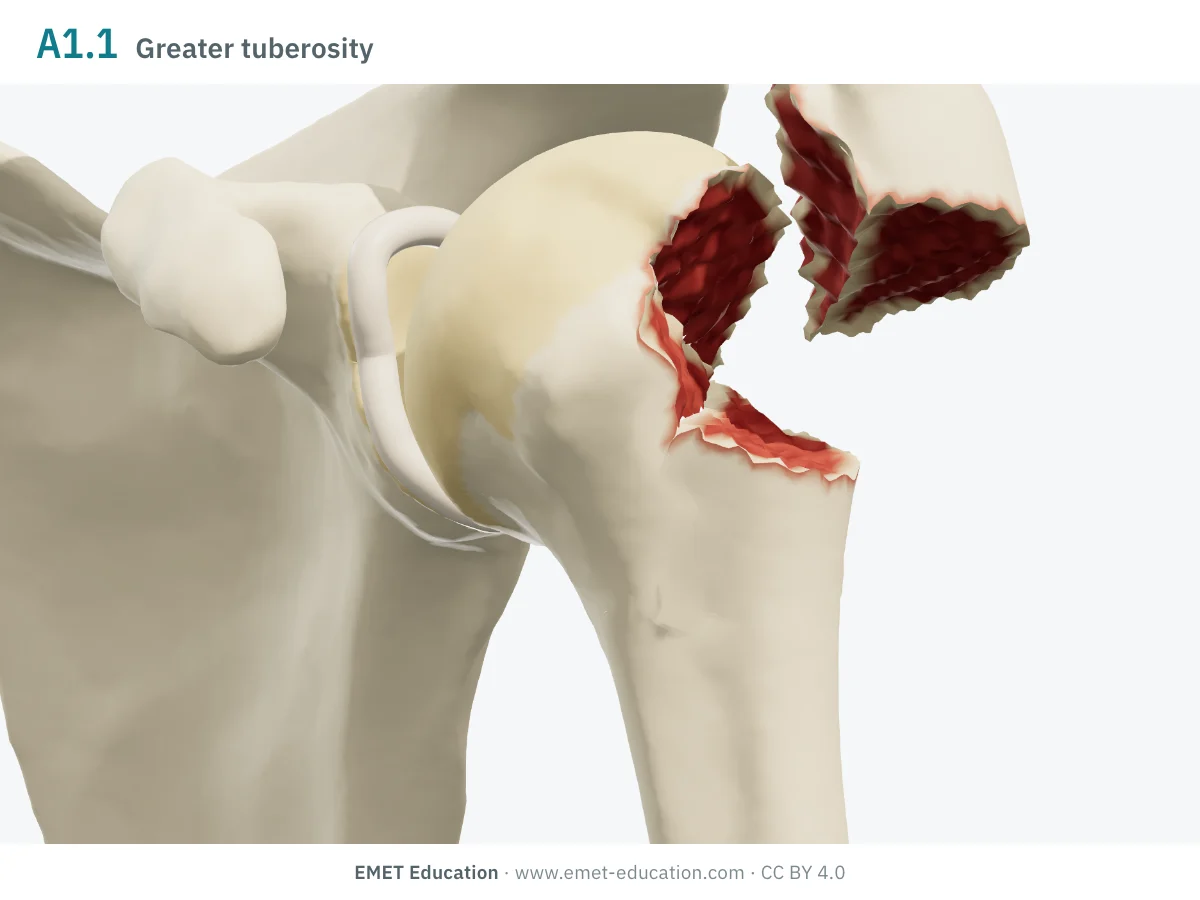
11A1.1Greater tuberosity
- Definition
- Type A, group 1 (tuberosity): extra-articular two-fragment fracture, the greater tuberosity detached from the rest of the humerus.
- Neer
- Two-part, greater tuberosity; with [5a], fracture-dislocation
- In the 3D model
- Greater tuberosity displaced 15 mm, tilted 20° backward and 30° upward by the cuff.
-
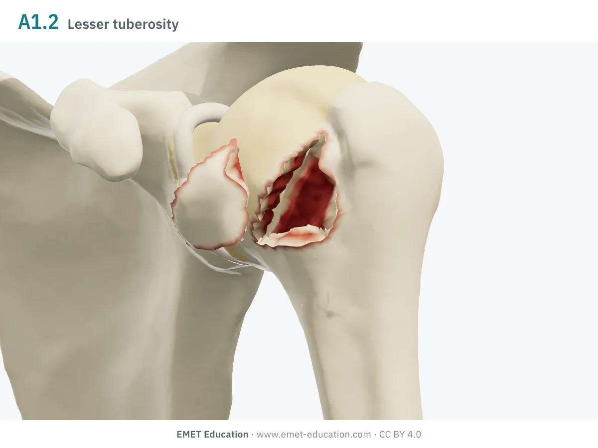
11A1.2Lesser tuberosity
- Definition
- Type A, group 1: extra-articular two-fragment fracture, the lesser tuberosity detached.
- Neer
- Two-part, lesser tuberosity
- In the 3D model
- Lesser tuberosity displaced 15 mm medially by the subscapularis, internally rotated 15°.
-
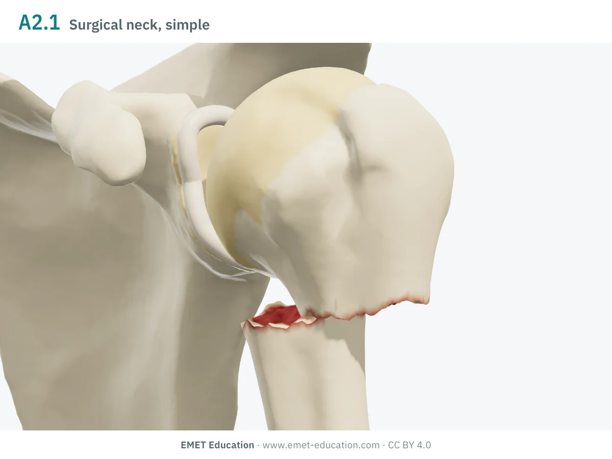
11A2.1Surgical neck, simple
- Definition
- Type A, group 2 (surgical neck): extra-articular two-fragment fracture through the surgical neck, along a simple line.
- Neer
- Two-part, surgical neck
- In the 3D model
- Shaft translated 15 mm medially, toward the scapula; the head keeps both tuberosities and stays in the glenoid.
-
11A2.2Surgical neck, wedge
- Definition
- Type A, group 2: the surgical neck fracture also detaches a metaphyseal wedge; once reduced, the two main fragments remain in contact.
- Neer
- Two-part, surgical neck
- In the 3D model
- Not drawn: the model does not cut a metaphyseal wedge.
-
11A2.3Surgical neck, multifragmentary
- Definition
- Type A, group 2: the metaphysis is fragmented at the surgical neck; once reduced, the main fragments no longer touch.
- Neer
- Two-part, surgical neck
- In the 3D model
- Not drawn: the model does not fragment the metaphysis.
-
11A3Vertical fracture
- Definition
- Type A, group 3: extra-articular two-fragment fracture, with a vertical line in the metaphysis. No subgroup.
- Neer
- Two-part (type A, unifocal)
- In the 3D model
- Not drawn in the model.
-
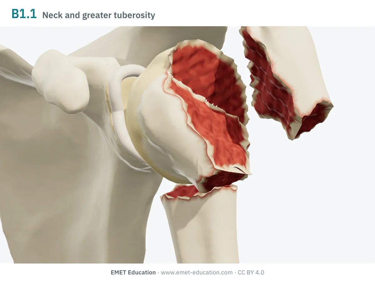
11B1.1Surgical neck and greater tuberosity
- Definition
- Type B, group 1: extra-articular bifocal three-fragment fracture, surgical neck and greater tuberosity. The qualification describes the neck wedge: intact (u) or fragmentary (v).
- Neer
- Three-part, neck and greater tuberosity
- In the 3D model
- Greater tuberosity displaced 15 mm up and back; shaft translated 10 mm; head in 30° of varus, rotated internally with the lesser tuberosity.
-
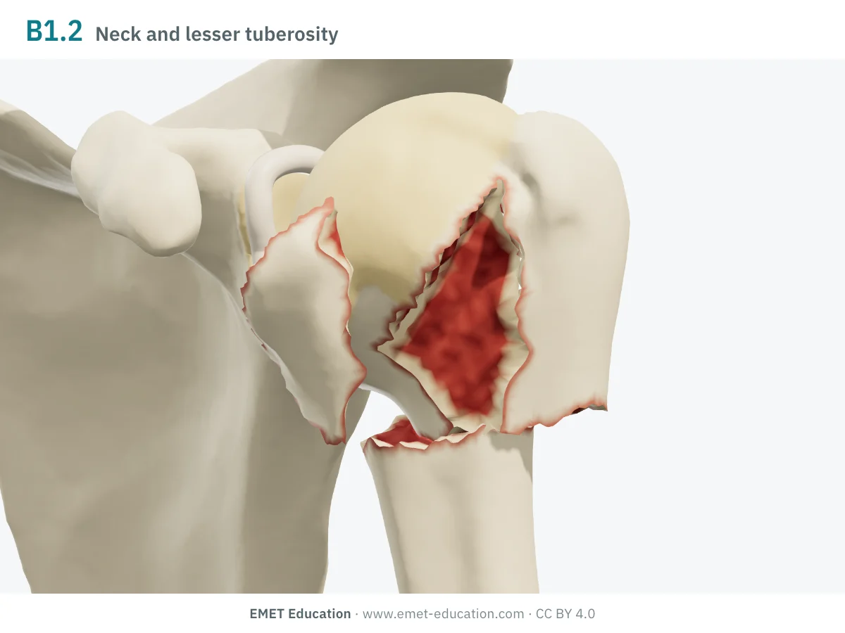
11B1.2Surgical neck and lesser tuberosity
- Definition
- Type B, group 1: extra-articular bifocal three-fragment fracture, surgical neck and lesser tuberosity; same qualifications (u, v).
- Neer
- Three-part, neck and lesser tuberosity
- In the 3D model
- Lesser tuberosity displaced 15 mm medially; shaft translated 10 mm; the head rotates externally with the greater tuberosity.
-
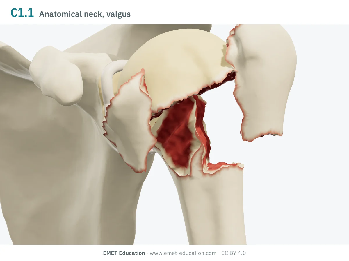
11C1.1Anatomical neck, valgus impacted
- Definition
- Type C, group 1 (anatomical neck): the head, detached at the anatomical neck, impacts into valgus. The qualification says which tuberosity is fractured: greater (n), lesser (o) or both (p).
- Neer
- Four-part, valgus impacted
- In the 3D model
- Head in 30° of valgus, shaft driven up beneath it; both tuberosities splayed 10 mm (qualification p).
-
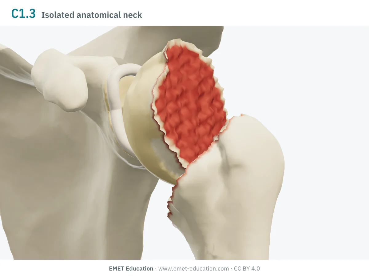
11C1.3Isolated anatomical neck
- Definition
- Type C, group 1: the head comes off alone at the anatomical neck, without fracture of the tuberosities or the metaphysis.
- Neer
- Two-part, anatomical neck
- In the 3D model
- The head tilts 45° into varus about its centre; the tuberosities and the shaft stay in one block.
-
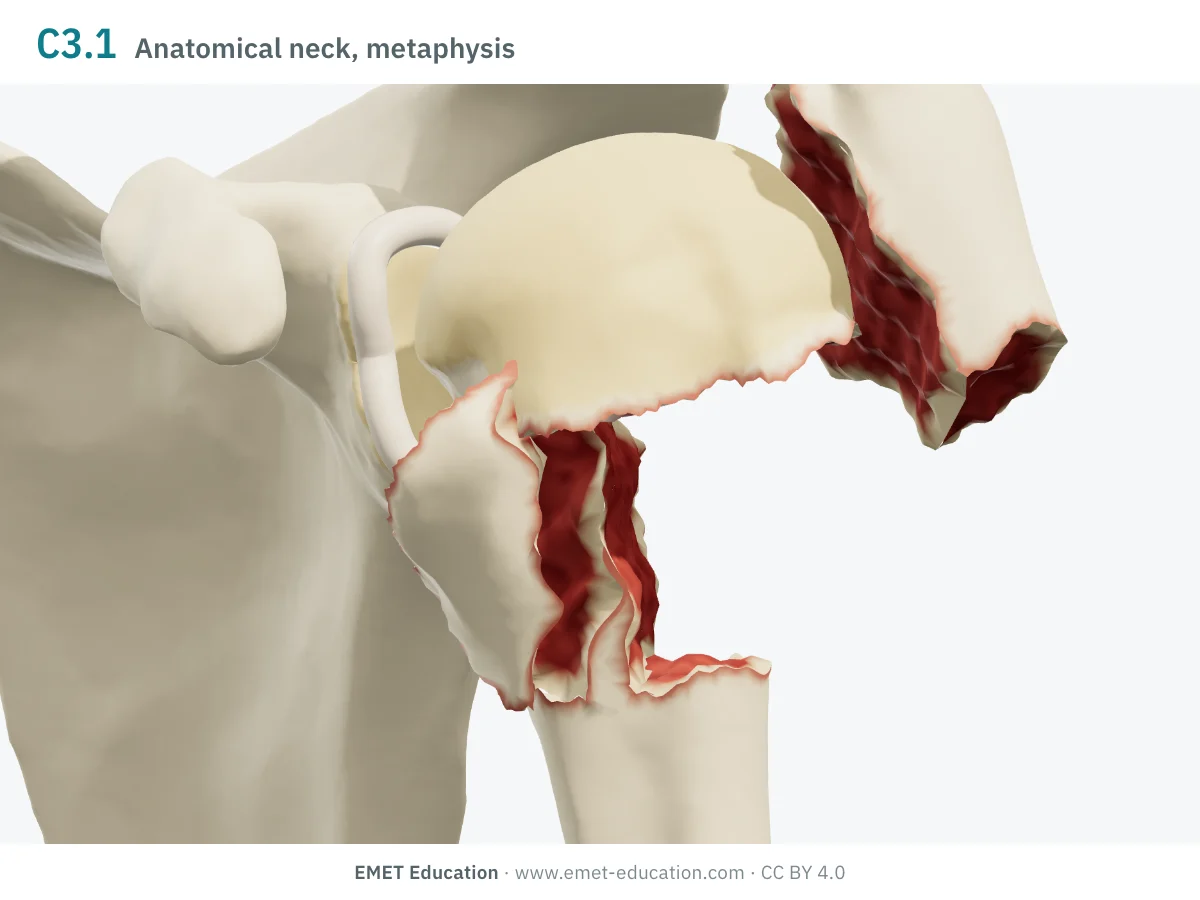
11C3.1Anatomical neck and multifragmentary metaphysis
- Definition
- Type C, group 3: anatomical neck fracture with a multifragmentary metaphysis; the articular surface is intact.
- Neer
- Four-part
- In the 3D model
- Head in 45° of valgus, both tuberosities separated, shaft translated 10 mm: the model shows the four main fragments, but keeps the metaphysis in one block with the shaft, without drawing its comminution.
-
11C3.2… and articular fracture
- Definition
- Type C, group 3: as 11C3.1, with in addition a line through the articular surface; qualification simple (x) or multifragmentary (y).
- Neer
- Four-part, with an articular surface fracture
- In the 3D model
- Not drawn as such: the model shows the head split on its own (Neer page), without a multifragmentary metaphysis.
-
11C3.3… diaphyseal extension and articular fracture
- Definition
- Type C, group 3: as 11C3.2, the metaphyseal fracture extending into the shaft; same qualifications (x, y).
- Neer
- Four-part, with an articular surface fracture
- In the 3D model
- Not drawn in the model.
Schematic rendering, for teaching: the model illustrates each code on a single shoulder, it does not reproduce a radiograph. Definitions reworded from the 2018 compendium (Meinberg et al.) and the AO Foundation's revision booklet; their figures and pictograms are not reproduced. The bar shows the type: one segment A, two B, three C.
Reading the code
From Müller (1990) to the 2018 compendium
The classification comes from Müller et al.'s classification of long-bone fractures, adopted by the Orthopaedic Trauma Association. The 2007 compendium divided the proximal humerus into 27 subgroups (3 types, 3 groups, 3 subgroups), by displacement, impaction or dislocation. The 2018 compendium cut this to 13, defined the types by Neer's parts and moved displacement, impaction and dislocation to modifiers shared by all bones.
The code reads from left to right:
- 11: bone 1, the humerus, and its segment 1, the proximal end;
- the type: A, extra-articular unifocal fracture (Neer two-part); B, extra-articular bifocal (three-part); C, articular, at the anatomical neck, or four-part. There is no "partial articular" type in the humerus (AO Foundation);
- the group and subgroup: A1 tuberosity, A2 surgical neck, A3 vertical fracture; B1 surgical neck and one tuberosity; C1 anatomical neck, C3 anatomical neck and metaphysis. The missing numbers (A1.3, C1.2, C2, B2, B3) were removed in 2018;
- qualifications, as lower-case letters in round brackets: neck wedge intact (u) or fragmentary (v) in 11B1; greater (n), lesser (o) or both tuberosities (p) in 11C1.1; simple (x) or multifragmentary (y) articular fracture in 11C3.2 and 11C3.3;
- universal modifiers, as numbers in square brackets: [1] undisplaced, [2] displaced, [3] impaction, [5a] to [5f] dislocation (anterior, posterior…), [6] subluxation, [7] diaphyseal extension, [8] cartilage injury, [9] poor bone quality. Thus 11A1.1[2,5a]: displaced greater tuberosity fracture with anterior dislocation.
In the series by Marongiu et al. (116 fractures coded with the 2018 version), the most common codes were 11A2.1 (a third of cases) and 11B1.1 (nearly a quarter), and the most common modifier was anterior dislocation [5a] (6%). In the Edinburgh series, coded with Müller's version, the most common fracture was valgus impacted, in one case in five, and type C accounted for only 6% of fractures (Court-Brown et al., 2001).
Reliability
How reproducible is the code?
The type is far more reproducible than the subgroup. As early as 1993, Siebenrock and Gerber observed that agreement worsened the further down groups and subgroups one went; on 3D-printed models, kappa fell from 0.30 for the type alone to 0.11 for the full code (Majed et al.). The 2018 version does at least as well as the 2007 one.
| Study | Measure | Value |
|---|---|---|
| Siebenrock and Gerber, 1993 | AO classification of the time, inter- then intraobserver kappa, 5 shoulder surgeons, 95 fractures (Neer: 0.40 · 0.60) | 0.53 · 0.58 |
| Sjödén et al., 1997 | Interobserver kappa, radiographs then CT, 10 readers, 26 fractures (Neer: 0.42 · 0.45) | 0.31 · 0.30 |
| Majed et al., 2011 | Interobserver kappa on 3D-printed models, 4 readers, 96 complex fractures: full code, then type only | 0.11 · 0.30 |
| Papakonstantinou et al., 2016 | Interobserver kappa, 3 surgeons, 104 registry fractures (Neer: 0.40–0.58) | 0.31–0.54 |
| Marongiu et al., 2020 | 2018 version, mean inter- then intraobserver kappa, 3 readers, 116 fractures (2007 version: 0.57 · 0.58; Neer: 0.67 · 0.85) | 0.67 · 0.75 |
| Diaz et al., 2022 | 2018 version, inter- then intraobserver kappa, 4 readers, 100 radiographs (Neer: 0.67 · 0.76) | 0.78 · 0.86 |
| Cocco et al., 2022 | Interobserver kappa, 4 readers, 75 fractures: CT, then 3D-printed models | 0.44 · 0.47 |
| Mitsuzawa et al., 2025 | Inter- then intraobserver kappa, 4 readers, 100 dislocated or severely displaced fractures (Neer: 0.49 · 0.57) | 0.56 · 0.67 |
In the study by Marmor et al. (2022), seven readers with radiographs and CT agreed better on the type alone (A, B, C) than on the full 2018 code, and all judged the 2018 code at least as descriptive as Neer. In practice: the type to communicate, the full code for research, with written thresholds.
Neer and AO/OTA
From one classification to the other
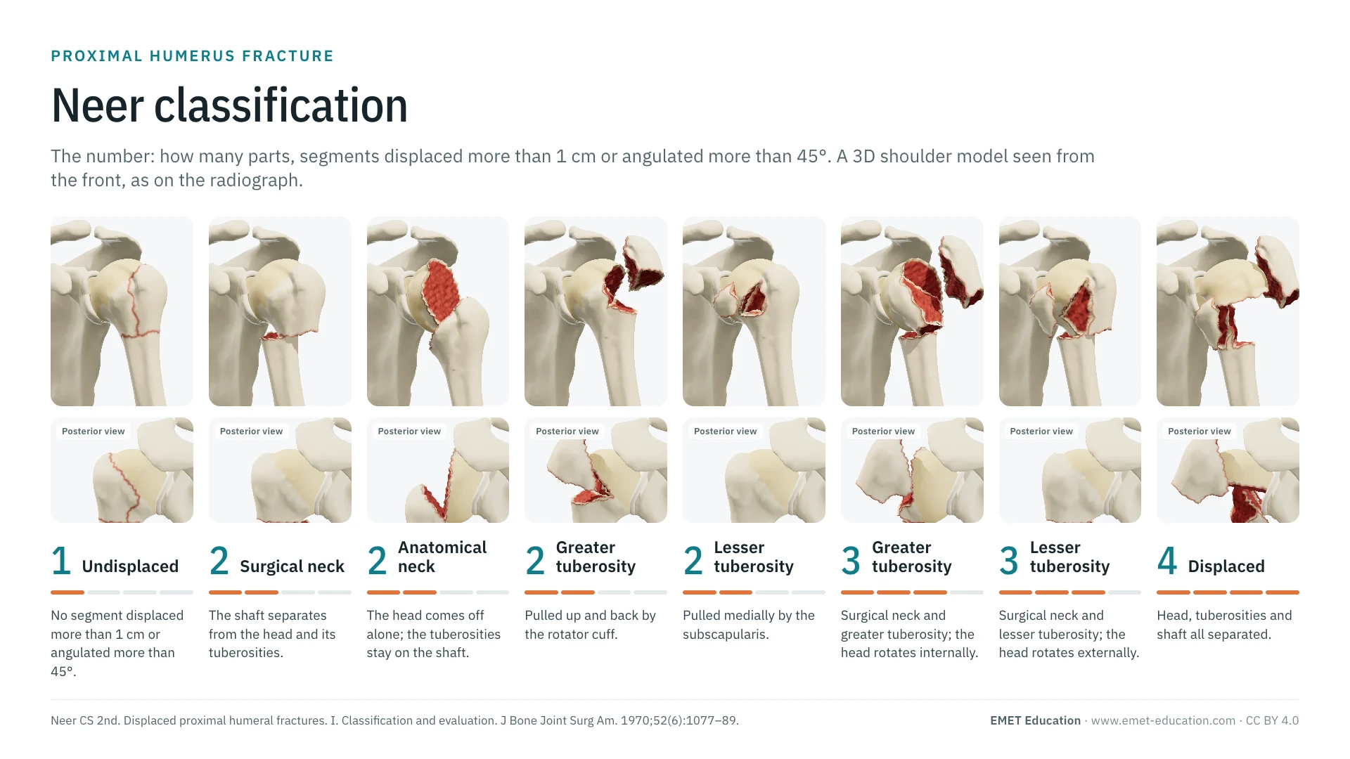
Until 2007, moving from Neer to AO was hazardous: of 432 possible combinations, Brorson et al. judged 151 implausible and 149 problematic. Since 2018, the AO/OTA types are defined by Neer's parts, and the correspondence has become almost direct.
Correspondence with the 2018 version:
| Neer | AO/OTA 2018 | Note |
|---|---|---|
| One-part | The code of its lines, with modifier [1] | The code describes the lines; the modifier says nothing is displaced. |
| Two-part, surgical neck | 11A2.1, 11A2.2 or 11A2.3 | Depending on whether the neck is simple, wedge or multifragmentary. |
| Two-part, anatomical neck | 11C1.3 | Type C: the anatomical neck line is articular. |
| Two-part, greater tuberosity | 11A1.1 | — |
| Two-part, lesser tuberosity | 11A1.2 | — |
| Three-part, greater tuberosity | 11B1.1 (u or v) | The qualification describes the neck wedge. |
| Three-part, lesser tuberosity | 11B1.2 (u or v) | — |
| Four-part, valgus impacted | 11C1.1 (n, o or p) | Added by Neer in 2002; described by Jakob et al. in 1991. |
| Four-part | 11C3.1 to 11C3.3 | The compendium has no subgroup for a four-part fracture without a multifragmentary metaphysis; group C3 is the closest. |
| Fracture-dislocation | The fracture code, with [5a] (anterior) or [5b] (posterior) | In 2007, separate subgroups (11A1.3, group B3…). |
| Articular surface (head split, impression) | 11C3.2 or 11C3.3 if the metaphysis is fragmented; [3a] for articular impaction | No code for an isolated head split. |
Use
In research and in clinical practice
In research
The AO/OTA code is the language of registries, databases and multicentre trauma studies: it fits on one line, sorts and groups. To read literature published before 2018, one needs to know which version was used: the same numbers do not designate the same fractures, and some have disappeared (Brorson et al., 2013).
In clinical practice
The type, A, B or C, sums up the essentials: extra-articular two- or three-fragment fracture, or a fracture that separates the head at the anatomical neck. Qualifications and modifiers say what the number of parts alone leaves out: the neck wedge, the fractured tuberosities of an impacted fracture, dislocation, articular impaction, bone quality.
Type C flags a head cut off from its attachments, exposed to ischaemia; Hertel's criteria (short calcar, disrupted medial hinge) refine this risk. The valgus impacted 11C1.1, which keeps its medial hinge, undergoes necrosis less often than other four-part fractures (Jakob et al.).
Grading pitfalls
Five pitfalls when coding a proximal humerus fracture
- Type first. Each level of the code adds disagreement between readers. For decisions and discussion, the type is often enough; the full code serves research, with written rules.
- Do not mix versions. 11A1.3, 11C1.2 and groups B2, B3 and C2 no longer exist since 2018; an older study that uses them refers to the 2007 or Müller version. State the version in any publication.
- Displacement is not in the code. The types follow Neer's parts, but the code itself does not define a displacement threshold (Brorson et al.): record [1] or [2], and state the threshold used (1 cm, 45°).
- 11B1.1 or 11C1.1? Surgical neck and greater tuberosity on one side, head impacted into valgus at the anatomical neck on the other: a head in valgus, driven between splayed tuberosities, should prompt a search for the anatomical neck line.
- Do not forget the dislocation. It no longer has a subgroup: without modifier [5a] or [5b], a fracture-dislocation is coded like a fracture in place. An isolated lesser tuberosity fracture should prompt a search for a posterior dislocation.
Self-test
Which code?
The same shoulder seen from the front at three-quarters, at the model's eight codes, in random order. Find the code of each one; the answer comes with the definition.
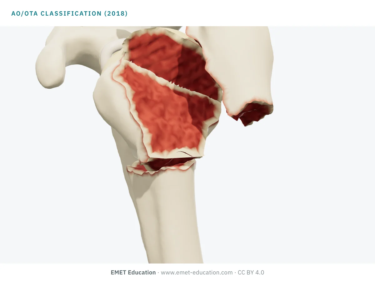
Which AO/OTA code is this?
Download and cite
Free infographics, for teaching and publication
Use them in a course, a presentation, a social media post or a scientific article, under the Creative Commons Attribution 4.0 license: free reuse, including commercial, provided EMET Education is credited with a link to this page. The license covers EMET Education's images, not the classification itself.
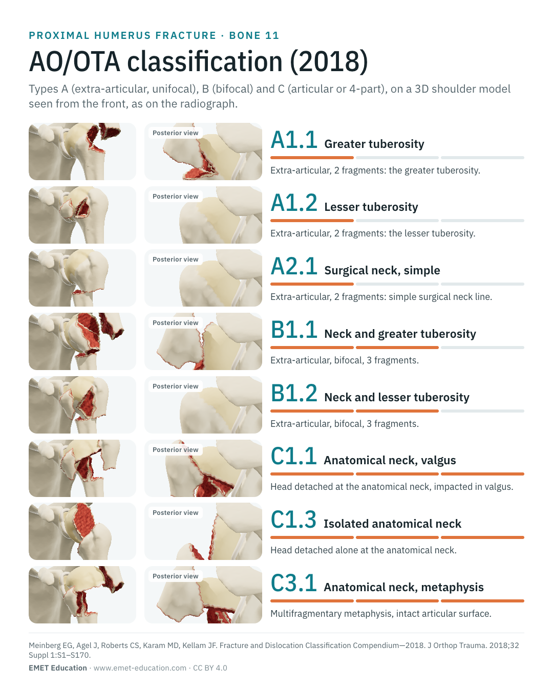
Cite this figure
Ready to paste. For a journal figure, add the original reference (Meinberg et al., 2018).
EMET Education. AO/OTA classification of proximal humerus fractures: 3D visualization [Internet]. 2026 [cited]. Available from: https://www.emet-education.com/en/classifications/ao-proximal-humerus/. CC BY 4.0.EMET Education. (2026). AO/OTA classification of proximal humerus fractures: 3D visualization [Infographic]. https://www.emet-education.com/en/classifications/ao-proximal-humerus/ (CC BY 4.0)<a href="https://www.emet-education.com/en/classifications/ao-proximal-humerus/"><img src="https://www.emet-education.com/en/classifications/ao-proximal-humerus/img/infographic.webp" alt="AO/OTA classification of proximal humerus fractures in 3D" width="960"></a><br>Figure: <a href="https://www.emet-education.com/en/classifications/ao-proximal-humerus/">EMET Education</a>, CC BY 4.0Questions
Frequently asked questions
What does "11" mean in the code?
The first digit is the bone (1: the humerus), the second its segment (1: the proximal end; 2: the shaft; 3: the distal end). Then come the type (A, B, C), the group and the subgroup.
Why is there no 11C1.2 or 11A1.3?
These codes existed in 2007 (11A1.3 was the greater tuberosity fracture-dislocation, 11C1.2 a varus impacted pattern) and were removed in 2018: dislocation is now recorded with a modifier, and the remaining subgroups were not renumbered.
How is a fracture-dislocation coded?
With the fracture code followed by the universal dislocation modifier in square brackets: [5a] anterior, [5b] posterior ([5a] to [5f] depending on direction). For example 11A1.1[5a] for a greater tuberosity fracture with anterior dislocation.
AO/OTA or Neer: which one to use?
They match since 2018: types A, B and C follow the two, three and four parts of the Neer classification. Neer remains the language of clinical discussion; the AO/OTA code, with its qualifications and modifiers, that of registries and research. Their reliability is similar.
Do the images reproduce the AO drawings?
No. The images are renderings of the EMET Education 3D model, and the definitions are reworded; no figure or pictogram from the compendium is reproduced. EMET Education is not affiliated with the AO Foundation or the Orthopaedic Trauma Association.
Can I reuse these images in a course or an article?
Yes. The images and infographics on this page are under the Creative Commons Attribution 4.0 license: free reuse, including commercial, with credit to EMET Education and a link to this page. The "Cite this figure" box gives ready-to-paste citations.
How were the 3D images made?
They are renderings of the EMET Education proximal humerus fracture model, set to each code: the fragments are cut from a humerus taken from CT, along the anatomical neck, the surgical neck and the bicipital groove, then displaced the way the muscles attached to them pull. It is a schematic model for teaching and patient explanation; it does not reproduce a radiograph.
Sources
Author and references
- Meinberg EG, Agel J, Roberts CS, Karam MD, Kellam JF. Fracture and Dislocation Classification Compendium—2018. J Orthop Trauma. 2018;32 Suppl 1:S1–S170. doi:10.1097/BOT.0000000000001063
- AO Foundation. AO/OTA Fracture and Dislocation Classification Compendium – 2018 Revisions. Information booklet, version 2.0; 2017. PDF
- Marsh JL, Slongo TF, Agel J, et al. Fracture and dislocation classification compendium – 2007: Orthopaedic Trauma Association classification, database and outcomes committee. J Orthop Trauma. 2007;21(10 Suppl):S1–133. doi:10.1097/00005131-200711101-00001
- Müller ME, Nazarian S, Koch P, Schatzker J. The Comprehensive Classification of Fractures of Long Bones. Berlin: Springer; 1990.
- Neer CS 2nd. Displaced proximal humeral fractures. I. Classification and evaluation. J Bone Joint Surg Am. 1970;52(6):1077–89. doi:10.2106/00004623-197052060-00001
- Brorson S, Eckardt H, Audigé L, Rolauffs B, Bahrs C. Translation between the Neer- and the AO/OTA-classification for proximal humeral fractures: do we need to be bilingual to interpret the scientific literature? BMC Res Notes. 2013;6:69. doi:10.1186/1756-0500-6-69
- Siebenrock KA, Gerber C. The reproducibility of classification of fractures of the proximal end of the humerus. J Bone Joint Surg Am. 1993;75(12):1751–5. doi:10.2106/00004623-199312000-00003
- Court-Brown CM, Garg A, McQueen MM. The epidemiology of proximal humeral fractures. Acta Orthop Scand. 2001;72(4):365–71. doi:10.1080/000164701753542023
- Sjödén GO, Movin T, Güntner P, et al. Poor reproducibility of classification of proximal humeral fractures. Additional CT of minor value. Acta Orthop Scand. 1997;68(3):239–42. doi:10.3109/17453679708996692
- Majed A, Macleod I, Bull AM, et al. Proximal humeral fracture classification systems revisited. J Shoulder Elbow Surg. 2011;20(7):1125–32. doi:10.1016/j.jse.2011.01.020
- Papakonstantinou MK, Hart MJ, Farrugia R, et al. Interobserver agreement of Neer and AO classifications for proximal humeral fractures. ANZ J Surg. 2016;86(4):280–4. doi:10.1111/ans.13451
- Marongiu G, Leinardi L, Congia S, Frigau L, Mola F, Capone A. Reliability and reproducibility of the new AO/OTA 2018 classification system for proximal humeral fractures: a comparison of three different classification systems. J Orthop Traumatol. 2020;21(1):4. doi:10.1186/s10195-020-0543-1
- Diaz LEP, Dada Neto F, Lofrano L, et al. Intra- and inter-observer agreement of proximal humeral fractures classifications in adults. Acta Ortop Bras. 2022;30(6):e257229. doi:10.1590/1413-785220223006e257229
- Cocco LF, Aihara AY, Lopes FPPL, et al. Three-dimensional printing models increase inter-rater agreement for classification and treatment of proximal humerus fractures. Patient Saf Surg. 2022;16(1):5. doi:10.1186/s13037-021-00312-7
- Mitsuzawa S, Takeuchi H, Ijiri K, et al. A new classification for dislocated and displaced proximal humeral fractures. J Orthop Surg Res. 2025;20(1):18. doi:10.1186/s13018-024-05423-2
- Marmor MT, Agel J, Dumpe J, et al. Comparison of the Neer classification to the 2018 update of the Orthopedic Trauma Association/AO fracture classification for classifying proximal humerus fractures. OTA Int. 2022;5(3):e200. doi:10.1097/OI9.0000000000000200
- Jakob RP, Miniaci A, Anson PS, Jaberg H, Osterwalder A, Ganz R. Four-part valgus impacted fractures of the proximal humerus. J Bone Joint Surg Br. 1991;73(2):295–8. doi:10.1302/0301-620X.73B2.2005159
- Hertel R, Hempfing A, Stiehler M, Leunig M. Predictors of humeral head ischemia after intracapsular fracture of the proximal humerus. J Shoulder Elbow Surg. 2004;13(4):427–33. doi:10.1016/j.jse.2004.01.034
3D models
Show the fracture to your patient
The images on this page come from an interactive model, the proximal humerus fracture 3D model. In clinic, choose the code, or switch to Neer without losing the fracture, set the displacement of each part from your patient's imaging, then show the proposed treatment: nonoperative care, nail, locking plate or reverse arthroplasty.
Explain the fracture in 3D
The proximal humerus fracture model opens in your browser, at the code you choose, with no sign-up.
Share this page with a colleague or a resident: