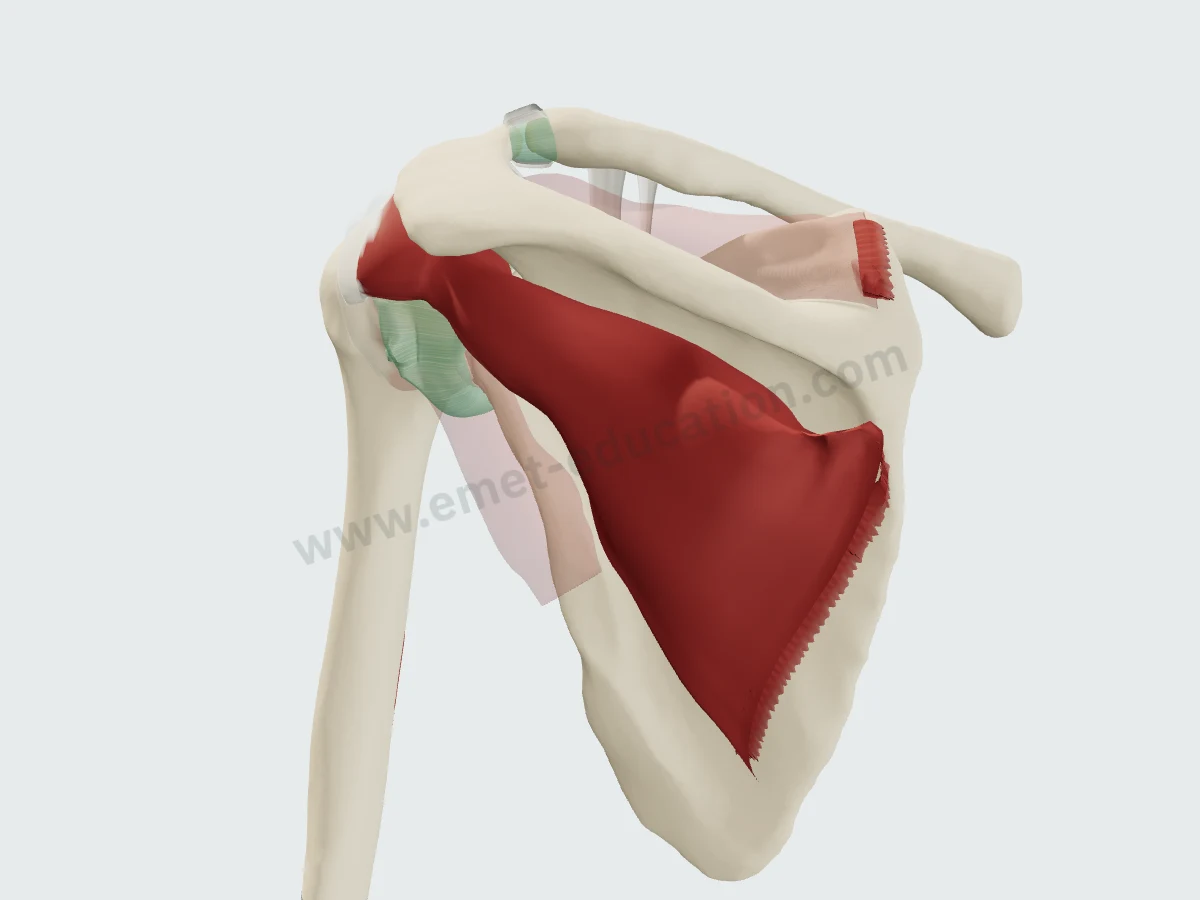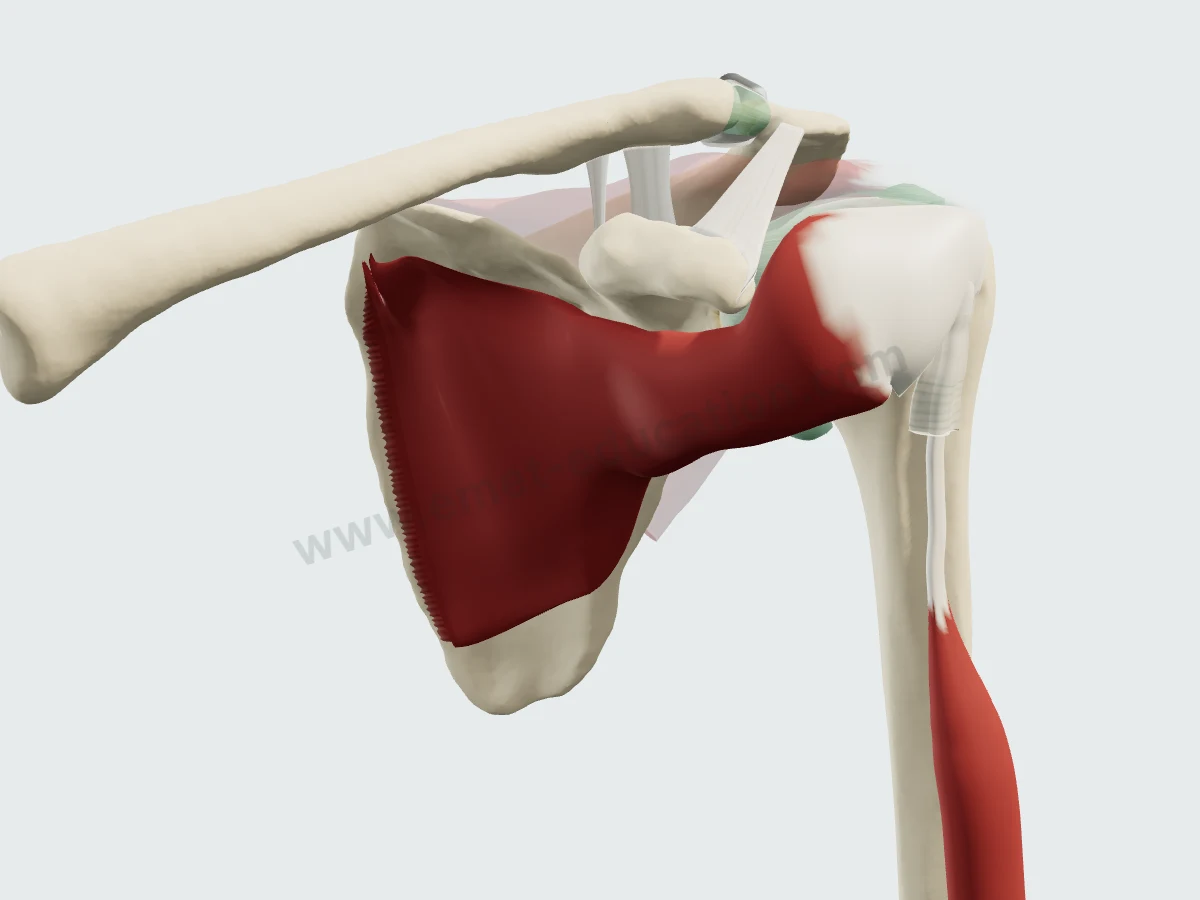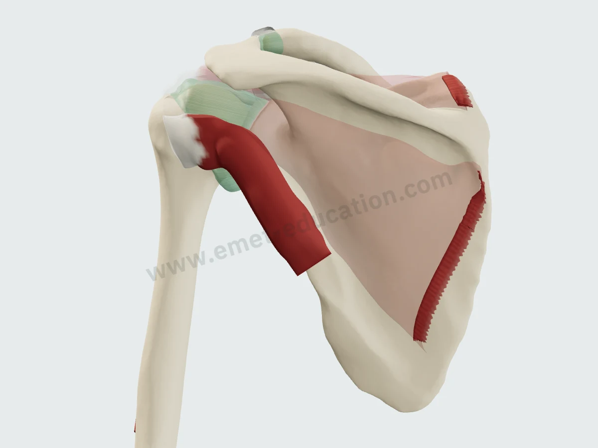
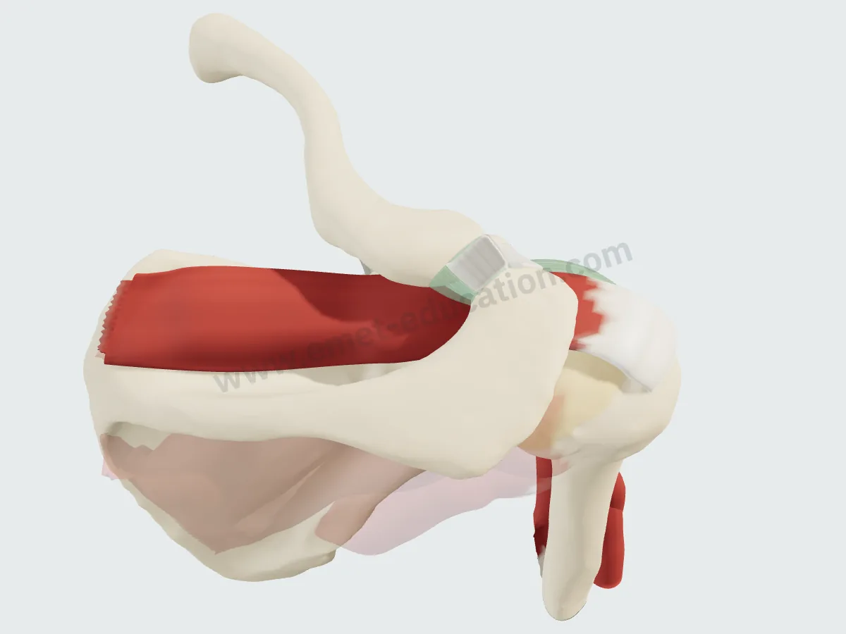
The Supraspinatus Muscle in 3D
Supraspinatus origin, insertion, innervation and relations in 3D, labeled MRI slices, its footprint in numbers, and how its description has changed.
3D Anatomy · Musculoskeletal
Each page follows one structure: origin, insertion, innervation and relations, the layers of the 3D model, labeled MRI and CT slices, published measurements and what they change in practice, with their sources.
Designed by an orthopedic surgeon
The slices are synthetic images computed from the 3D model: they locate the structures; nothing is measured on them.
By joint
Grouped by joint. Each page links to the 3D models and classifications it relates to.
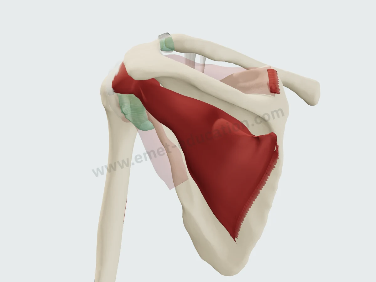
Supraspinatus origin, insertion, innervation and relations in 3D, labeled MRI slices, its footprint in numbers, and how its description has changed.
Infraspinatus origin, insertion, innervation and relations in 3D, labeled MRI slices, its footprint in numbers, and how its description has changed.
Subscapularis origin, insertion, innervation and relations in 3D, labeled MRI slices, its nerves in numbers, and how its description has changed.
Teres minor origin, insertion, innervation and relations in 3D, labeled MRI slices, its nerve in numbers, its atrophy, and how its description has changed.
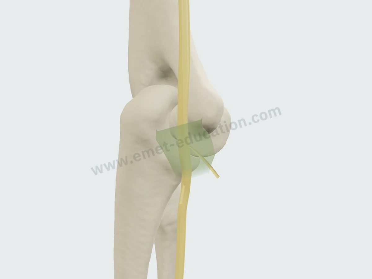
Course, cubital tunnel, compression sites, branches and mobility of the ulnar nerve at the elbow in 3D, labeled MRI slices, measurements and history.
The 3D models open in your browser, no sign-up needed.