- EMET Education
- 3D Anatomy
- Shoulder
- Supraspinatus muscle
Anatomy · Shoulder · Rotator cuff
The Supraspinatus Muscle in 3D
The supraspinatus in 3D, layer by layer, then on labeled MRI slices. With its measured footprint, what anatomic studies have found over thirty years, and the landmarks for reading its atrophy.
Designed by an orthopedic surgeon
Latin musculus supraspinatus · French supra-épineux, sus-épineux
- 12.6 mmAnteroposterior width of the footprint on the greater tuberosity (26 shoulders)
- 2.45Physiological cross-sectional area of the anterior belly relative to the posterior one (25 shoulders)
- 0.70Share of the fossa filled by the muscle on the Y-view MRI, in controls
- 2.59Rotator cable thickness relative to the crescent it protects
At a glance
Origin, insertion, innervation
| Origin | Medial two-thirds of the supraspinous fossa and supraspinous fascia. |
|---|---|
| Course | Beneath the coracoacromial arch (acromion, coracoacromial ligament), separated from it by the subacromial-subdeltoid bursa, above the glenohumeral joint. |
| Insertion | Superior facet of the greater tuberosity and upper half of the middle facet; inconsistently, the superior lesser tuberosity (21% of specimens). |
| Innervation | Suprascapular nerve (C5, C6). |
| Blood supply | Suprascapular artery. |
| Action | Arm abduction with the deltoid; compression of the humeral head into the glenoid. |
| Relations | Anteriorly, the rotator interval, long head of biceps, and subscapularis; posteriorly, the infraspinatus, whose tendon partly covers it; superiorly, the coracoacromial arch and bursa; deep, the capsule. |
Layer by layer
From the trapezius to the tendon
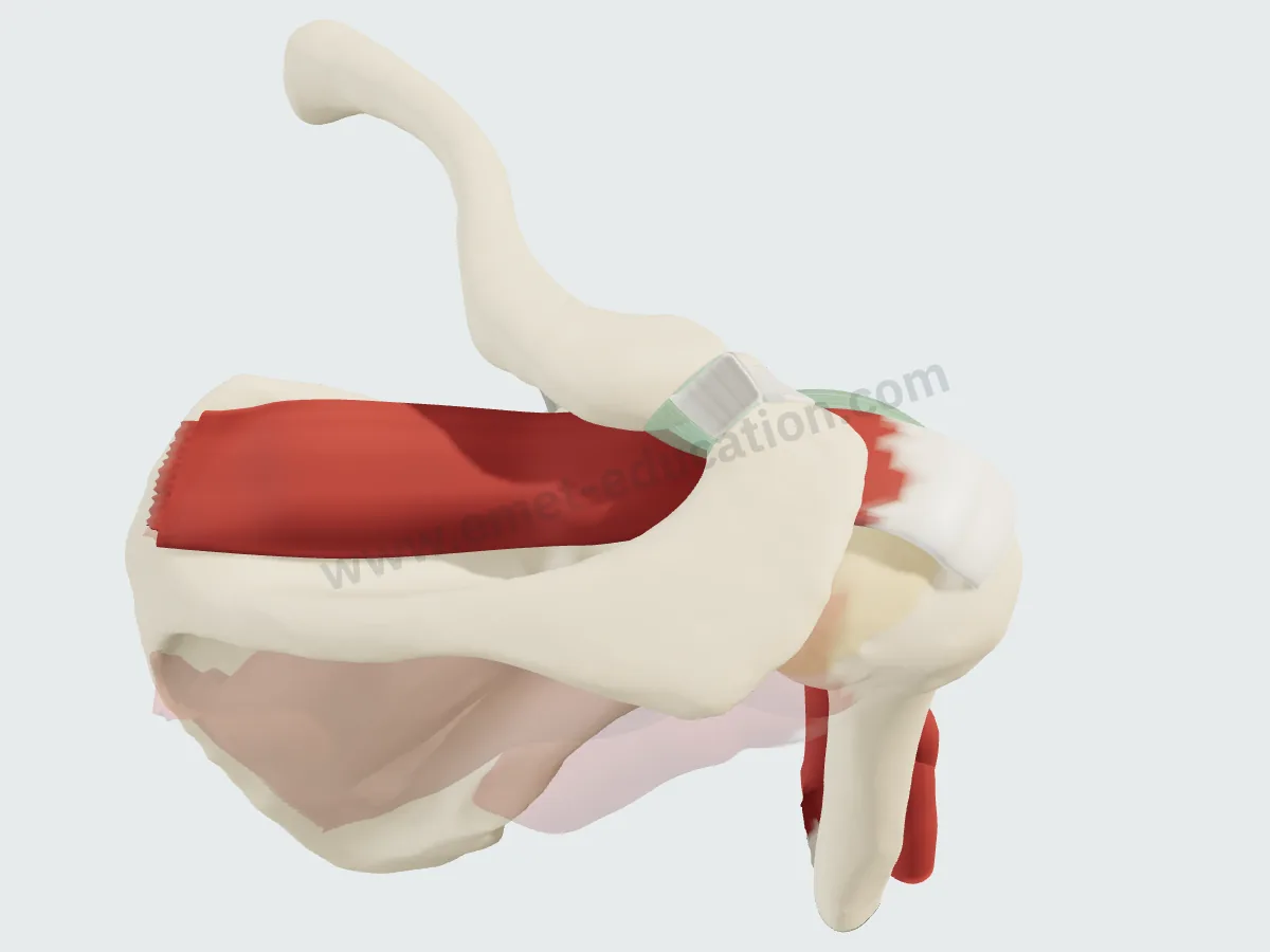
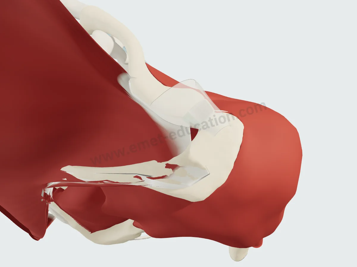
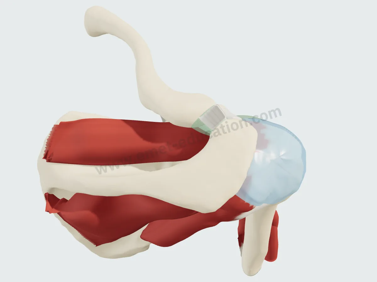
The same view, from above and behind the shoulder, removing one layer per image.
Trapezius and deltoidThe supraspinatus is hidden: the trapezius covers the fossa, the deltoid the humeral head.
The bursaTrapezius and deltoid removed: the subacromial-subdeltoid bursa caps the tendon.
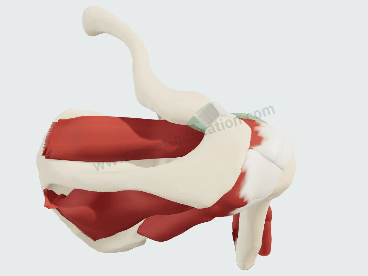
The cuffBursa removed: the supraspinatus in its fossa above the spine, the infraspinatus below.
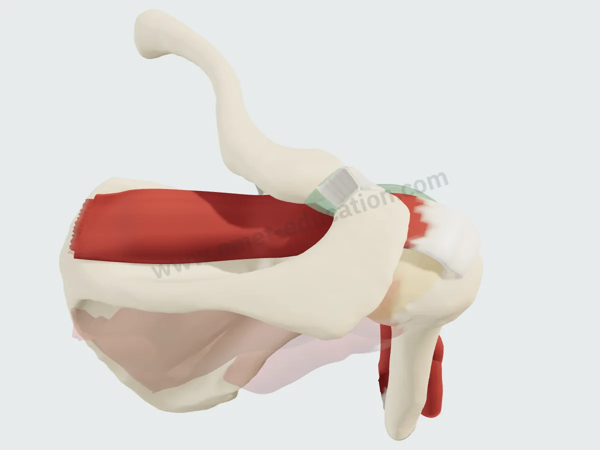
The supraspinatusOther muscles faded: from the fossa to the greater tuberosity, under the acromion.
Imaging
The supraspinatus on MRI and CT
Synthetic images. These slices do not come from a patient: they are computed from the 3D model, by cutting each structure and giving it the signal it would have in the chosen sequence, on MRI or CT. They are for learning where the muscle lies on a slice; nothing is measured on them.
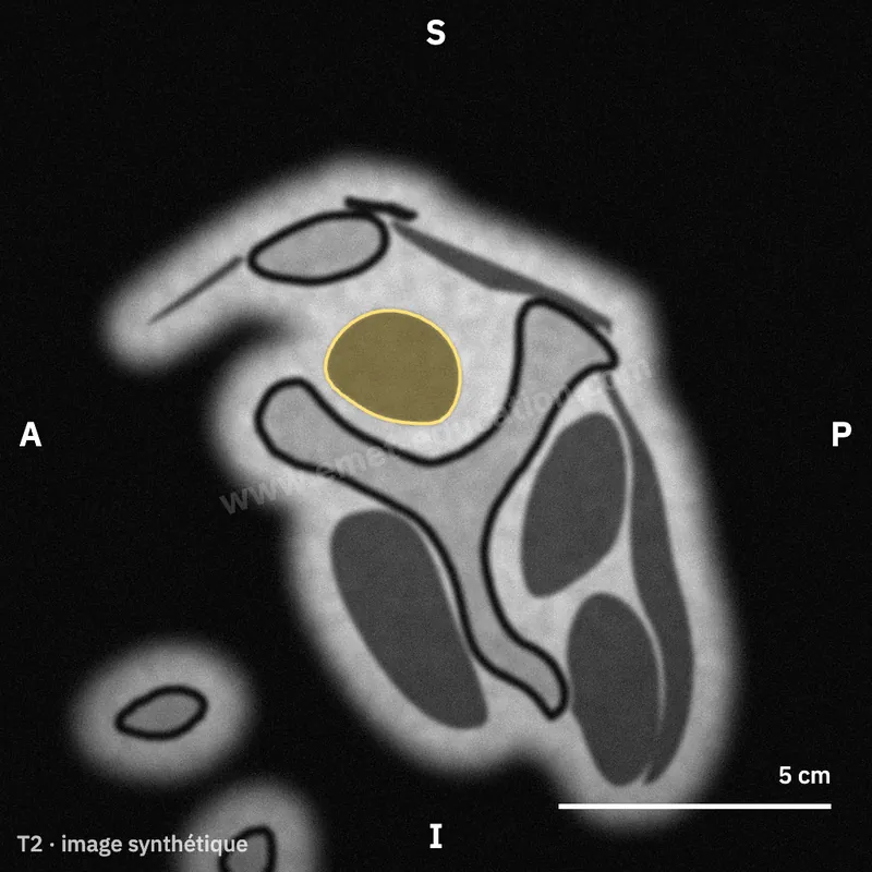
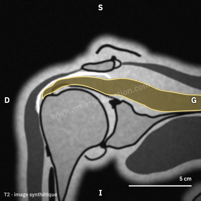
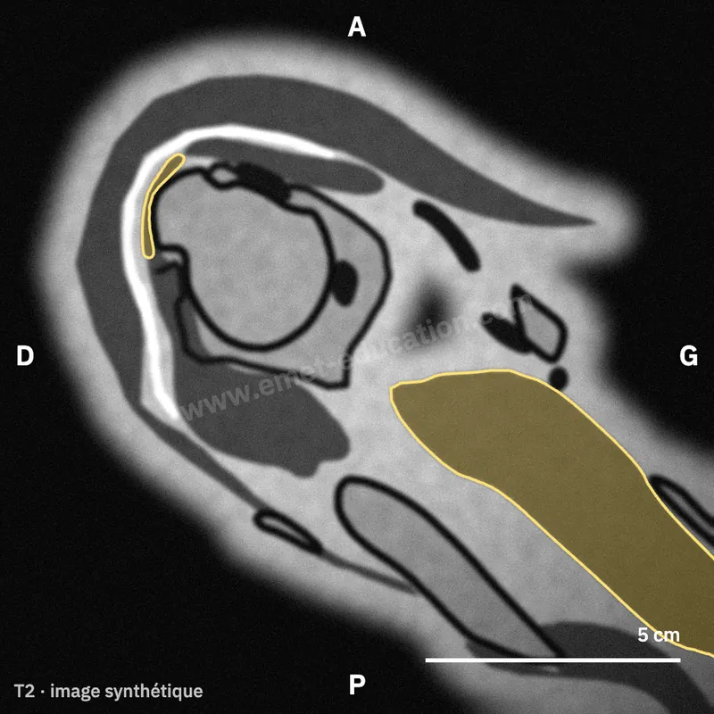
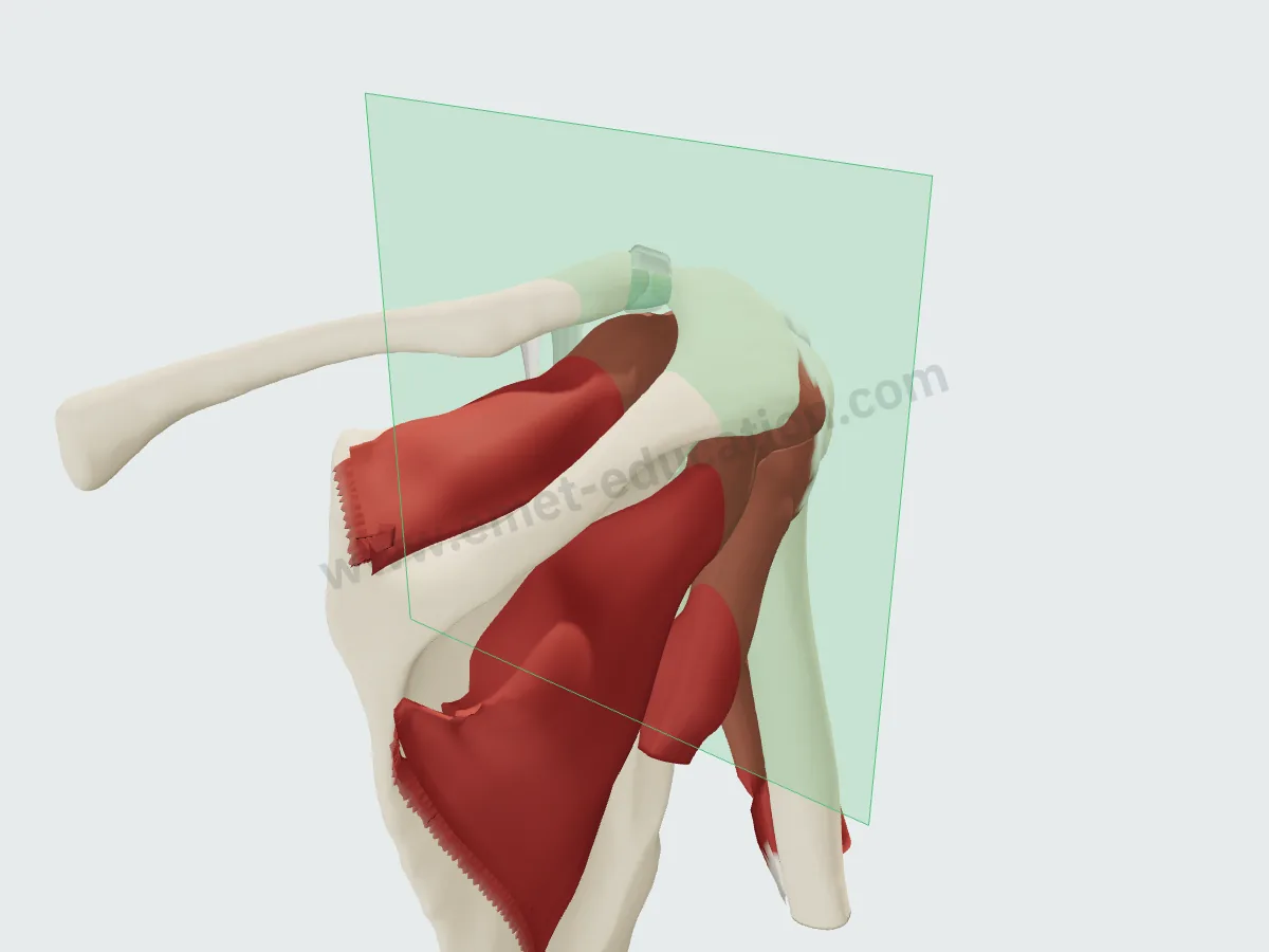
Where the Y view cuts
The oblique sagittal plane (green) on the 3D model: parallel to the glenoid, it crosses the scapula where the spine still meets the body. Hover over a slice and click “Scroll” to page through it as on a workstation: the muscle tapers into tendon laterally.
In numbers
The footprint and the muscle, measured
Published mean values, with sample size and source under each figure.
| Value | Reference | Year | Sample | Note |
|---|---|---|---|---|
| 12.6 mm | Mochizuki | 2008 | 26 shoulders | Mean maximum; triangular footprint, part of the surface being occupied by infraspinatus |
| 23 mm | Curtis | 2006 | 20 cadavers | Mean maximum length (23 × 16 mm); orientation to be confirmed in the full text to be confirmed |
| 25 mm | Ruotolo | 2004 | 17 specimens | Mean; 17 intact specimens kept out of 48, mean age 70 years |
| Value | Reference | Year | Sample | Note |
|---|---|---|---|---|
| 6.9 mm | Mochizuki | 2008 | 26 shoulders | Mean maximum |
| 12.1 mm | Ruotolo | 2004 | 17 specimens | Mean, at mid-tendon (11.6 at the rotator interval, 12 at the posterior edge) |
| 14.7 mm | Dugas | 2002 | 10 specimens | Minimum transverse dimension of the whole cuff insertion at mid supraspinatus (± 3.2); lower bound of the calculation |
| 16 mm | Curtis | 2006 | 20 cadavers | Mean maximum width (23 × 16 mm); orientation to be confirmed in the full text to be confirmed |
| Value | Reference | Year | Sample | Note |
|---|---|---|---|---|
| 1.55 cm² | Dugas | 2002 | 10 specimens | Mean (± 0.66; 0.68 to 2.64), 3D digitizer mapping |
Published means, from smallest to largest. Sample size is as each study reports it (shoulders, cadavers or specimens); measurement protocols differ from one study to the next.
Maximum dimensions, to scale: a bounding box, not the shape of the footprint.
- 12.6 mmAnterior half of the superior cuff tendon, supraspinatus only (± 1.1)Mean, 10 shoulders · Minagawa 1998
- 140 mm²Anterior belly physiological cross-sectional area (± 43), 2.45 times the posteriorMean, 25 shoulders · Roh 2000
- 3.5 mmSuperior capsule, minimum attachment width on the greater tuberosityNimura 2012
- 2.59Cable thickness relative to crescent thicknessMean, 20 shoulders · Burkhart 1993
- 0.70Fossa occupation ratio on the Y view, controlsMean, 15 shoulders · Thomazeau 1996
Published insertion dimensions vary from study to study. Mochizuki found a smaller footprint than previously described, with the infraspinatus occupying part of the area; Nimura showed that the capsule also attaches to the greater tuberosity, and that studies not separating it overestimated the footprint.
History
How its description has changed since 1992
Classic anatomy describes an insertion on the superior facet of the greater tuberosity. Anatomic studies published since have refined it, step by step.
- 1992
A common insertion
The cuff tendons do not insert separately: they interdigitate into a continuous, five-layered insertion. Clark and Harryman
- 1993
The suspension bridge
A thick cable borders the crescent, the thin distal part of the supraspinatus and infraspinatus, and shields it from stress. Burkhart
- 2000
Two bellies
The anterior belly, with the larger physiological cross-sectional area, pulls through a thinner tendon: stress is higher in the anterior tendon, which should be included in any repair. Roh
- 2002
Right at the cartilage
Along its first 2 cm, the cuff insertion lies within 1 mm of the articular margin. Dugas
- 2008
A small triangle
The supraspinatus footprint is a triangle at the front of the greater tuberosity, smaller than described; the infraspinatus occupies much of the area once attributed to it. Mochizuki
- 2012
The capsule too
The capsule also attaches to the greater tuberosity: by not separating it, earlier studies overestimated the footprint. Its thinnest point lies at the posterior edge of the supraspinatus insertion. Nimura
In practice
What anatomy changes in practice
- Reading atrophy. On the Y view: fossa occupation ratio (Thomazeau) and the tangent sign, positive in 9 of 10 patients with large tears and in none of 70 asymptomatic subjects (Zanetti).
- Grading fatty infiltration. On CT (Goutallier) and on MRI, each method is reproducible between observers, but agreement between them is only fair to moderate: the two gradings are not interchangeable (Fuchs).
- After repair. Moderate supraspinatus fatty infiltration regressed in 6 of 14 patients; infraspinatus infiltration weighed heavily on the outcome (Goutallier).
- Repairing the right tendon. The anterior tendon is the muscle's primary contractile unit (Roh).
Your turn
Where is the supraspinatus?
Six unlabeled synthetic slices, in random order: click the supraspinatus. The answer shows its outline.
Click the supraspinatus.
Conditions
Related conditions
Classifications
Related classifications
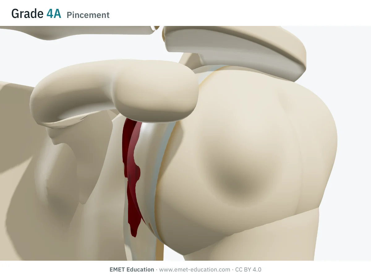 HamadaMassive rotator cuff tears: grades 1 to 5, in 3D
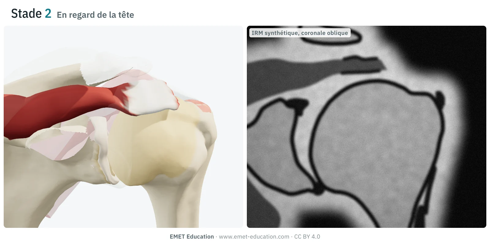 PatteSupraspinatus tendon retraction: stages 1 to 3, in 3D and in section
- SoonGoutallierFatty infiltration of the cuff muscles, on CT (1994)
- SoonThomazeauSupraspinatus atrophy on the Y-view MRI (1996)
Neighbors
Its neighbors
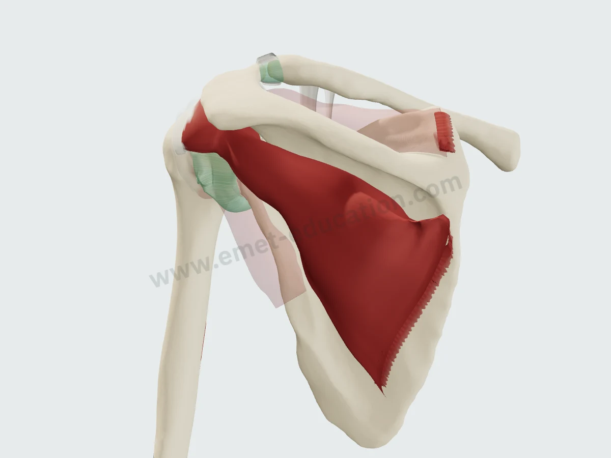 InfraspinatusPosterior
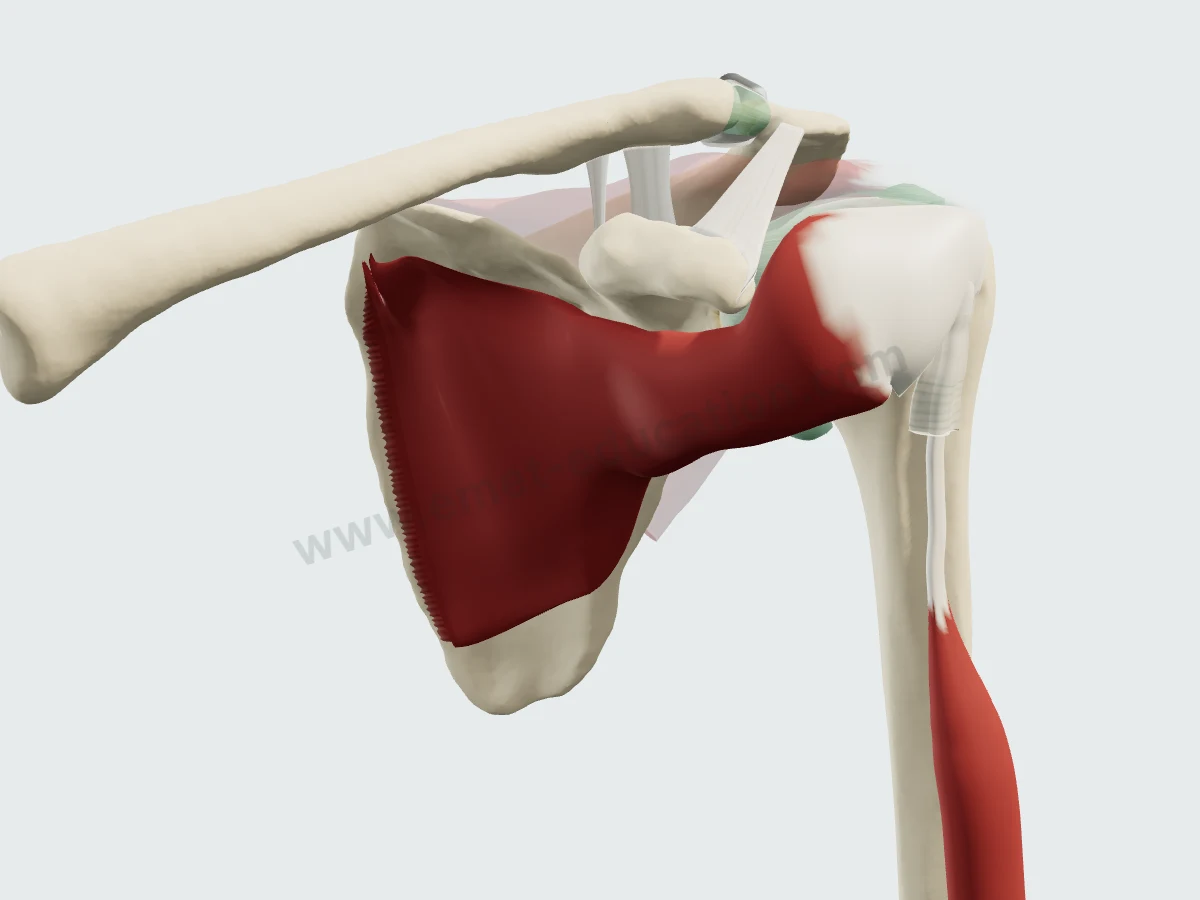 SubscapularisAnterior
- SoonLong head of bicepsAnterior
- SoonCoracoacromial ligamentSuperior
- SoonSubacromial bursaSuperficial
- SoonDeltoidSuperficial
- SoonTrapeziusSuperficial
- SoonJoint capsuleDeep
Questions
Frequently asked questions
Where does the supraspinatus insert?
On the superior facet of the greater tuberosity and the upper half of the middle facet. Its tendinous anterior portion also reaches the superior lesser tuberosity in 21% of specimens. Its footprint averages 12.6 mm wide and 6.9 mm long (Mochizuki, 26 shoulders).
What nerve innervates the supraspinatus?
The suprascapular nerve, from the C5 and C6 roots.
How do you assess supraspinatus atrophy on MRI?
On the oblique sagittal Y view: fossa occupation ratio (mean 0.70 in controls per Thomazeau) and the tangent sign (Zanetti).
What is the rotator cable?
A thick bundle bordering the thin distal part of the supraspinatus and infraspinatus, the crescent. On average 2.59 times thicker, it takes up the crescent's stress like a suspension bridge (Burkhart).
Why does it have two bellies?
By fiber insertion, the muscle divides into an anterior and a posterior belly. The anterior one has a 2.45 times larger physiological cross-sectional area and pulls through a thinner tendon (Roh).
Are the MRI images on this page real?
No: they are synthetic, computed from the 3D model. They show where the muscle lies on a slice; they do not replace an actual exam.
Sources
Author and references
- Standring S, ed. Gray's Anatomy: The Anatomical Basis of Clinical Practice. 42nd ed. Elsevier; 2020.
- Clark JM, Harryman DT 2nd. Tendons, ligaments, and capsule of the rotator cuff. Gross and microscopic anatomy. J Bone Joint Surg Am. 1992;74(5):713–725. PMID 1624486
- Burkhart SS, Esch JC, Jolson RS. The rotator crescent and rotator cable: an anatomic description of the shoulder's "suspension bridge". Arthroscopy. 1993;9(6):611–616. doi:10.1016/s0749-8063(05)80496-7
- Goutallier D, Postel JM, Bernageau J, Lavau L, Voisin MC. Fatty muscle degeneration in cuff ruptures. Pre- and postoperative evaluation by CT scan. Clin Orthop Relat Res. 1994;(304):78–83. PMID 8020238
- Thomazeau H, Rolland Y, Lucas C, Duval JM, Langlais F. Atrophy of the supraspinatus belly. Assessment by MRI in 55 patients with rotator cuff pathology. Acta Orthop Scand. 1996;67(3):264–268. doi:10.3109/17453679608994685
- Minagawa H, Itoi E, Konno N, et al. Humeral attachment of the supraspinatus and infraspinatus tendons: an anatomic study. Arthroscopy. 1998;14(3):302–306. doi:10.1016/s0749-8063(98)70147-1
- Zanetti M, Gerber C, Hodler J. Quantitative assessment of the muscles of the rotator cuff with magnetic resonance imaging. Invest Radiol. 1998;33(3):163–170. doi:10.1097/00004424-199803000-00006
- Fuchs B, Weishaupt D, Zanetti M, Hodler J, Gerber C. Fatty degeneration of the muscles of the rotator cuff: assessment by computed tomography versus magnetic resonance imaging. J Shoulder Elbow Surg. 1999;8(6):599–605. doi:10.1016/s1058-2746(99)90097-6
- Roh MS, Wang VM, April EW, Pollock RG, Bigliani LU, Flatow EL. Anterior and posterior musculotendinous anatomy of the supraspinatus. J Shoulder Elbow Surg. 2000;9(5):436–440. doi:10.1067/mse.2000.108387
- Dugas JR, Campbell DA, Warren RF, Robie BH, Millett PJ. Anatomy and dimensions of rotator cuff insertions. J Shoulder Elbow Surg. 2002;11(5):498–503. doi:10.1067/mse.2002.126208
- Curtis AS, Burbank KM, Tierney JJ, Scheller AD, Curran AR. The insertional footprint of the rotator cuff: an anatomic study. Arthroscopy. 2006;22(6):609.e1. doi:10.1016/j.arthro.2006.04.001
- Ward SR, Hentzen ER, Smallwood LH, et al. Rotator cuff muscle architecture: implications for glenohumeral stability. Clin Orthop Relat Res. 2006;448:157–163. doi:10.1097/01.blo.0000194680.94882.d3
- Mochizuki T, Sugaya H, Uomizu M, et al. Humeral insertion of the supraspinatus and infraspinatus. New anatomical findings regarding the footprint of the rotator cuff. J Bone Joint Surg Am. 2008;90(5):962–969. doi:10.2106/JBJS.G.00427
- Nimura A, Kato A, Yamaguchi K, et al. The superior capsule of the shoulder joint complements the insertion of the rotator cuff. J Shoulder Elbow Surg. 2012;21(7):867–872. doi:10.1016/j.jse.2011.04.034
- Patte D. Classification of rotator cuff lesions. Clin Orthop Relat Res. 1990;(254):81–86. PMID 2323151
- Ruotolo C, Fow JE, Nottage WM. The supraspinatus footprint: an anatomic study of the supraspinatus insertion. Arthroscopy. 2004;20(3):246–249. doi:10.1016/j.arthro.2004.01.002
In clinic
Show the rotator cuff to your patient
The images on this page come from an interactive model. In clinic, show subacromial impingement, the cuff tear, and its repair.
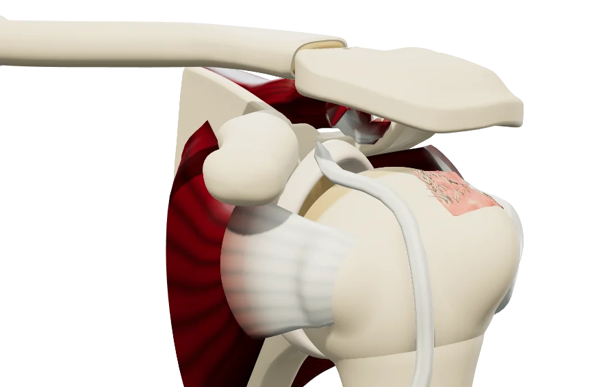
The supraspinatus, in 3D, for your patient
Open the rotator cuff model in your browser, no sign-up needed.
A colleague preparing a lecture on the cuff?