- EMET Education
- 3D Anatomy
- Shoulder
- Subscapularis muscle
Anatomy · Shoulder · Rotator cuff
The Subscapularis Muscle in 3D
The subscapularis in 3D, layer by layer, then on labeled MRI slices. With its trochlea-like insertion around the long head of biceps, its nerves and their distance from the coracoid, and the landmarks for examining and repairing its tendon.
Designed by an orthopedic surgeon
Latin musculus subscapularis · French subscapulaire, sous-scapulaire
- 40 × 20 mmMaximum dimensions of the insertion on the lesser tuberosity (20 cadavers)
- 18 mmDistance of the insertion from the cartilage at its inferior border (20 cadavers)
- 27.4%Subscapularis tears in a series of 435 arthroscopic cuff repairs
- 11 mmClosest distance between a subscapular nerve's entry and the coracoid base (20 specimens)
At a glance
Origin, insertion, innervation
| Origin | Medial two-thirds of the subscapular fossa (costal surface of the scapula) and intramuscular septa; the medial border itself receives the serratus anterior. |
|---|---|
| Course | Anterior to the scapular neck and the capsule, beneath the coracoid process; the subscapular bursa, under its upper tendon, communicates with the joint. |
| Insertion | Lesser tuberosity, adjacent to the bicipital groove, at the edge of the articular surface, tapering away 18 mm at its inferior border. The superiormost insertion lies on the upper margin of the lesser tuberosity, from which a thin tendinous slip reaches the fovea capitis. The lower, muscular part attaches to the surgical neck. |
| Innervation | Upper and lower subscapular nerves (C5, C6), often with a middle branch: the supply is variable. The branches enter on average more than 2 cm medial to the musculotendinous junction. |
| Blood supply | Subscapular artery. |
| Action | Medial rotation of the arm; anterior stabilization of the humeral head. Greatest force-producing capacity of the cuff by physiological cross-sectional area; contributes passive tension at maximum abduction and lateral rotation, the apprehension position. |
| Relations | Superiorly, the rotator interval and the supraspinatus; laterally, the long head of biceps in its groove, held by a trochlea-like structure; anteriorly, the coracoid and the conjoint tendon, then the deltoid and the pectoralis major; posteriorly, the capsule and the scapular neck. |
Layer by layer
From the pectoralis major to the tendon
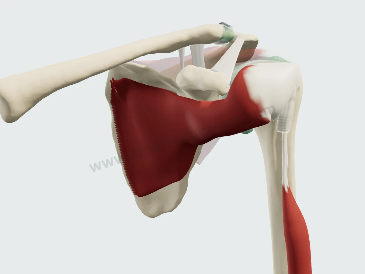
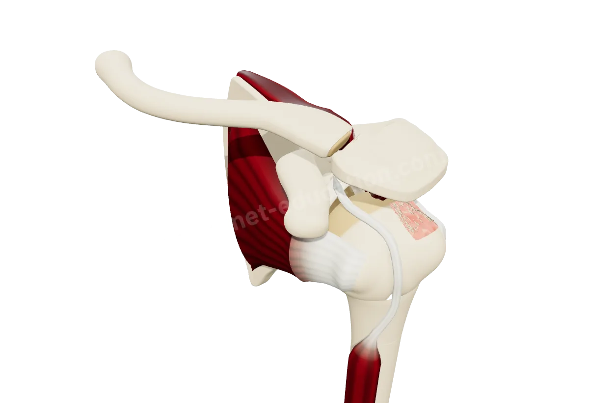
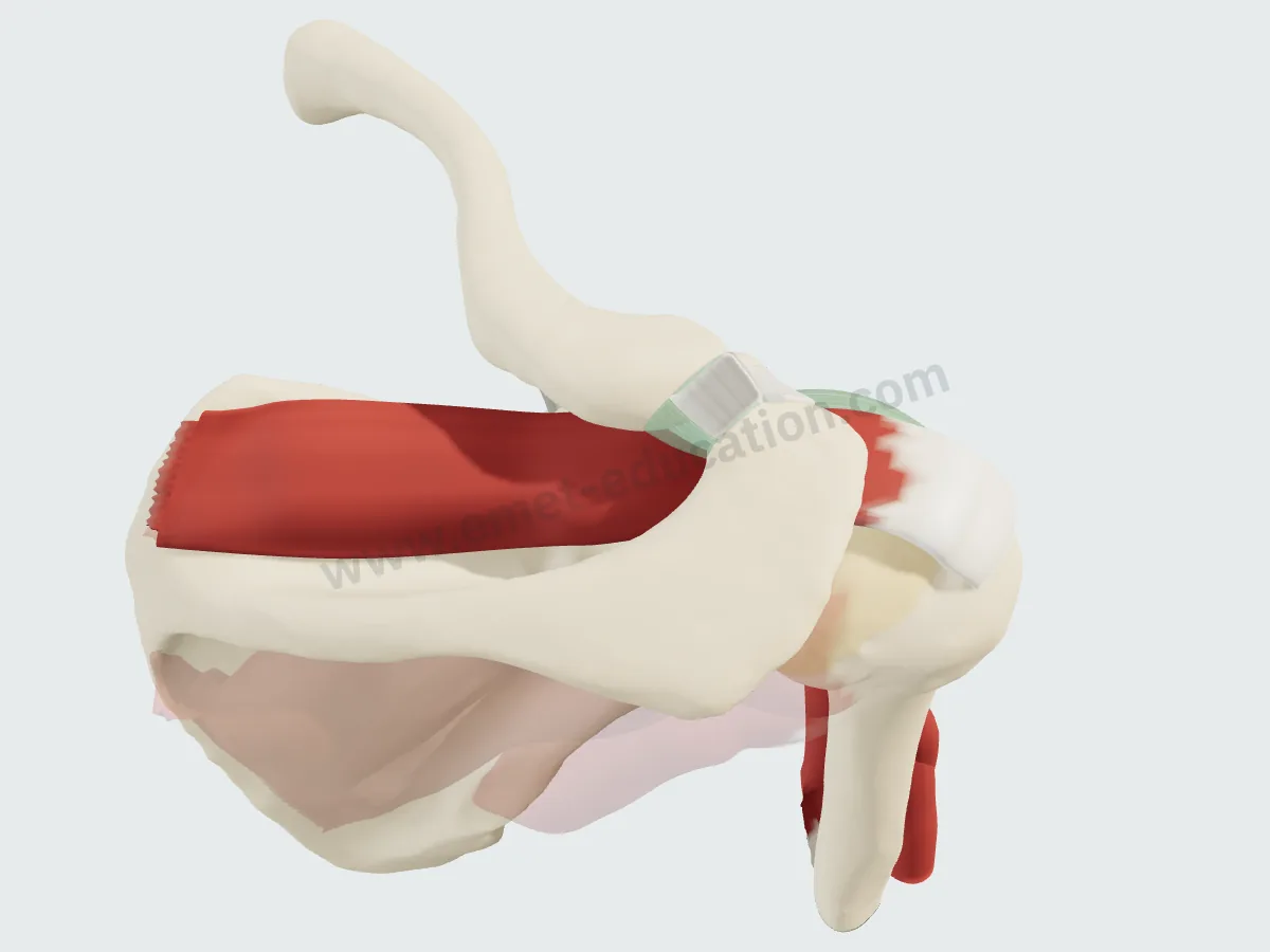
The same view, from the front and slightly medial to the shoulder, removing one layer per image. In the last two, the thorax is removed: the muscle lies on the costal surface of the scapula.
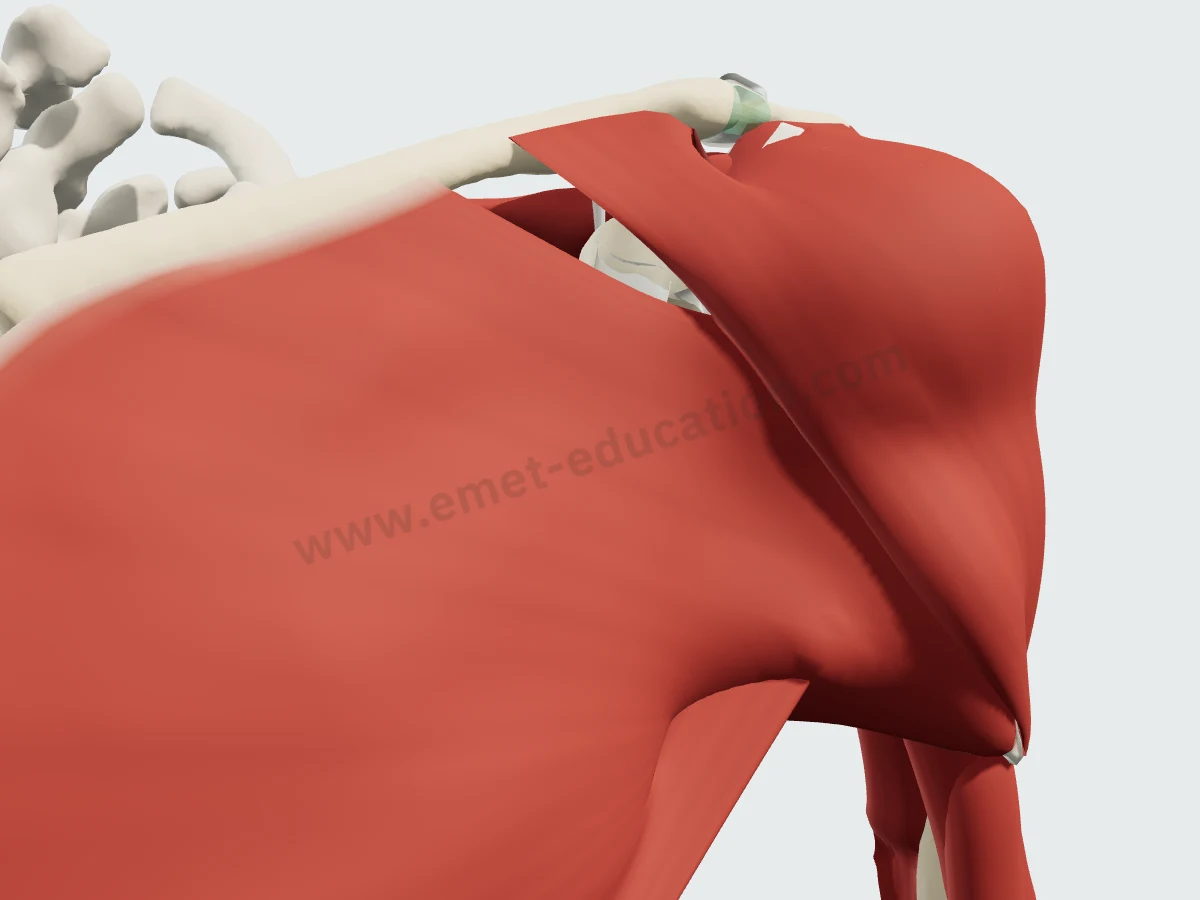
Pectorals and deltoidThe subscapularis is hidden: the pectoralis major covers the region, the deltoid the humeral head.
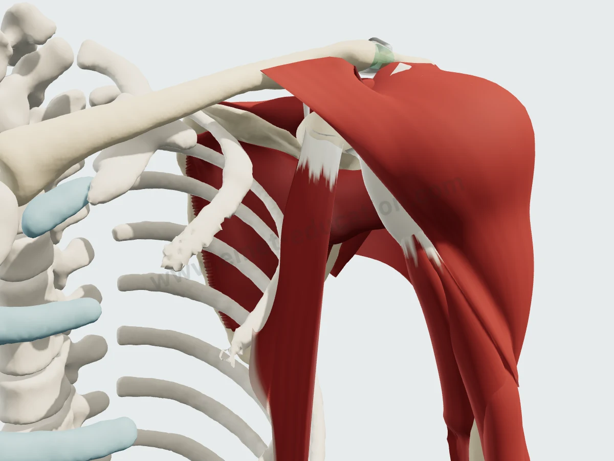
The conjoint tendonPectoralis major removed: the pectoralis minor, the conjoint tendon and the deltoid still screen the muscle.
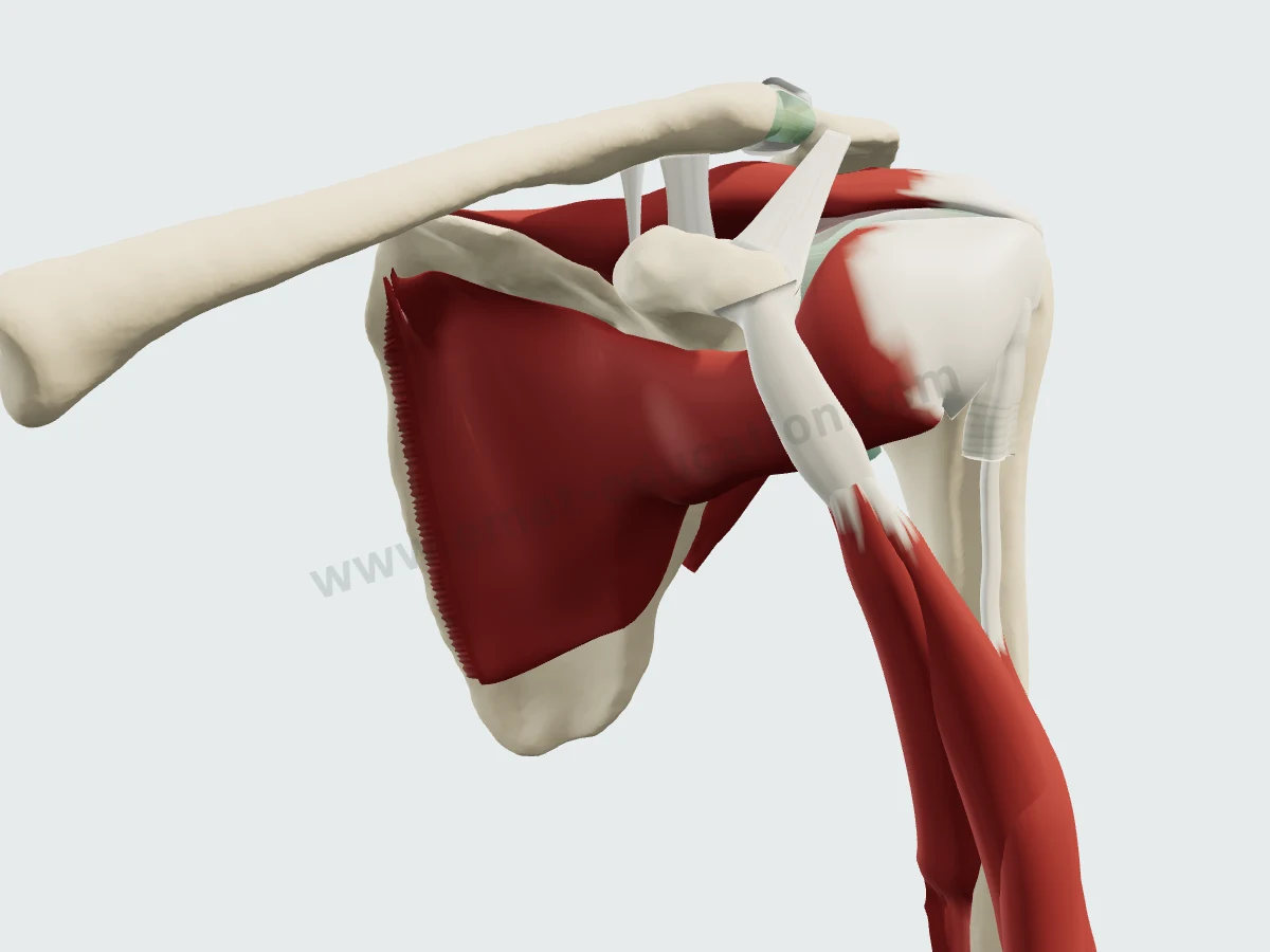
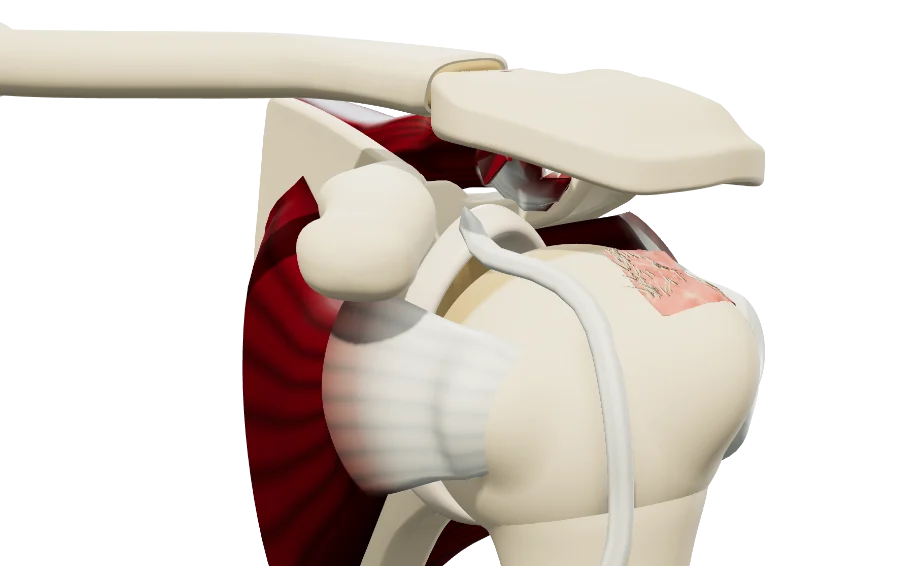
The anterior cuffDeltoid, bursa and pectoralis minor removed, thorax hidden: the subscapularis on the scapula, below the coracoid and its conjoint tendon.
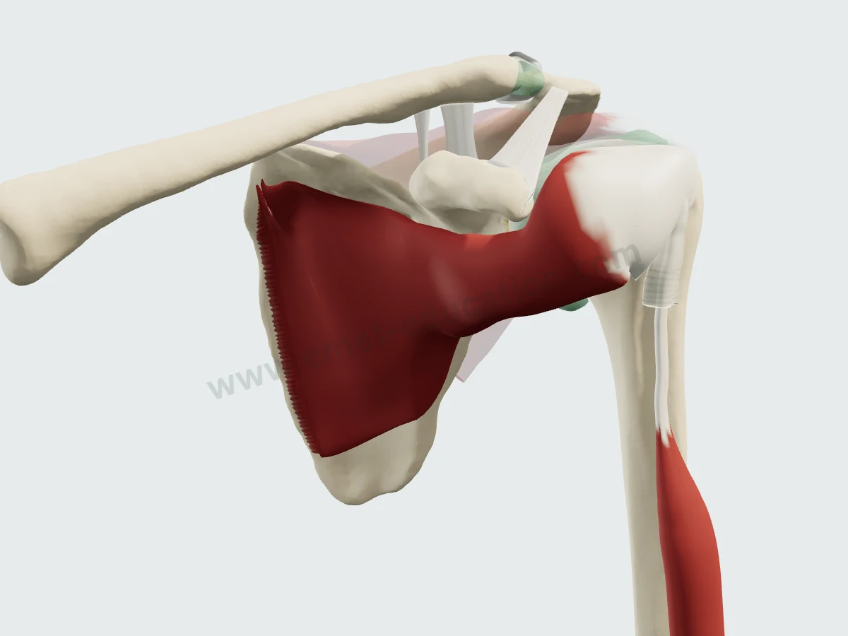
The subscapularisOther muscles faded: from the subscapular fossa to the lesser tuberosity, below the coracoid.
Imaging
The subscapularis on MRI and CT
Synthetic images. These slices do not come from a patient: they are computed from the 3D model, by cutting each structure and giving it the signal it would have in the chosen sequence, on MRI or CT. They are for learning where the muscle lies on a slice; nothing is measured on them.
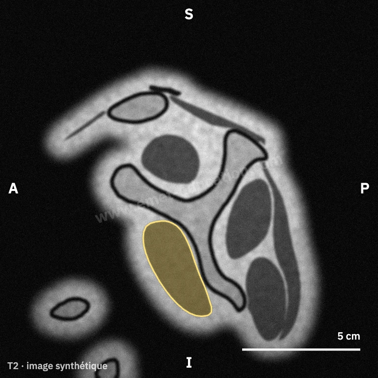
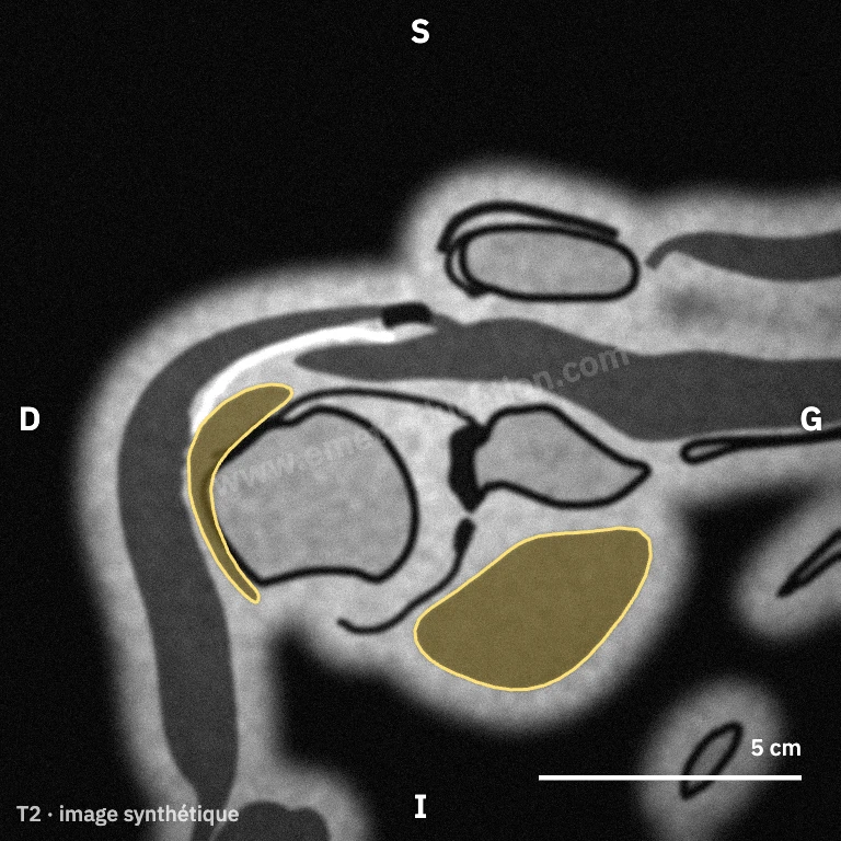
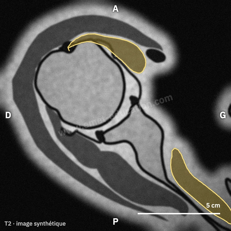
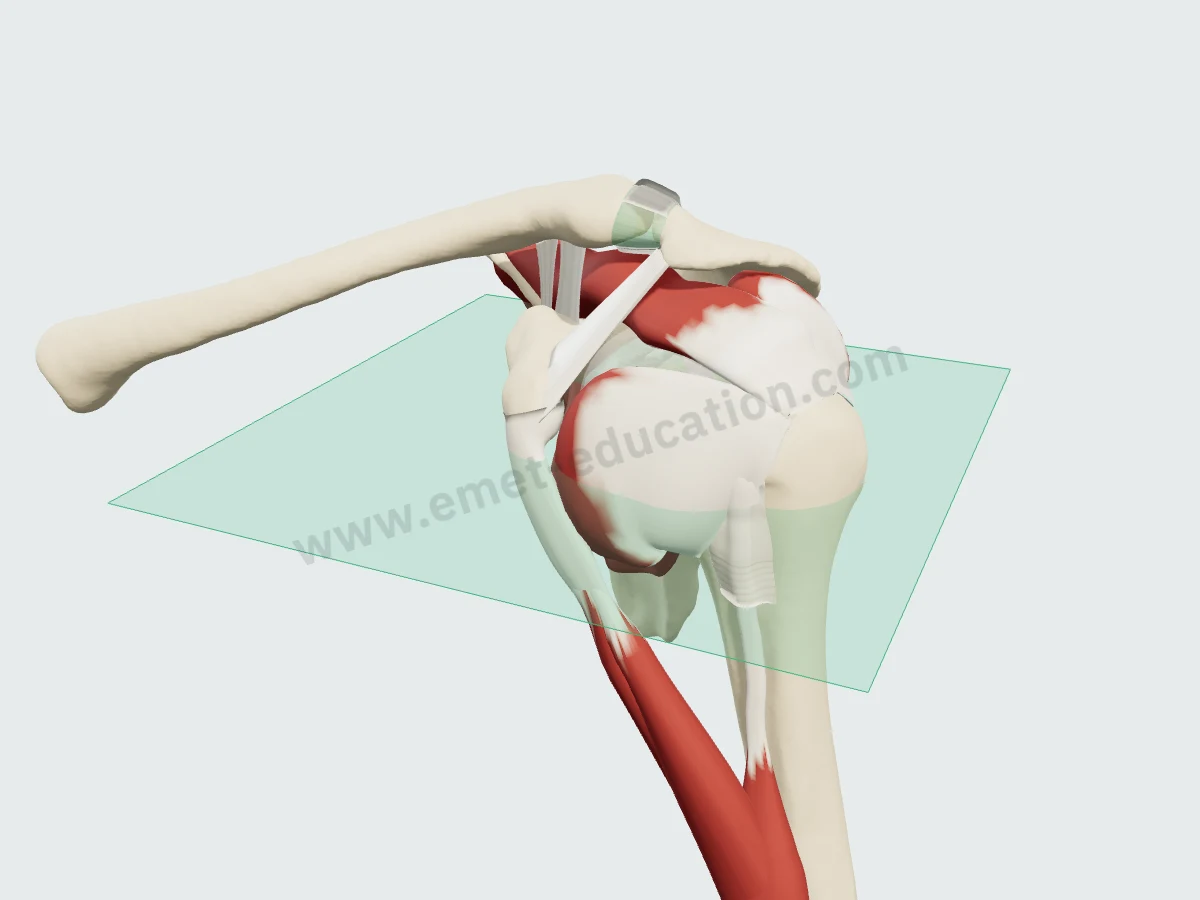
Where the axial view cuts
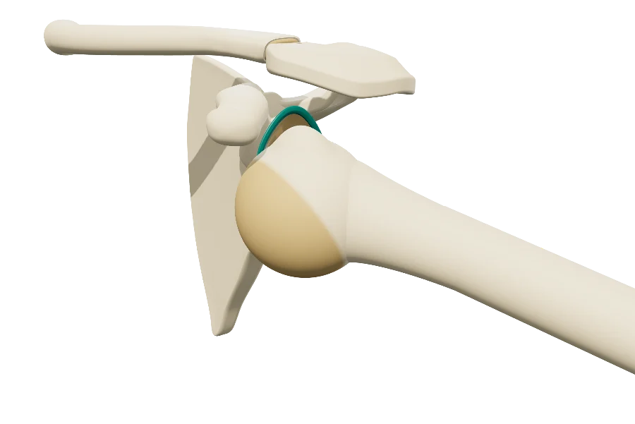
The axial plane (green) on the 3D model: horizontal, at the level of the center of the humeral head. Hover over a slice and click “Scroll” to page through it as on a workstation: upward, the muscle thins into a narrow tendon; downward, its belly widens.
In numbers
The insertion and the nerves, measured
Published mean values, with sample size and source under each figure.
| Value | Reference | Year | Sample | Note |
|---|---|---|---|---|
| 20 mm | Curtis | 2006 | 20 cadavers | Mean maximum width (40 × 20 mm); orientation to be confirmed in the full text to be confirmed |
| 40 mm | Curtis | 2006 | 20 cadavers | Mean maximum length (40 × 20 mm); orientation to be confirmed in the full text to be confirmed |
| Value | Reference | Year | Sample | Note |
|---|---|---|---|---|
| 7 mm | Leschinger | 2017 | 20 specimens | Upper branch, vertical distance from the medial base of the coracoid |
| 22 mm | Leschinger | 2017 | 20 specimens | Middle branch, vertical distance |
| 31.6 mm | Denard | 2013 | 20 specimens | Upper subscapular nerve (± 6.6; 22 to 45) |
| 42.6 mm | Denard | 2013 | 20 specimens | Lower subscapular nerve (± 7.6; 25 to 55) |
| 44 mm | Leschinger | 2017 | 20 specimens | Lower branch, vertical distance |
Published means, from smallest to largest. Sample size is as each study reports it (shoulders, cadavers or specimens); measurement protocols differ from one study to the next.
Maximum dimensions, to scale: a bounding box, not the shape of the footprint. The orientation of the two sides is to be confirmed in the full text.
- 31.6 mmUpper subscapular nerve entry, from the medial base of the coracoid, arm in neutral rotation (± 6.6); 24.2 mm in maximal lateral rotationMean, 20 specimens · Denard 2013
- 42.6 mmLower subscapular nerve entry, from the same base, neutral rotation (± 7.6); 33.9 mm in maximal lateral rotationMean, 20 specimens · Denard 2013
- 11 mmClosest distance between a nerve's entry and the coracoid base: releasing adhesions below the coracoid does not appear to risk denervation20 specimens · Denard 2013
- > 2 cmMean distance of the nerve branches from the musculotendinous junction, medially; at the closest, 1.1 to 1.3 cm20 specimens · Leschinger 2017
- 1stForce-producing capacity in the cuff by physiological cross-sectional area, ahead of the infraspinatus, supraspinatus and teres minor10 specimens · Ward 2006
Nerve distances are measured from different landmarks across studies (coracoid base, musculotendinous junction): they cannot be compared with each other. The nerve moves 7 to 9 mm closer to the coracoid when the humerus is laterally rotated (Denard).
History
How its description has changed since 1991
Classic anatomy places the subscapularis on the lesser tuberosity. Studies published since have described its isolated rupture, its superiormost insertion and its variable nerve supply.
- 1991
An isolated rupture
The tendon can tear on its own, from forced hyperextension or lateral rotation of the adducted arm: anterior pain, increased lateral rotation, decreased medial rotation strength. The lift-off test makes or excludes the diagnosis. Gerber
- 1992
A common insertion
The cuff tendons interdigitate into a continuous insertion, and the long head of biceps is ensheathed by fibres of the subscapularis and supraspinatus. Clark and Harryman
- 1996
The internal rotation lag
As specific as the lift-off test and more sensitive, the internal rotation lag sign detects partial tears the lift-off test can miss. Hertel
- 2006
A large footprint
A 40 × 20 mm insertion against the bicipital groove, at the cartilage edge, tapering away 18 mm at its inferior border. Curtis
- 2008
A pulley for the biceps
The superiormost insertion and a tendinous slip to the fovea capitis form a trochlea-like structure against the long head of biceps, holding it in the groove; transverse tears always involve this insertion. Arai
- 2017
A variable nerve
Often three branches, upper, middle and lower, entering on average more than 2 cm medial to the musculotendinous junction, but as close as 1.1 cm. Leschinger
In practice
What anatomy changes in practice
- Examining it. The lift-off test makes or excludes a clinically relevant tear (Gerber); the internal rotation lag sign is as specific and more sensitive, detecting partial tears the lift-off test can miss (Hertel).
- Locating the tear. A tear can involve the superior third of the tendon, its superior two-thirds or the whole insertion (Lafosse); transverse tears involve the superiormost insertion and were accompanied by intra-articular biceps dislocation in 44.8% of full-thickness tears (Arai).
- Repairing it. In 17 isolated tears repaired arthroscopically, the relative Constant score rose from 58% to 96%, the repair was intact in 15 of 17 patients on CT arthrography, and fatty infiltration did not progress (Lafosse).
- Reading the muscle. It rarely degenerates, and then only moderately, even when its tendon is not torn (Goutallier). In massive tears, loss of active medial rotation is related to its involvement; pseudoparalysis was present in 80% of cases combining supraspinatus and complete subscapularis tears (Collin).
- Protecting the nerves. In anterior approaches, avoid dissecting or releasing more than 1 cm medial to the musculotendinous junction; to split the muscle horizontally, go 3.2–3.6 cm vertically from the coracoid base (Leschinger). Below the coracoid, releasing adhesions does not appear to risk denervation: the closest is 11 mm, and the nerves move closer in lateral rotation (Denard).
Your turn
Where is the subscapularis?
Six unlabeled synthetic slices, in random order: click the subscapularis. The answer shows its outline.
Click the subscapularis.
Conditions
Related conditions
Neighbors
Its neighbors
 SupraspinatusSuperior
- SoonLong head of bicepsLateral
- SoonConjoint tendonAnterior
- SoonDeltoidSuperficial
- SoonJoint capsuleDeep
Questions
Frequently asked questions
Where does the subscapularis insert?
On the lesser tuberosity, against the bicipital groove; its lower, muscular part attaches to the surgical neck. Its superiormost insertion lies on the upper margin of the lesser tuberosity, from which a tendinous slip reaches the fovea capitis (Arai). It averages 40 × 20 mm (Curtis, 20 cadavers).
What nerves innervate the subscapularis?
The upper and lower subscapular nerves (C5, C6). The supply is variable: three branches, upper, middle and lower, in 14 of 20 shoulders (Leschinger).
How do you examine the subscapularis?
By the lift-off test (Gerber) and the internal rotation lag sign, which is more sensitive: it detects partial tears the lift-off test can miss (Hertel).
Why does a subscapularis tear involve the long head of biceps?
Because the muscle's superiormost insertion and its tendinous slip form, with the cranial intramuscular tendons, a trochlea-like structure holding the long head of biceps in the groove. Transverse tears involve this insertion and were accompanied by intra-articular biceps dislocation in 44.8% of full-thickness tears (Arai).
How do you avoid injuring its nerves in an anterior approach?
By avoiding dissection or release more than 1 cm medial to the musculotendinous junction and, to split the muscle horizontally, by going 3.2–3.6 cm vertically from the coracoid base (Leschinger).
Are the MRI images on this page real?
No: they are synthetic, computed from the 3D model. They show where the muscle lies on a slice; they do not replace an actual exam.
Sources
Author and references
- Standring S, ed. Gray's Anatomy: The Anatomical Basis of Clinical Practice. 42nd ed. Elsevier; 2020.
- Gerber C, Krushell RJ. Isolated rupture of the tendon of the subscapularis muscle. Clinical features in 16 cases. J Bone Joint Surg Br. 1991;73(3):389–394. doi:10.1302/0301-620X.73B3.1670434
- Clark JM, Harryman DT 2nd. Tendons, ligaments, and capsule of the rotator cuff. Gross and microscopic anatomy. J Bone Joint Surg Am. 1992;74(5):713–725. PMID 1624486
- Hertel R, Ballmer FT, Lombert SM, Gerber C. Lag signs in the diagnosis of rotator cuff rupture. J Shoulder Elbow Surg. 1996;5(4):307–313. doi:10.1016/s1058-2746(96)80058-9
- Goutallier D, Postel JM, Bernageau J, Lavau L, Voisin MC. Fatty muscle degeneration in cuff ruptures. Pre- and postoperative evaluation by CT scan. Clin Orthop Relat Res. 1994;(304):78–83. PMID 8020238
- Curtis AS, Burbank KM, Tierney JJ, Scheller AD, Curran AR. The insertional footprint of the rotator cuff: an anatomic study. Arthroscopy. 2006;22(6):609.e1. doi:10.1016/j.arthro.2006.04.001
- Lafosse L, Jost B, Reiland Y, Audebert S, Toussaint B, Gobezie R. Structural integrity and clinical outcomes after arthroscopic repair of isolated subscapularis tears. J Bone Joint Surg Am. 2007;89(6):1184–1193. doi:10.2106/JBJS.F.00007
- Arai R, Sugaya H, Mochizuki T, Nimura A, Moriishi J, Akita K. Subscapularis tendon tear: an anatomic and clinical investigation. Arthroscopy. 2008;24(9):997–1004. doi:10.1016/j.arthro.2008.04.076
- Ward SR, Hentzen ER, Smallwood LH, et al. Rotator cuff muscle architecture: implications for glenohumeral stability. Clin Orthop Relat Res. 2006;448:157–163. doi:10.1097/01.blo.0000194680.94882.d3
- Denard PJ, Duey RE, Dai X, Hanypsiak B, Burkhart SS. Relationship of the subscapular nerves to the base of the coracoid. Arthroscopy. 2013;29(6):986–989. doi:10.1016/j.arthro.2013.02.005
- Leschinger T, Hackl M, Zeifang F, Scaal M, Müller LP, Wegmann K. Nerve supply of the subscapularis during anterior shoulder surgery: definition of a potential risk area. Arch Orthop Trauma Surg. 2017;137(1):135–140. doi:10.1007/s00402-016-2585-7
- Collin P, Matsumura N, Lädermann A, Denard PJ, Walch G. Relationship between massive chronic rotator cuff tear pattern and loss of active shoulder range of motion. J Shoulder Elbow Surg. 2014;23(8):1195–1202. doi:10.1016/j.jse.2013.11.019
In clinic
Show the rotator cuff to your patient
The images on this page come from an interactive model. In clinic, show the cuff tear, subscapularis included, and its repair.
The subscapularis, in 3D, for your patient
Open the rotator cuff model in your browser, no sign-up needed.
A colleague preparing a lecture on the cuff?