
- EMET Education
- 3D Anatomy
- Shoulder
- Teres minor muscle
Anatomy · Shoulder · Rotator cuff
The Teres Minor Muscle in 3D
The teres minor in 3D, layer by layer, then on labeled MRI slices. With its insertion below that of the infraspinatus, its nerve and fascial sling, its atrophy, and what it becomes when the cuff fails.
Designed by an orthopedic surgeon
Latin musculus teres minor · French petit rond
- 29 × 21 mmMaximum dimensions of the humeral insertion (20 cadavers)
- 44 mmEntry of the primary nerve beneath the fascia, medial to the insertion (31 shoulders)
- 90.8%Teres minor of normal appearance in 1,572 images of rotator cuff tears
- 6.7%Atrophy on MRI at ages 60–69, versus 2.1% at ages 10–39
At a glance
Origin, insertion, innervation
| Origin | Upper two-thirds of the dorsal surface of the lateral border of the scapula. |
|---|---|
| Structure | Two distinct muscular bundles: fatty degeneration of the medial-dorsal bundle exceeds that of the lateral-ventral bundle in most cases (Chambers). A fascia separates it from the infraspinatus in half of shoulders and envelops both together in the other half (Chafik). |
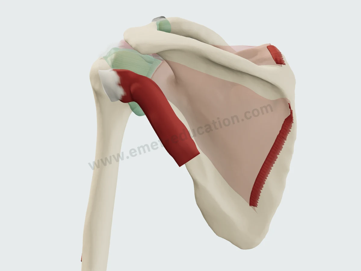
| Insertion | Inferior facet of the greater tuberosity and, below it, the surgical neck of the humerus. The cuff footprint is horseshoe-shaped and tapers away from the cartilage from superior to inferior: the teres minor inserts below the infraspinatus, away from the cartilage. |
| Innervation | Axillary nerve (C5, C6), posterior branch. The muscle's primary nerve travels around a fascial sling, becoming sub-fascial on average 44 mm medial to the insertion; accessory branches begin at 30 mm on average and remain extra-fascial. The course, length and branching of the nerve vary from one shoulder to another. |
| Blood supply | Circumflex scapular and posterior circumflex humeral arteries. |
| Action | Lateral rotation of the arm; posterior stabilization of the humeral head. It has the smallest force-producing capacity of the cuff by physiological cross-sectional area; when the infraspinatus is torn, it becomes the only lateral rotator. |
| Relations | It forms the superior border of the quadrangular space, bounded inferiorly by the teres major, medially by the long head of the triceps and laterally by the surgical neck: the axillary nerve and the posterior circumflex humeral artery cross it. |
Layer by layer
From the trapezius to the tendon
The same view, from behind, slightly lateral to and below the shoulder, removing one layer per image.
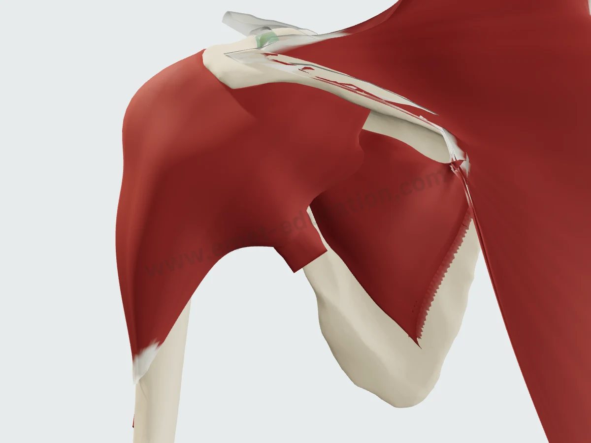
Trapezius and deltoidThe teres minor is hidden: the posterior deltoid covers the humeral head and its insertion, the trapezius the upper scapula.
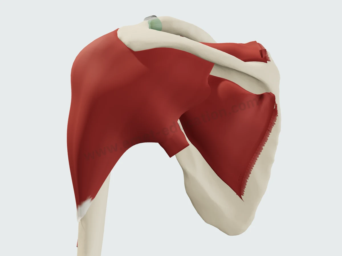
The posterior deltoidTrapezius removed: the posterior deltoid still covers the teres minor insertion on the humerus.
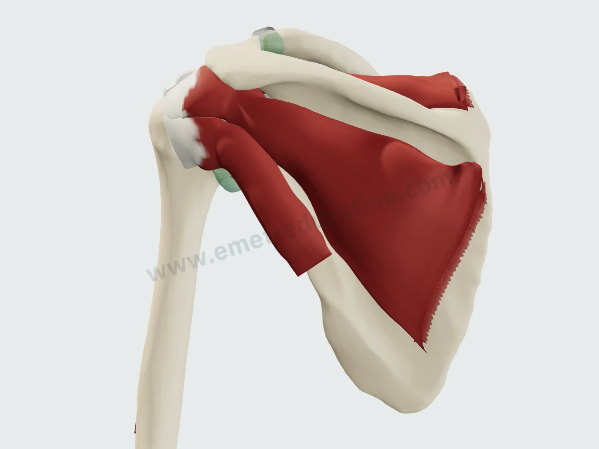
The posterior cuffDeltoid removed: the infraspinatus in its fossa and, below it, the teres minor, along the lateral border of the scapula.
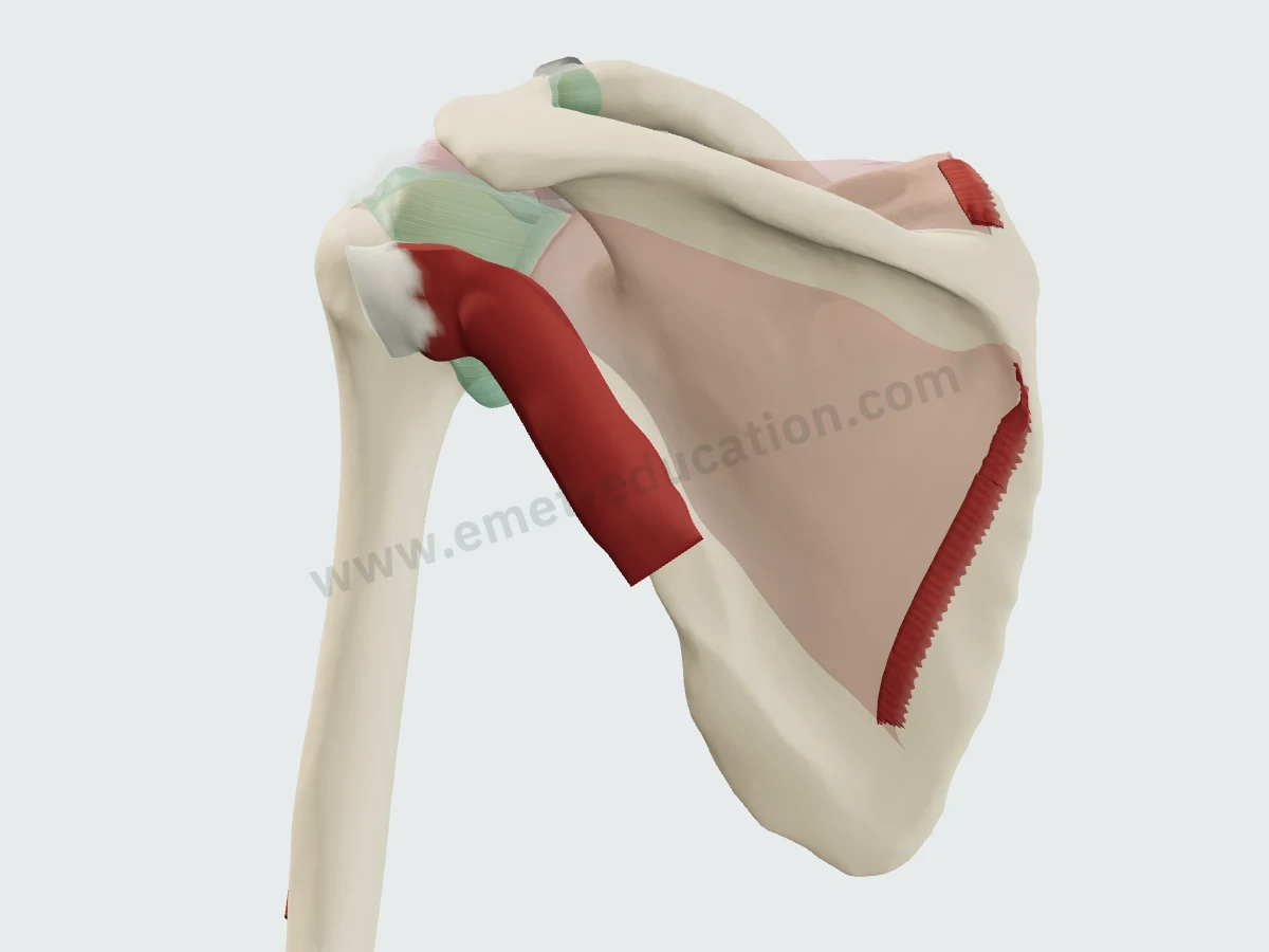
The teres minorOther muscles faded: from the lateral border of the scapula to the inferior facet of the greater tuberosity.
Imaging
The teres minor on MRI and CT
Synthetic images. These slices do not come from a patient: they are computed from the 3D model, by cutting each structure and giving it the signal it would have in the chosen sequence, on MRI or CT. They are for learning where the muscle lies on a slice; nothing is measured on them.
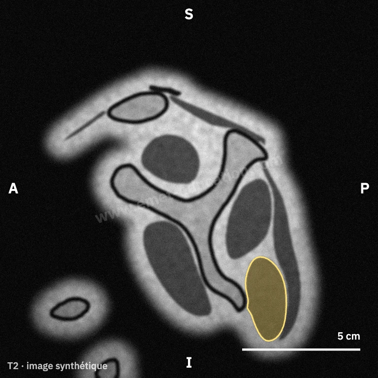
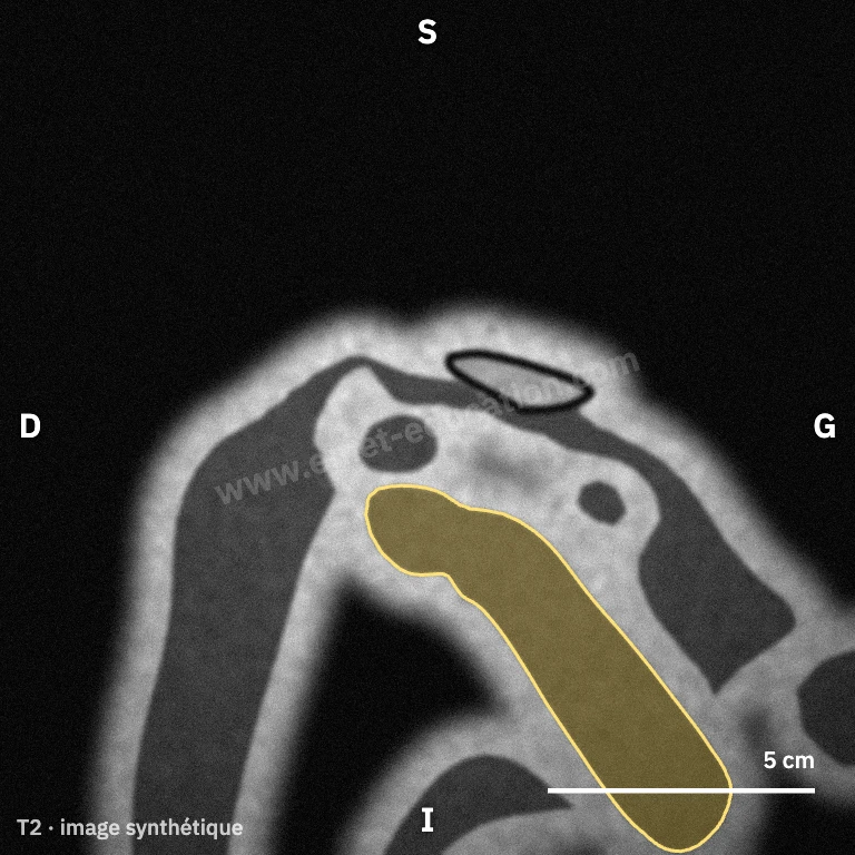
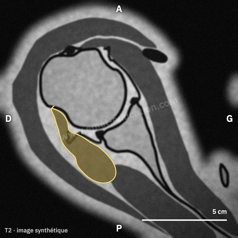
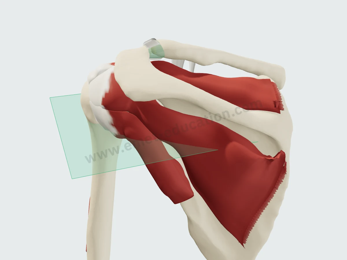
Where the axial view cuts
The axial plane (green) on the 3D model: horizontal, below the center of the humeral head, it crosses the teres minor from its belly to its tendon. Hover over a slice and click “Scroll” to page through it as on a workstation: the infraspinatus appears upward, the teres major downward.
In numbers
The insertion, the nerve and the atrophy, measured
Published mean values, with sample size and source under each figure.
| Value | Reference | Year | Sample | Note |
|---|---|---|---|---|
| 21 mm | Curtis | 2006 | 20 cadavers | Mean maximum width (29 × 21 mm); orientation to be confirmed in the full text to be confirmed |
| 29 mm | Curtis | 2006 | 20 cadavers | Mean maximum length (29 × 21 mm); orientation to be confirmed in the full text to be confirmed |
Published mean values, from smallest to largest. Sample size is as each study reports it (shoulders, cadavers or images); populations and protocols differ from one study to the next.
Maximum dimensions, to scale: a bounding box, not the shape of the footprint. The orientation of the two sides is to be confirmed in the full text.
- 44 mmEntry of the primary nerve beneath the fascia, medial to the insertion (25–68): the fascial sling is a probable site of compression and tetheringMean, 31 shoulders · Chafik 2012
- 30 mmOrigin of the accessory motor branches, medial to the insertion (15–48): extra-fascial, with a less angulated courseMean, 31 shoulders · Chafik 2012
- 2.1 → 6.7%Teres minor atrophy on MRI: ages 10–39, then 60–69; cuff intact in 64% of atrophies in young patients and 36% in older ones (difference not significant)528 and 884 shoulders · Takeuchi 2022
- + 70 to 90%Teres minor torque in a massive posterosuperior tear with an intact teres minor (Collin type D), compared with the healthy shoulder; activity up to 7-fold higherMusculoskeletal model · Menze 2024
- 4thForce-producing capacity in the cuff by physiological cross-sectional area: the smallest of the four muscles, behind the subscapularis, infraspinatus and supraspinatus10 specimens · Ward 2006
Atrophy rates depend on the population (cuff tear, age, sport) and on how the image is read: they cannot be compared directly. The incidence of isolated atrophy is reported at 3% to 6.2% (Chambers).
History
How its description has changed since 2006
Classic anatomy describes it as the smallest lateral rotator. Studies published since have clarified its insertion, its nerve, its imaging appearance and its role when the cuff fails.
- 2006
Below the infraspinatus
The cuff insertion is a horseshoe; the teres minor's, at most 29 × 21 mm, lies below the infraspinatus, away from the cartilage. Curtis
- 2006
The smallest of the cuff
Its physiological cross-sectional area is the smallest of the four cuff muscles: it is the least powerful. Ward
- 2009
A variable nerve, a distinct atrophy
The nerve to the teres minor varies in course, length and branching; isolated atrophy is distinct from quadrilateral space syndrome. The latissimus dorsi transfer mostly compensates a deficient teres minor. Friend, Nové-Josserand
- 2011
A morphological classification
Normal, hypertrophic, atrophic or absent, in 1,572 images: the teres minor is normal in nine cases out of ten, hypertrophic mostly in anterior tears, atrophic in posterosuperior tears. Melis
- 2012
A fascial sling
Two equally common fascial variants and a sling where the primary nerve changes plane, on average 44 mm from the insertion: the probable site of compression. Chafik
- 2018
A decisive muscle when the cuff fails
Overlooked when normal, the teres minor becomes the main lateral rotator in a massive tear; its state informs transfers and reverse arthroplasty, and its two bundles atrophy unequally. Williams, Chambers
In practice
What anatomy changes in practice
- Examining it. The Patte test, the Neer drop sign, the external rotation lag sign and the Hertel drop sign test lateral rotation when the infraspinatus no longer provides it (Williams). After latissimus dorsi transfer, the hornblower sign was corrected in 6 of the 15 patients who had it (Nové-Josserand).
- Reading it on imaging. It is classed as normal, hypertrophic, atrophic or absent (Melis); fatty infiltration is graded by Goutallier (Goutallier). It affects the medial-dorsal bundle more than the lateral-ventral one (Chambers), and can occur with an intact cuff, even in young patients, where sport is suggested as a factor (Takeuchi).
- Weighing the prognosis. In massive tears, an intact teres minor becomes the only lateral rotator (Williams). Isolated fatty infiltration did not prevent a cuff repair from succeeding: 51 of 816 patients (Kim). Before reverse arthroplasty, its hypertrophy is a negative predictor of the 2-year ASES score (73.3 versus 84.2) (Hung).
- Respecting its nerve. The primary nerve changes plane under a fascial sling on average 44 mm from the insertion, with accessory branches at 30 mm (Chafik); intramuscular branches concentrate between 1/5 and 3/5 of the muscle, from the axillary border of the scapula to the insertion, which its authors use to guide a botulinum toxin injection (Yi).
- Looking for a neighboring lesion. Teres minor atrophy can come from the cuff, from quadrilateral space syndrome (Dalagiannis) or remain isolated (Friend). In 105 MRIs showing denervation, 82.9% had a cuff tear and 82.3% a posterior labrocapsular complex tear (Lee).
Your turn
Where is the teres minor?
Six unlabeled synthetic slices, in random order: click the teres minor. The answer shows its outline.
Click the teres minor.
Conditions
Related conditions
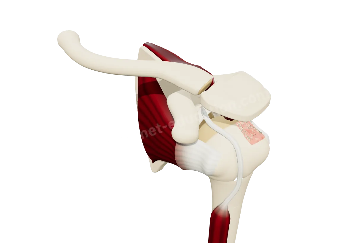
Neighbors
Its neighbors
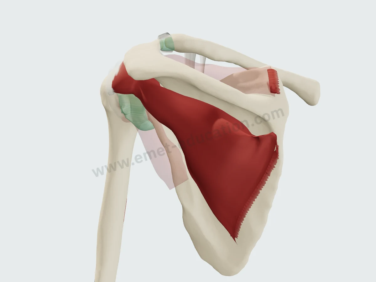 InfraspinatusSuperior
- SoonTeres majorInferior
- SoonLong head of tricepsMedial
- SoonDeltoidSuperficial
- SoonJoint capsuleDeep
Questions
Frequently asked questions
Where does the teres minor insert?
On the inferior facet of the greater tuberosity and, below it, on the surgical neck of the humerus (Gray's Anatomy). Its insertion lies below that of the infraspinatus, away from the cartilage; it measures at most 29 × 21 mm (Curtis, 20 cadavers).
What nerve innervates the teres minor?
The posterior branch of the axillary nerve (C5, C6). Its primary nerve travels around a fascial sling and becomes sub-fascial on average 44 mm medial to the insertion; accessory branches begin at 30 mm (Chafik, 31 shoulders). Its course varies from one shoulder to another (Friend).
Why does the teres minor matter when the cuff is torn?
Because once the infraspinatus is torn, it becomes the only lateral rotator: from insignificant, it takes the main role. Its presence or absence informs the prognosis of tendon transfers and reverse arthroplasty (Williams).
How common is teres minor atrophy?
Isolated atrophy is reported in 3% to 6.2% of cases (Chambers). On MRI it affects 2.1% of 10–39-year-olds and 6.7% of 60–69-year-olds, with or without an intact cuff (Takeuchi). In rotator cuff tears, the muscle is atrophic in 3.2% of cases (Melis).
Does teres minor atrophy always come from axillary nerve compression?
No. Quadrilateral space syndrome can cause it (Dalagiannis), but isolated atrophy is clinically distinct from that syndrome (Friend) and often accompanies a rotator cuff tear, especially after age 60 (Takeuchi).
Are the MRI images on this page real?
No: they are synthetic, computed from the 3D model. They show where the muscle lies on a slice; they do not replace an actual exam.
Sources
Author and references
- Standring S, ed. Gray's Anatomy: The Anatomical Basis of Clinical Practice. 42nd ed. Elsevier; 2020.
- Curtis AS, Burbank KM, Tierney JJ, Scheller AD, Curran AR. The insertional footprint of the rotator cuff: an anatomic study. Arthroscopy. 2006;22(6):609.e1. doi:10.1016/j.arthro.2006.04.001
- Ward SR, Hentzen ER, Smallwood LH, et al. Rotator cuff muscle architecture: implications for glenohumeral stability. Clin Orthop Relat Res. 2006;448:157–163. doi:10.1097/01.blo.0000194680.94882.d3
- Chafik D, Galatz LM, Keener JD, Kim HM, Yamaguchi K. Teres minor muscle and related anatomy. J Shoulder Elbow Surg. 2013;22(1):108–114. doi:10.1016/j.jse.2011.12.005
- Friend J, Francis S, McCulloch J, Ecker J, Breidahl W, McMenamin P. Teres minor innervation in the context of isolated muscle atrophy. Surg Radiol Anat. 2010;32(3):243–249. doi:10.1007/s00276-009-0605-9
- Yi KH, Kim SB, Lee K, Hu H, Lee JH, Lee HJ. Intramuscular neural distribution of the teres minor muscle using Sihler's stain: application to botulinum neurotoxin injection. Anat Cell Biol. 2023;56(3):322–327. doi:10.5115/acb.23.087
- Melis B, DeFranco MJ, Lädermann A, Barthelemy R, Walch G. The teres minor muscle in rotator cuff tendon tears. Skeletal Radiol. 2011;40(10):1335–1344. doi:10.1007/s00256-011-1178-3
- Chambers MM, Khan AZ, Namdari S. Teres minor muscle atrophy: anatomy, patterns, and clinical manifestations. JBJS Rev. 2022;10(12). doi:10.2106/JBJS.RVW.22.00130
- Takeuchi Y, Sugaya H, Takahashi N, et al. Incidence of teres minor muscle atrophy in young and middle-aged populations. JSES Int. 2022;6(3):459–462. doi:10.1016/j.jseint.2022.01.003
- Kim JK, Yoo HJ, Jeong JH, Kim SH. Effect of teres minor fatty infiltration on rotator cuff repair outcomes. Arthroscopy. 2016;32(4):552–558. doi:10.1016/j.arthro.2015.10.021
- Williams MD, Edwards TB, Walch G. Understanding the importance of the teres minor for shoulder function: functional anatomy and pathology. J Am Acad Orthop Surg. 2018;26(5):150–161. doi:10.5435/JAAOS-D-15-00258
- Nové-Josserand L, Costa P, Liotard JP, Safar JF, Walch G, Zilber S. Results of latissimus dorsi tendon transfer for irreparable cuff tears. Orthop Traumatol Surg Res. 2009;95(2):108–113. doi:10.1016/j.otsr.2008.10.002
- Hung LW, Wu S, Lee A, et al. Teres minor muscle hypertrophy is a negative predictor of outcomes after reverse total shoulder arthroplasty: an evaluation of preoperative magnetic resonance imaging and postoperative implant position. J Shoulder Elbow Surg. 2021;30(10):e636–e645. doi:10.1016/j.jse.2020.12.020
- Menze J, Rojas JT, Ferguson SJ, et al. Lower trapezius and latissimus dorsi transfer relieve teres minor activity into the physiological range in Collin D irreparable posterosuperior massive rotator cuff tears: a biomechanical analysis. J Shoulder Elbow Surg. 2024;33(12):2586–2595. doi:10.1016/j.jse.2024.03.019
- Lee JY, Yoo YS, Shon K. Teres minor denervation and pathologies resulting in shoulder joint instability and rotator cuff tears: a retrospective cross-sectional MRI study. Medicine (Baltimore). 2024;103(8):e37232. doi:10.1097/MD.0000000000037232
- Dalagiannis N, Tranovich M, Ebraheim N. Teres minor and quadrilateral space syndrome: a review. J Orthop. 2020;20:144–146. doi:10.1016/j.jor.2020.01.021
- Goutallier D, Postel JM, Bernageau J, Lavau L, Voisin MC. Fatty muscle degeneration in cuff ruptures. Pre- and postoperative evaluation by CT scan. Clin Orthop Relat Res. 1994;(304):78–83. PMID 8020238
- Collin P, Matsumura N, Lädermann A, Denard PJ, Walch G. Relationship between massive chronic rotator cuff tear pattern and loss of active shoulder range of motion. J Shoulder Elbow Surg. 2014;23(8):1195–1202. doi:10.1016/j.jse.2013.11.019
In clinic
Show the rotator cuff to your patient
The images on this page come from an interactive model. In clinic, show the cuff tear, teres minor included, and its repair.
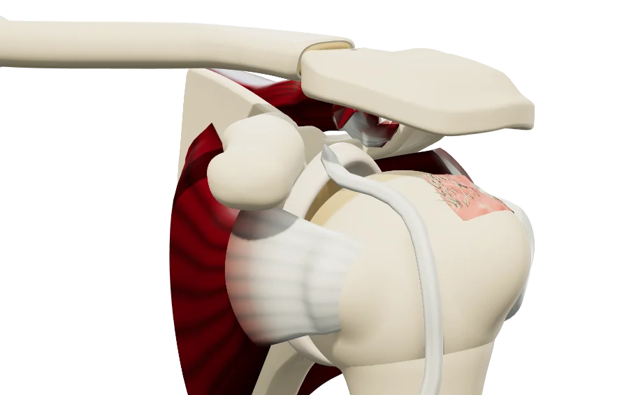
The teres minor, in 3D, for your patient
Open the rotator cuff model in your browser, no sign-up needed.
A colleague preparing a lecture on the cuff?