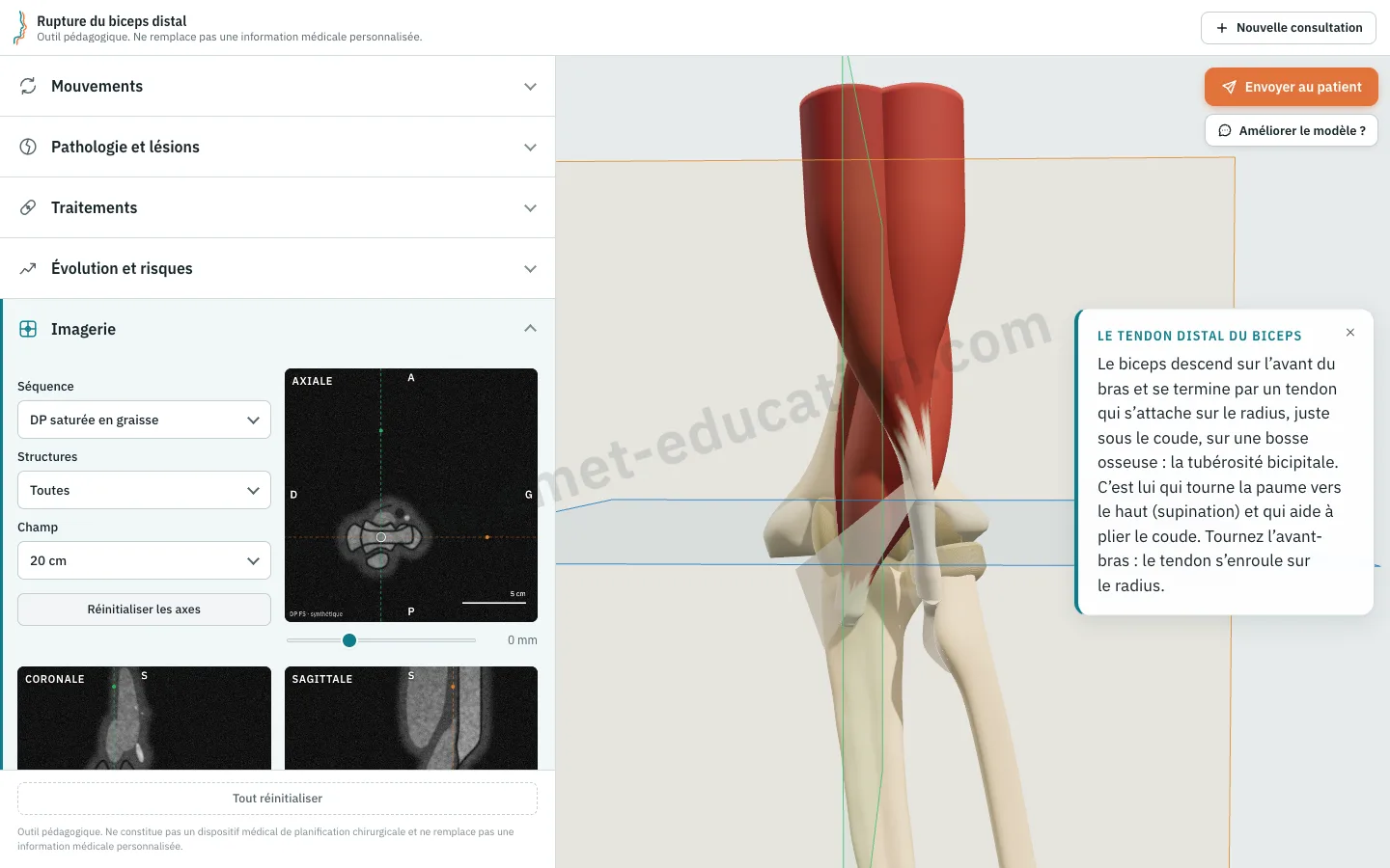
The Imaging panel slices the 3D model in its current state (lesion, stage, treatment) along three orthogonal planes, rendered as MRI sequences (PD fat-sat, T1, T2) or CT (bone window, soft-tissue window). Clicking a structure tracks it in yellow on the model and on all three slices. The images are synthetic: they help patients link their own scan to the model, and nothing is measured on their data.
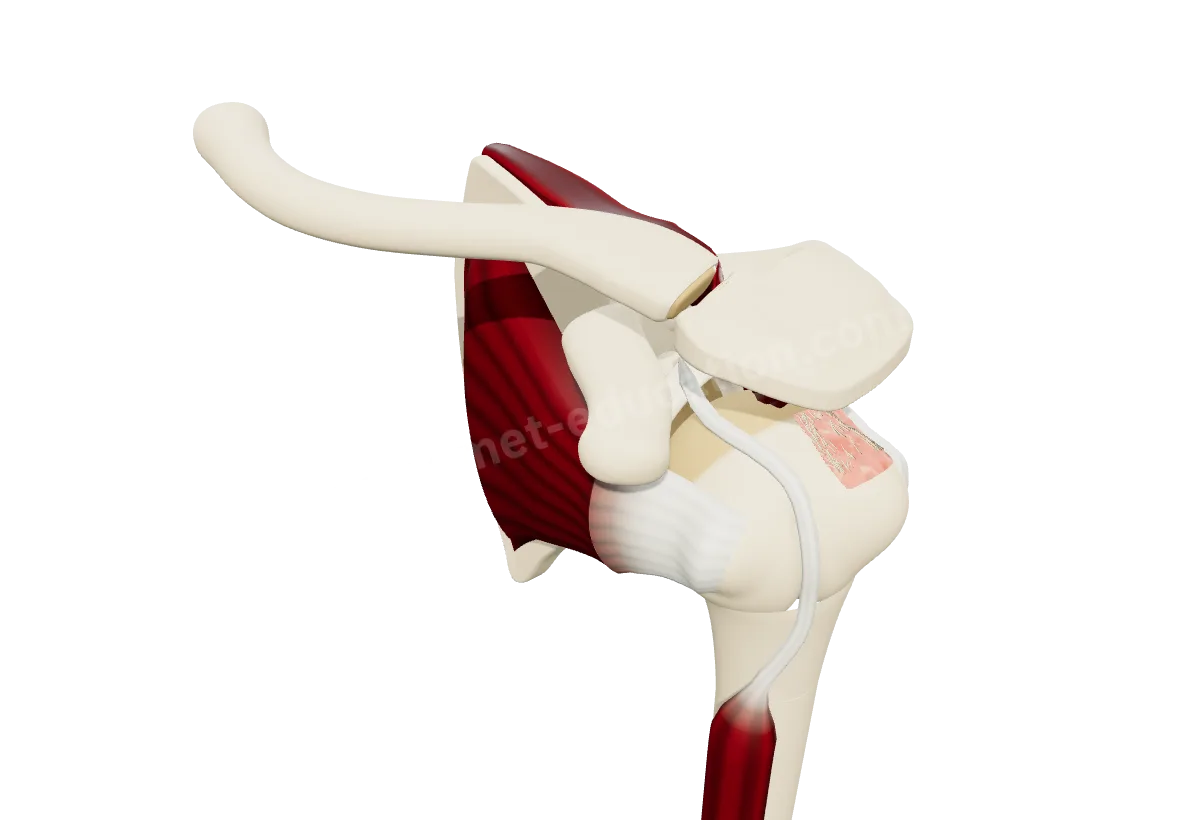
Supraspinatus retraction is set by Patte stages 1 to 3, subscapularis tears by the Lafosse classification (Ia to IV). Each step is named above the gauges, with the selected one highlighted. The arm now elevates in scapular-plane abduction up to 70° and in flexion up to 85°.
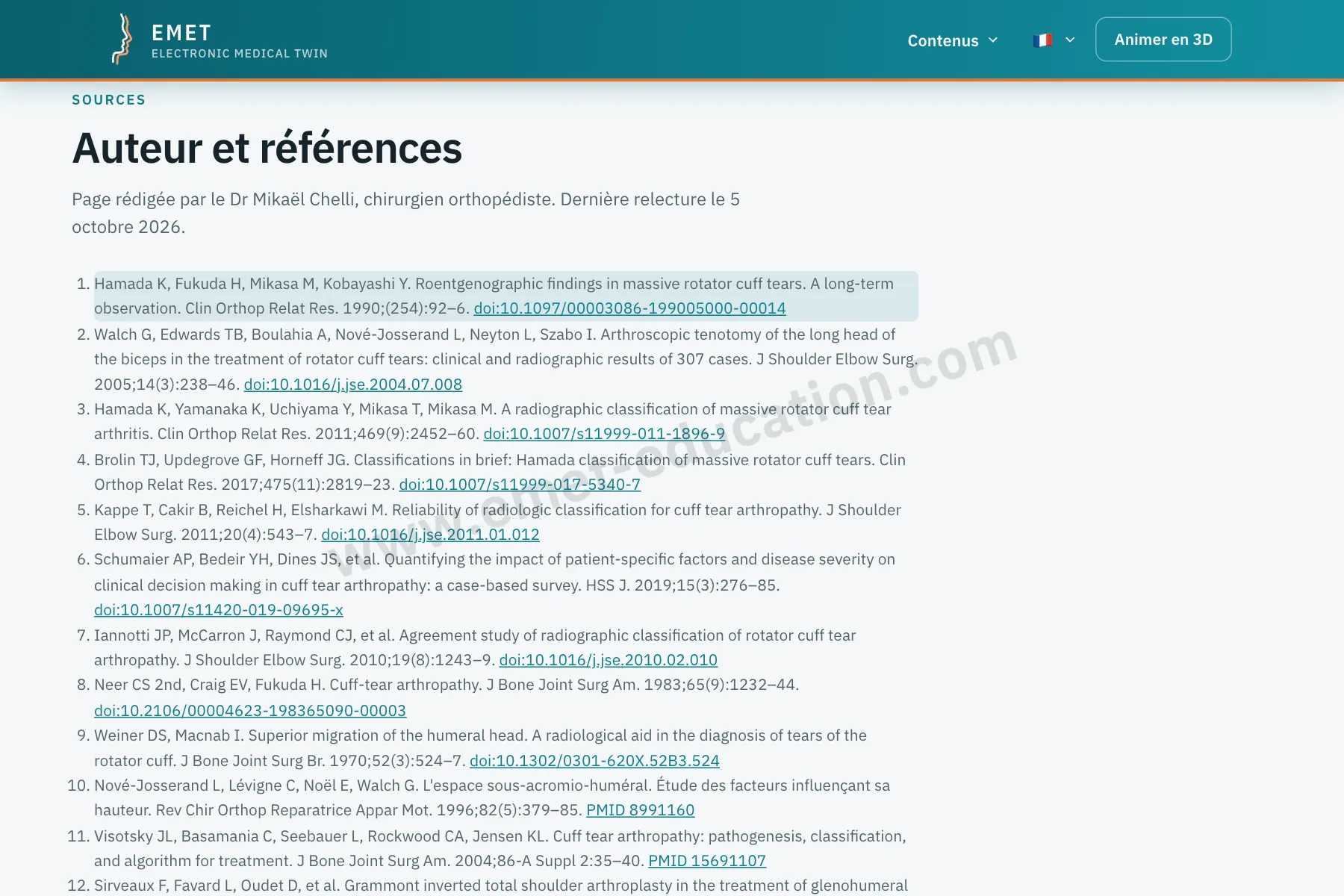
On the classification pages and the shoulder and elbow model pages, every cited author links to the matching reference, and every reference carries a verified DOI or PubMed link (title, journal, year, first author).
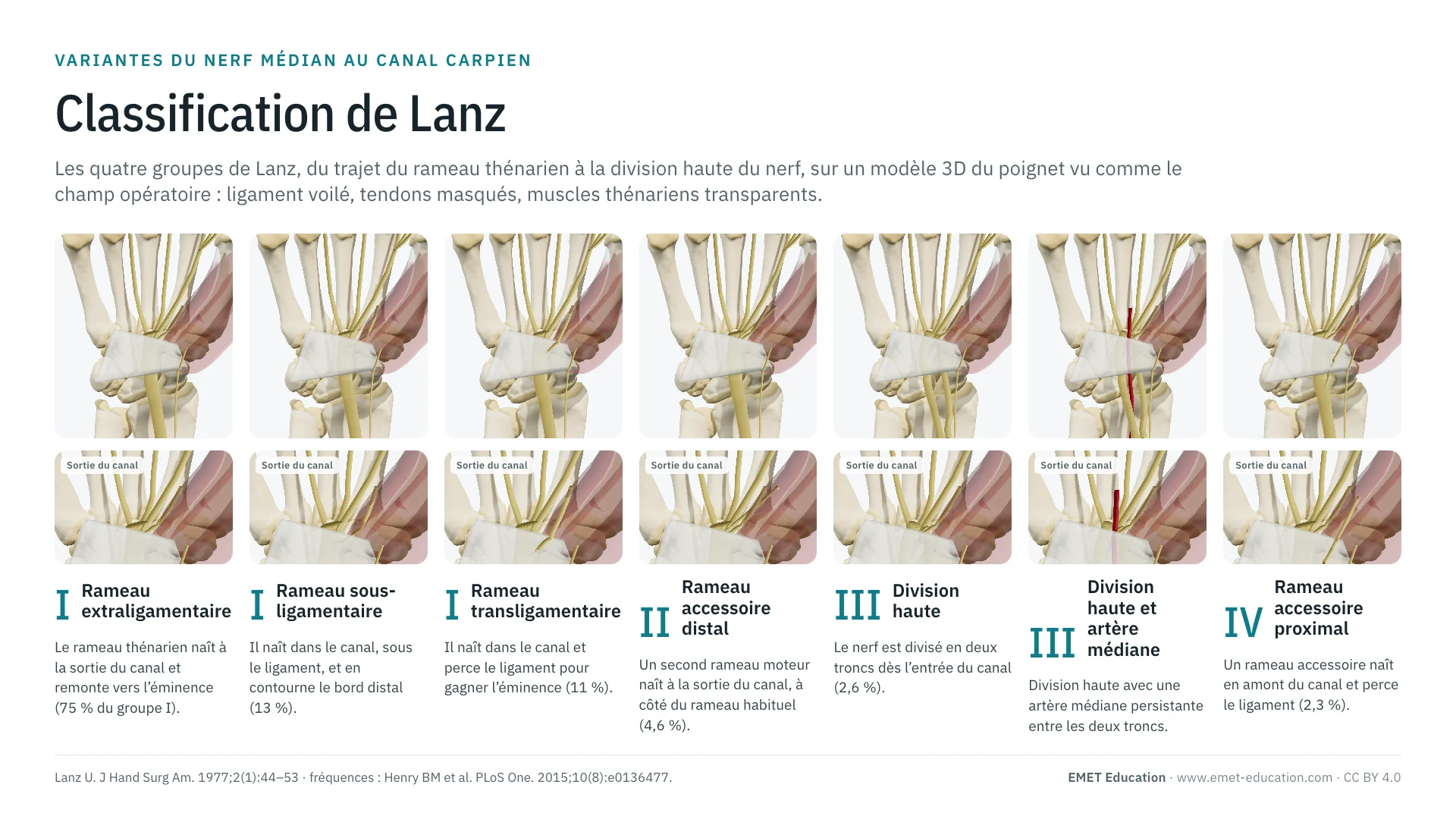
The four groups of median nerve variations in the carpal tunnel, from the course of the thenar branch to high division, shown as in the surgical field, with the original article, the frequency of each form and a free infographic (CC BY 4.0).
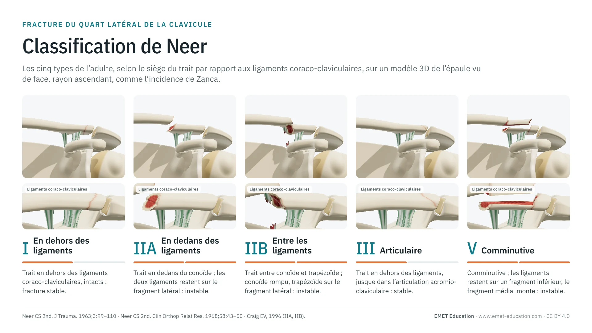
Types I to V by the position of the fracture line relative to the coracoclavicular ligaments, stable or unstable, with a zoom on the ligaments and a link that opens the model at the type shown.
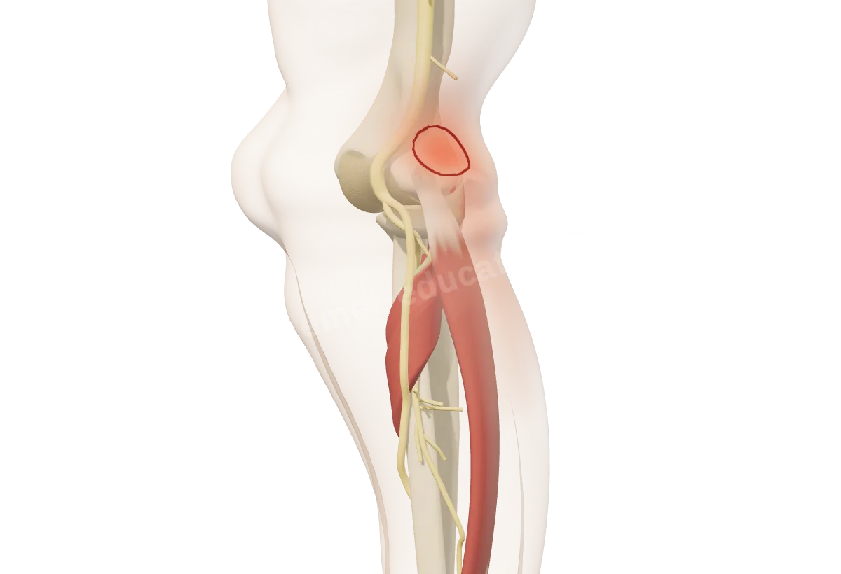
The risks and complications of the proposed treatment now have their own box, below the treatment, on the consultation sheet and in the patient view. First model: distal biceps rupture, with posterior interosseous nerve palsy, heterotopic ossification and re-rupture.
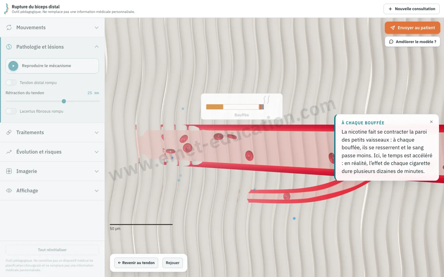
Under “Course & risks”, “Effect of smoking” dives into the tendon down to its arterioles: vasoconstriction with each puff, hypoxia from carbon monoxide, wall thickening over the years, deteriorating collagen fibers; quitting reopens the flow. Once played, the sequence adds “Stop smoking” to the proposed treatment, flagged in red.
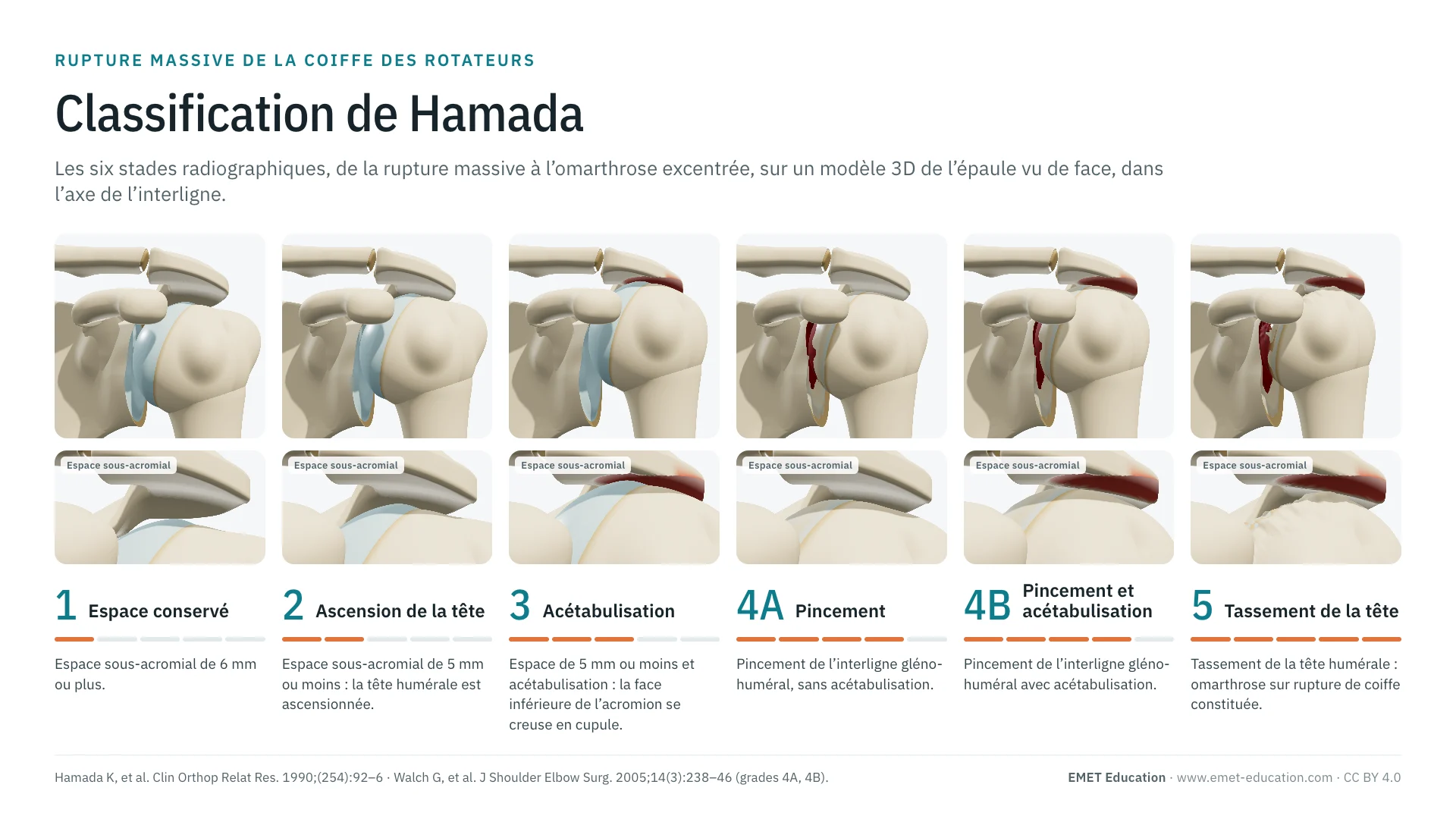
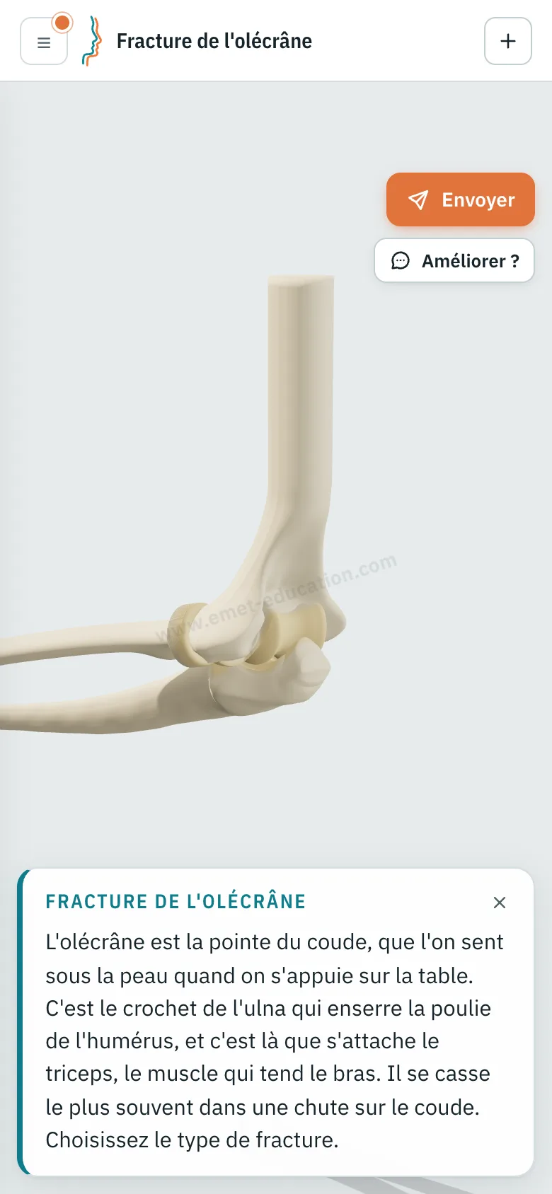
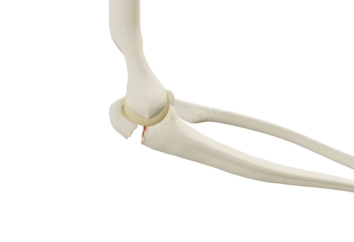
Three new pages: the stages of massive rotator cuff tears up to cuff tear arthropathy (Hamada), the grades of concentric shoulder osteoarthritis (Samilson-Prieto) and the types of olecranon fractures (Mayo), each animated on the 3D model.
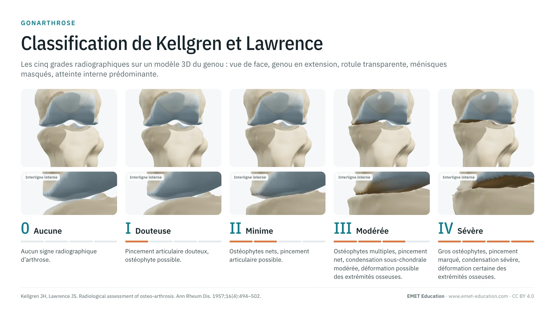
Reference classifications, grade by grade, on the 3D model, with the original article, published reliability, grading pitfalls, a quiz and free infographics for teaching and publication. First pages: Kellgren-Lawrence and McGowan. The section is in the “Resources” menu.
On every model, the ☰ button draws attention until the drawer has been opened once. Choosing a movement, pathology, treatment or course closes the drawer at once so the animation can be seen in full; a gauge keeps it open while you adjust it.
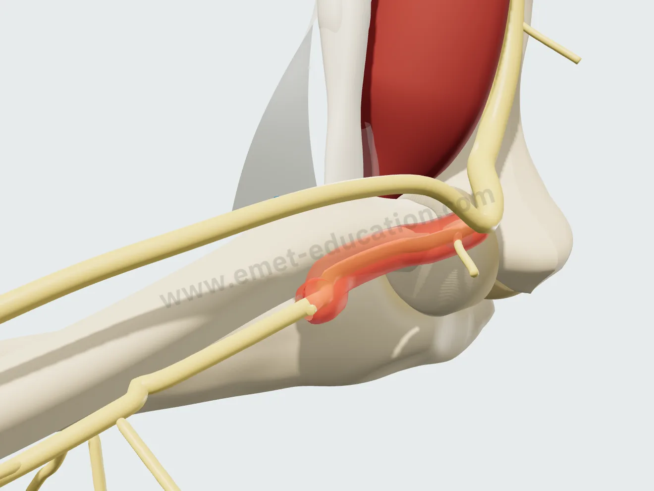
New four-step treatment: nerve exposure, release of the arcade of Frohse, opening of the proximal supinator fibers over 2 cm, then recovery as the nerve regains its calibre. Each step is now named above the tendon and nerve gauges.