- EMET Education
- 3D Anatomy
- Shoulder
- Infraspinatus muscle
Anatomy · Shoulder · Rotator cuff
The Infraspinatus Muscle in 3D
The infraspinatus in 3D, layer by layer, then on labeled MRI slices. With its footprint, wider and more anterior than once described, its innervation at the spinoglenoid notch, and the landmarks for reading its fatty infiltration.
Designed by an orthopedic surgeon
Latin musculus infraspinatus · French infra-épineux, sous-épineux
- 32.7 mmAnteroposterior width of the footprint on the greater tuberosity (26 shoulders)
- 1.76 cm²Insertion area, larger than the supraspinatus's (10 specimens)
- 2 cmFrom the posterior glenoid rim to the muscle's motor branches (31 shoulders)
- 2.5 yearsMean time from symptom onset to stage 2 fatty infiltration (1,688 patients)
At a glance
Origin, insertion, innervation
| Origin | Medial two-thirds of the infraspinous fossa and infraspinous fascia. |
|---|---|
| Course | Converges laterally, behind the scapular neck and the capsule, beneath the posterior deltoid, to the posterosuperior aspect of the humeral head. |
| Insertion | Middle facet of the greater tuberosity. Its superior tendinous portion curves anteriorly to the anterolateral highest impression, covering the posterior border of the supraspinatus; inferiorly, the insertion tapers away from the cartilage, framing the bare area. Trapezoidal footprint. |
| Innervation | Suprascapular nerve (C5, C6), after the spinoglenoid notch; three or four motor branches of similar size in about half of shoulders. Its transverse part receives branches arising proximal to the notch. |
| Blood supply | Suprascapular and circumflex scapular arteries. |
| Action | Lateral rotation of the arm; posterior stabilization of the humeral head, under passive tension at rest with the supraspinatus. |
| Relations | Superiorly, the scapular spine and the supraspinatus; inferiorly, the teres minor, with no gap between the two tendons; superficially, the posterior deltoid laterally and the trapezius medially; deep, the fossa, the posterior capsule and, at the spinoglenoid notch, the suprascapular nerve. |
Layer by layer
From the trapezius to the tendon
The same view, from behind, slightly above and lateral to the shoulder, removing one layer per image.
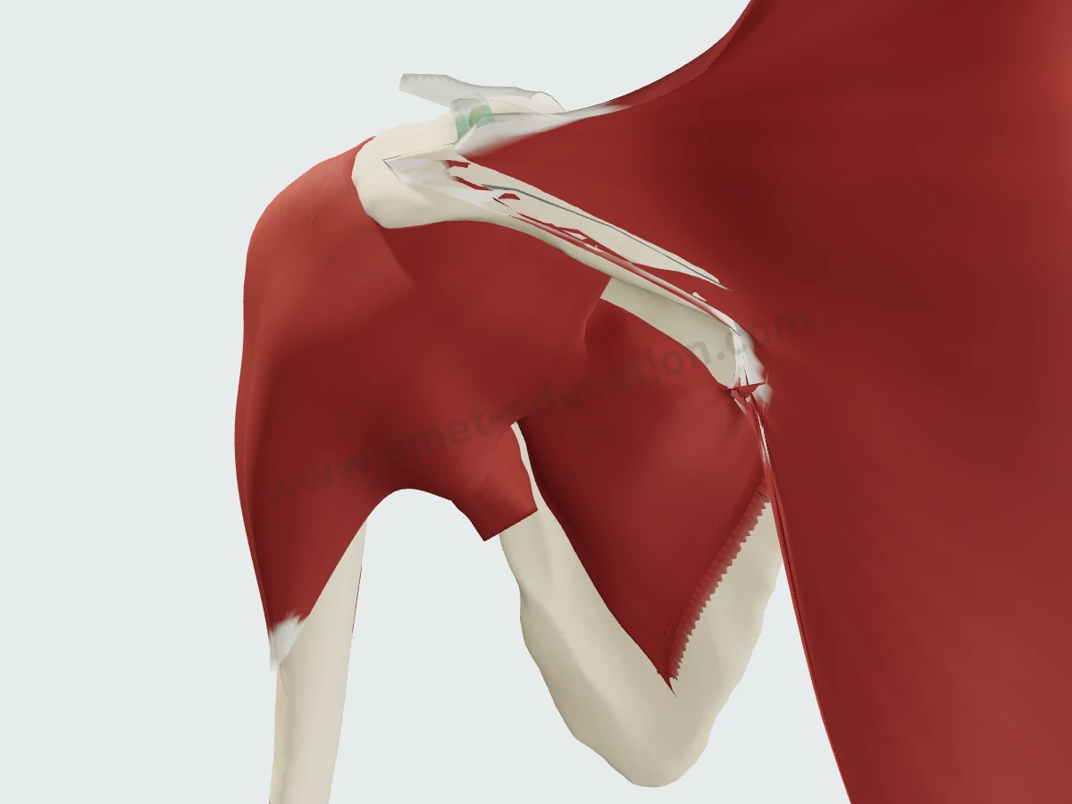
Trapezius and deltoidThe infraspinatus shows only between the trapezius, medially, and the deltoid, laterally.
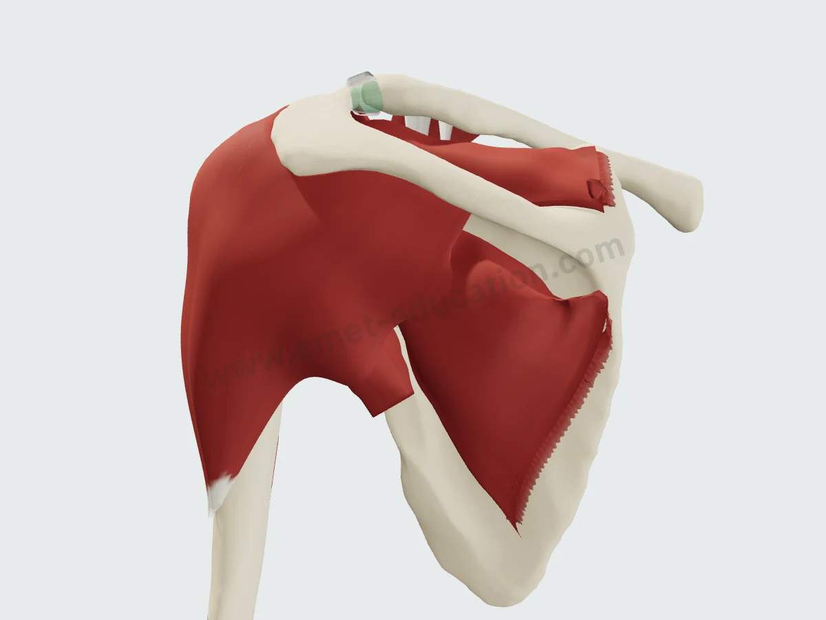
The posterior deltoidTrapezius removed: the posterior deltoid covers the lateral part of the muscle and the humeral head.
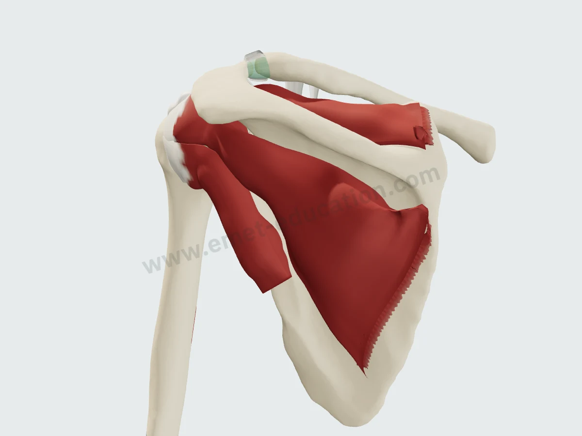
The posterior cuffDeltoid removed: the supraspinatus above the spine, the infraspinatus in its fossa, the teres minor below it.
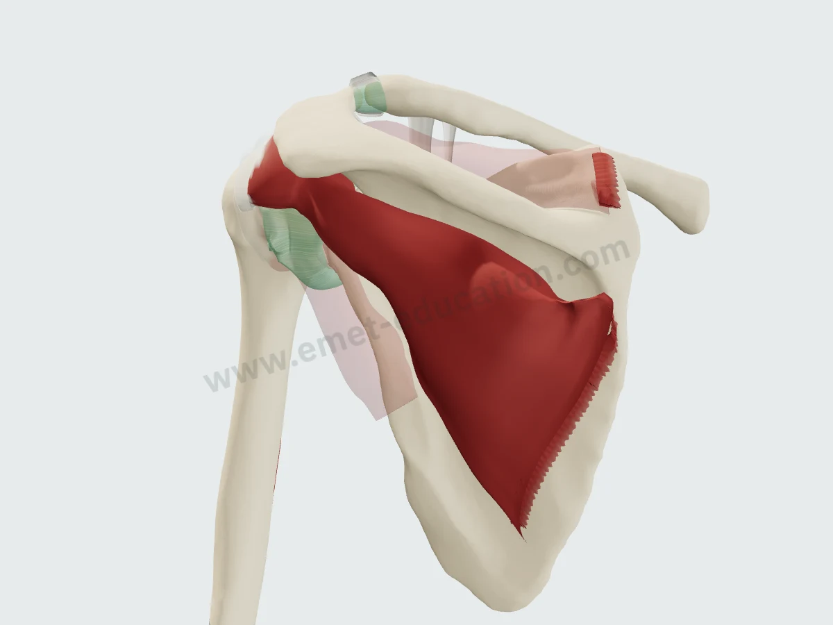
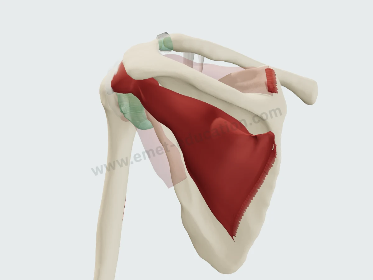
The infraspinatusOther muscles faded: from the infraspinous fossa to the middle facet of the greater tuberosity.
Imaging
The infraspinatus on MRI and CT
Synthetic images. These slices do not come from a patient: they are computed from the 3D model, by cutting each structure and giving it the signal it would have in the chosen sequence, on MRI or CT. They are for learning where the muscle lies on a slice; nothing is measured on them.
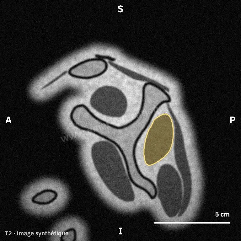
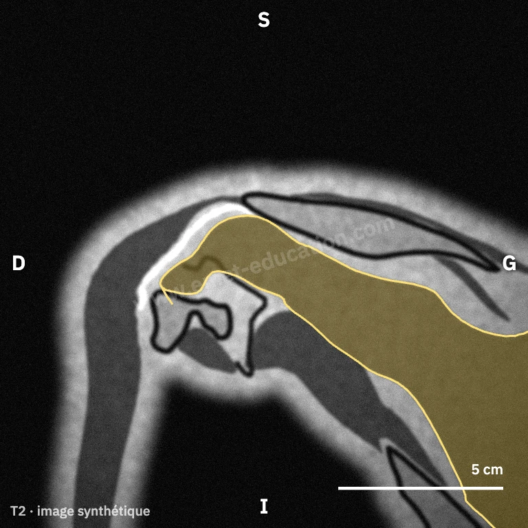
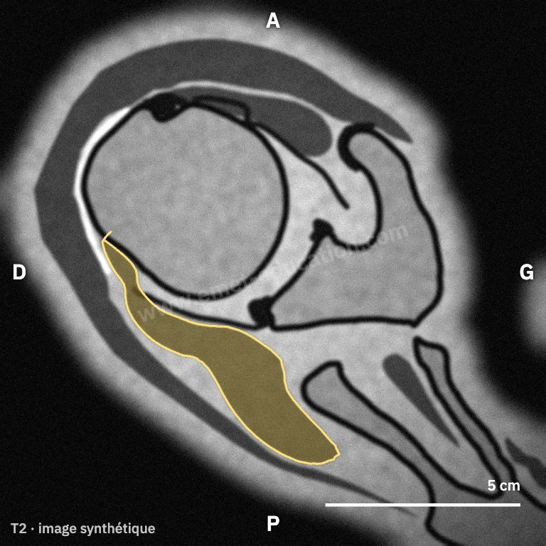
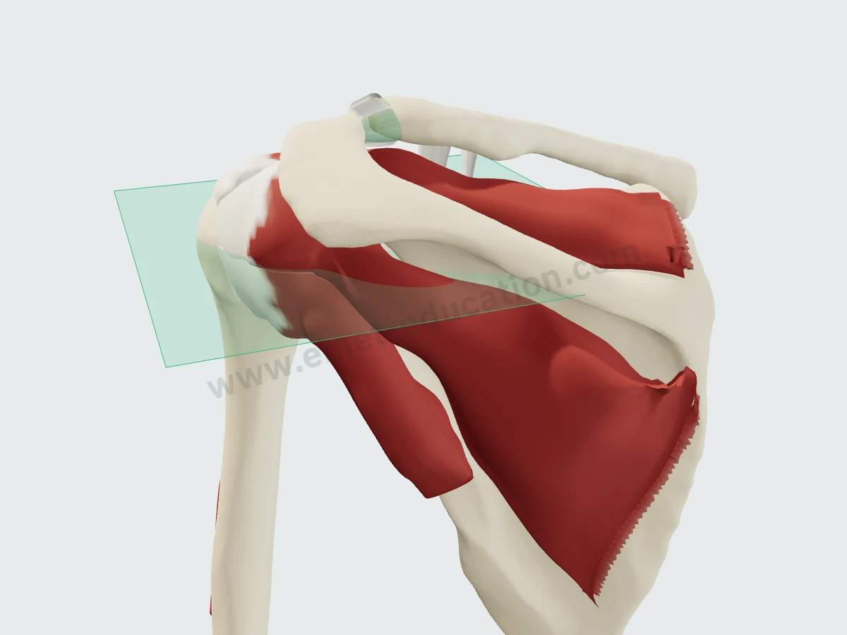
Where the axial view cuts
The axial plane (green) on the 3D model: horizontal, above the center of the humeral head, it crosses the infraspinatus from its fossa to its tendon. Hover over a slice and click “Scroll” to page through it as on a workstation: the supraspinatus appears upward, the teres minor downward.
In numbers
The footprint and the nerve, measured
Published mean values, with sample size and source under each figure.
| Value | Reference | Year | Sample | Note |
|---|---|---|---|---|
| 29 mm | Curtis | 2006 | 20 cadavers | Mean maximum length (29 × 19 mm); orientation to be confirmed in the full text to be confirmed |
| 32.7 mm | Mochizuki | 2008 | 26 shoulders | Mean maximum; trapezoidal footprint, extending anteriorly to the highest impression |
| Value | Reference | Year | Sample | Note |
|---|---|---|---|---|
| 1.76 cm² | Dugas | 2002 | 10 specimens | Mean (± 0.40; 1.23 to 2.53), 3D digitizer mapping; 1.55 cm² for the supraspinatus |
Published means, from smallest to largest. Sample size is as each study reports it (shoulders, cadavers or specimens); measurement protocols differ from one study to the next.
Maximum dimensions, to scale: a bounding box, not the shape of the footprint. Anteroposterior width shown vertically.
- 9.8 mmPosterior half of the superior cuff tendon, made of supraspinatus and infraspinatus (± 3.2)Mean, 10 shoulders · Minagawa 1998
- 15–16 mmPosterior to the biceps tendon: the most frequent site of degenerative tears, near the supraspinatus–infraspinatus junctionUltrasound, 360 shoulders · Kim 2010
- 1 to 3 cmLateral advancement of the tendon before the motor branches tighten (anterosuperior approach, then Debeyre technique)31 shoulders · Warner 1992
- 12 of 96Elite volleyball players with isolated, asymptomatic paralysis of the dominant-side infraspinatusFerretti 1987
- 2ndForce-producing capacity in the cuff by physiological cross-sectional area, after the subscapularis10 specimens · Ward 2006
Published footprint dimensions vary from study to study. Mochizuki described a superior tendinous portion curving anteriorly to the highest impression of the greater tuberosity: his 32.7 mm width includes that extension, an area classic descriptions attributed to the supraspinatus.
History
How its description has changed since 1992
Classic anatomy places the infraspinatus on the middle facet of the greater tuberosity, behind the supraspinatus. Studies published since have shown a wider, more anterior insertion, and a muscle in two parts.
- 1992
A common insertion
The cuff tendons do not insert separately: the infraspinatus interdigitates with the supraspinatus into a continuous insertion. Clark and Harryman
- 1993
The suspension bridge
The crescent, the thin distal part of the supraspinatus and infraspinatus, is bordered by a thick cable that shields it from stress. Burkhart
- 2002
A large area
Mapped with a digitizer, its insertion averages 1.76 cm², more than the supraspinatus's (1.55 cm²). Dugas
- 2006
A horseshoe
Superiorly, it wraps the posterior border of the supraspinatus at the cartilage; inferiorly, it tapers away and frames the bare area. Curtis
- 2008
To the front of the tuberosity
Its superior tendon curves anteriorly to the highest impression of the greater tuberosity: it occupies much of the area attributed to the supraspinatus, which explains its frequent atrophy in small to medium tears. Mochizuki
- 2012
Two parts
A main oblique part and a partly independent transverse part, supplied by branches arising proximal to the spinoglenoid notch. Kato
In practice
What anatomy changes in practice
- Examining it. The external rotation lag sign tests the supraspinatus and infraspinatus, the drop sign the infraspinatus: less sensitive than the Jobe sign but more specific, and the lag grows with tear size (Hertel).
- Grading its fatty infiltration. It can occur in a large anterosuperior tear with its own tendon intact, does not regress after repair, and weighs heavily on the outcome (Goutallier). On axial slices, stage 2 appears on average 2.5 years after symptom onset, stages 3 and 4 at 4 years (Melis).
- Thinking of the nerve. Beyond the spinoglenoid notch, the suprascapular nerve supplies only the infraspinatus: a lesion there gives isolated atrophy, as in 12 of 96 elite volleyball players examined, asymptomatic despite 22% less lateral rotation strength (Ferretti).
- Mobilizing a retracted tear. The motor branches arise 2 cm from the posterior glenoid rim; lateral advancement of the tendon is limited to 1 cm through the anterosuperior approach, up to 3 cm with the Debeyre technique (Warner).
- Filling a Hill-Sachs lesion. Remplissage is an arthroscopic capsulotenodesis of the posterior capsule and infraspinatus tendon into the defect (Purchase and Wolf).
Your turn
Where is the infraspinatus?
Six unlabeled synthetic slices, in random order: click the infraspinatus. The answer shows its outline.
Click the infraspinatus.
Conditions
Related conditions
Classifications
Related classifications
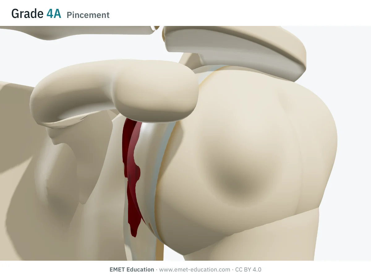 HamadaMassive rotator cuff tears: grades 1 to 5, in 3D
- SoonGoutallierFatty infiltration of the cuff muscles, on CT (1994)
- SoonCollinMassive tears: the five cuff components and loss of active motion (2014)
Neighbors
Its neighbors
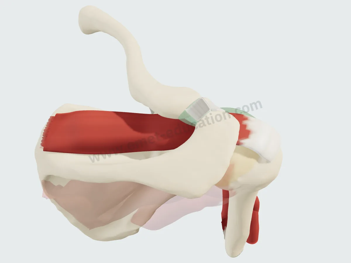 SupraspinatusSuperior
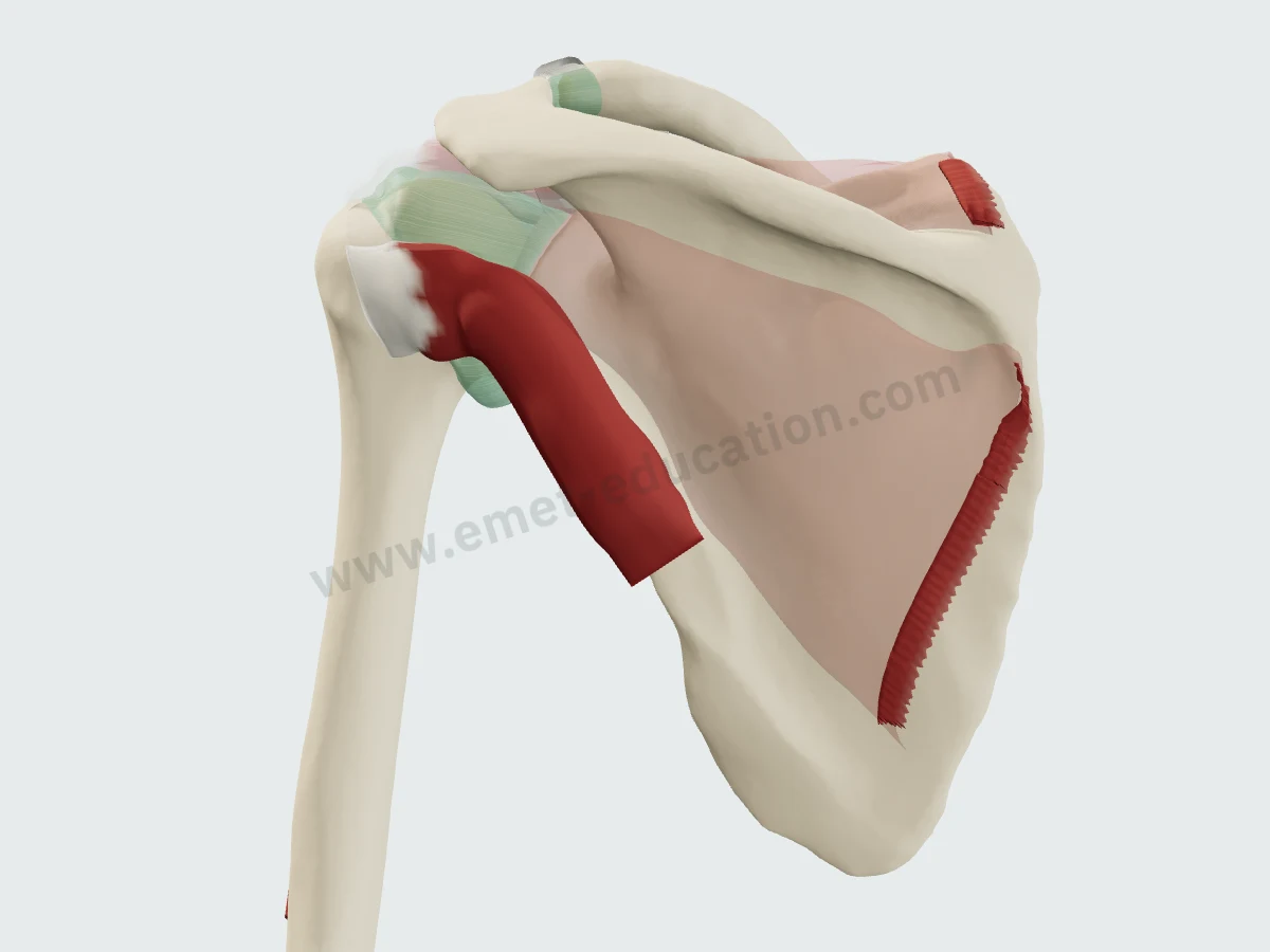 Teres minorInferior
- SoonDeltoidSuperficial
- SoonTrapeziusSuperficial
- SoonJoint capsuleDeep
- SoonSuprascapular nerveDeep
Questions
Frequently asked questions
Where does the infraspinatus insert?
On the middle facet of the greater tuberosity. Its superior tendinous portion curves anteriorly to the anterolateral highest impression. Its trapezoidal footprint averages 32.7 mm wide and 10.2 mm long (Mochizuki, 26 shoulders), with an area of 1.76 cm² (Dugas).
What nerve innervates the infraspinatus?
The suprascapular nerve (C5, C6), after it passes through the spinoglenoid notch. Its transverse part receives branches arising proximal to the notch (Kato).
How do you examine the infraspinatus?
By lateral rotation strength, the external rotation lag sign and the drop sign, less sensitive than the Jobe sign but more specific (Hertel).
Why does the infraspinatus atrophy in tears that seem to involve only the supraspinatus?
Because it occupies much of the highest impression of the greater tuberosity, long attributed to the supraspinatus (Mochizuki). Its fatty infiltration can occur with its own tendon intact and does not regress after repair (Goutallier).
What is remplissage?
An arthroscopic capsulotenodesis: the posterior capsule and infraspinatus tendon are fixed into the Hill-Sachs lesion to fill it (Purchase and Wolf).
Are the MRI images on this page real?
No: they are synthetic, computed from the 3D model. They show where the muscle lies on a slice; they do not replace an actual exam.
Sources
Author and references
- Standring S, ed. Gray's Anatomy: The Anatomical Basis of Clinical Practice. 42nd ed. Elsevier; 2020.
- Ferretti A, Cerullo G, Russo G. Suprascapular neuropathy in volleyball players. J Bone Joint Surg Am. 1987;69(2):260–263. PMID 3805088
- Clark JM, Harryman DT 2nd. Tendons, ligaments, and capsule of the rotator cuff. Gross and microscopic anatomy. J Bone Joint Surg Am. 1992;74(5):713–725. PMID 1624486
- Warner JP, Krushell RJ, Masquelet A, Gerber C. Anatomy and relationships of the suprascapular nerve: anatomical constraints to mobilization of the supraspinatus and infraspinatus muscles in the management of massive rotator-cuff tears. J Bone Joint Surg Am. 1992;74(1):36–45. PMID 1734012
- Burkhart SS, Esch JC, Jolson RS. The rotator crescent and rotator cable: an anatomic description of the shoulder's "suspension bridge". Arthroscopy. 1993;9(6):611–616. doi:10.1016/s0749-8063(05)80496-7
- Goutallier D, Postel JM, Bernageau J, Lavau L, Voisin MC. Fatty muscle degeneration in cuff ruptures. Pre- and postoperative evaluation by CT scan. Clin Orthop Relat Res. 1994;(304):78–83. PMID 8020238
- Hertel R, Ballmer FT, Lombert SM, Gerber C. Lag signs in the diagnosis of rotator cuff rupture. J Shoulder Elbow Surg. 1996;5(4):307–313. doi:10.1016/s1058-2746(96)80058-9
- Minagawa H, Itoi E, Konno N, et al. Humeral attachment of the supraspinatus and infraspinatus tendons: an anatomic study. Arthroscopy. 1998;14(3):302–306. doi:10.1016/s0749-8063(98)70147-1
- Zanetti M, Gerber C, Hodler J. Quantitative assessment of the muscles of the rotator cuff with magnetic resonance imaging. Invest Radiol. 1998;33(3):163–170. doi:10.1097/00004424-199803000-00006
- Dugas JR, Campbell DA, Warren RF, Robie BH, Millett PJ. Anatomy and dimensions of rotator cuff insertions. J Shoulder Elbow Surg. 2002;11(5):498–503. doi:10.1067/mse.2002.126208
- Curtis AS, Burbank KM, Tierney JJ, Scheller AD, Curran AR. The insertional footprint of the rotator cuff: an anatomic study. Arthroscopy. 2006;22(6):609.e1. doi:10.1016/j.arthro.2006.04.001
- Ward SR, Hentzen ER, Smallwood LH, et al. Rotator cuff muscle architecture: implications for glenohumeral stability. Clin Orthop Relat Res. 2006;448:157–163. doi:10.1097/01.blo.0000194680.94882.d3
- Mochizuki T, Sugaya H, Uomizu M, et al. Humeral insertion of the supraspinatus and infraspinatus. New anatomical findings regarding the footprint of the rotator cuff. J Bone Joint Surg Am. 2008;90(5):962–969. doi:10.2106/JBJS.G.00427
- Purchase RJ, Wolf EM, Hobgood ER, Pollock ME, Smalley CC. Hill-Sachs "remplissage": an arthroscopic solution for the engaging Hill-Sachs lesion. Arthroscopy. 2008;24(6):723–726. doi:10.1016/j.arthro.2008.03.015
- Kim HM, Dahiya N, Teefey SA, et al. Location and initiation of degenerative rotator cuff tears: an analysis of three hundred and sixty shoulders. J Bone Joint Surg Am. 2010;92(5):1088–1096. doi:10.2106/JBJS.I.00686
- Melis B, Wall B, Walch G. Natural history of infraspinatus fatty infiltration in rotator cuff tears. J Shoulder Elbow Surg. 2010;19(5):757–763. doi:10.1016/j.jse.2009.12.002
- Kato A, Nimura A, Yamaguchi K, Mochizuki T, Sugaya H, Akita K. An anatomical study of the transverse part of the infraspinatus muscle that is closely related with the supraspinatus muscle. Surg Radiol Anat. 2012;34(3):257–265. doi:10.1007/s00276-011-0872-0
- Collin P, Matsumura N, Lädermann A, Denard PJ, Walch G. Relationship between massive chronic rotator cuff tear pattern and loss of active shoulder range of motion. J Shoulder Elbow Surg. 2014;23(8):1195–1202. doi:10.1016/j.jse.2013.11.019
In clinic
Show the rotator cuff to your patient
The images on this page come from an interactive model. In clinic, show the cuff tear and its repair, or remplissage of a Hill-Sachs lesion.
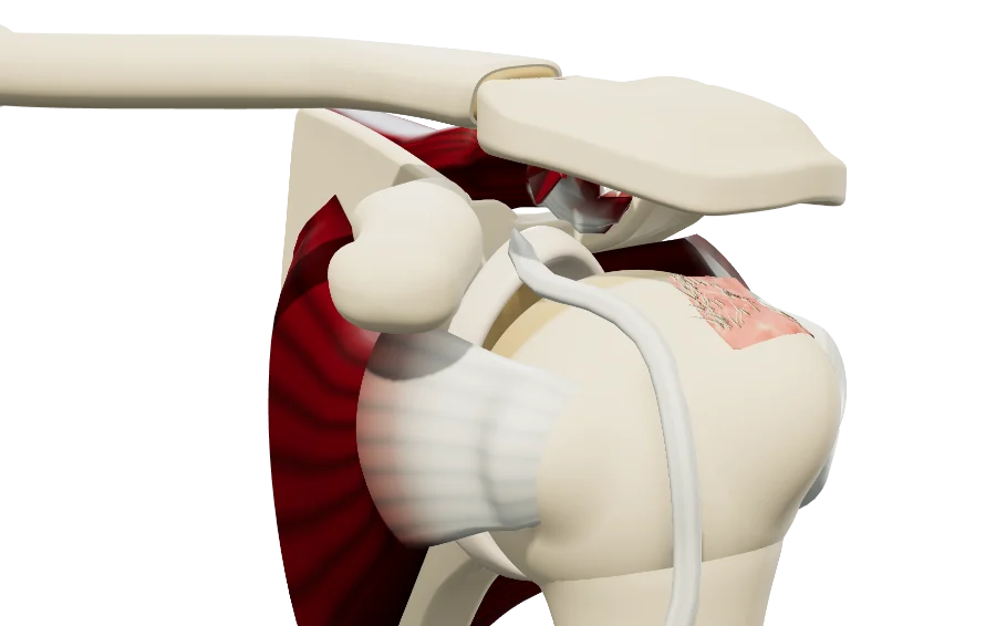
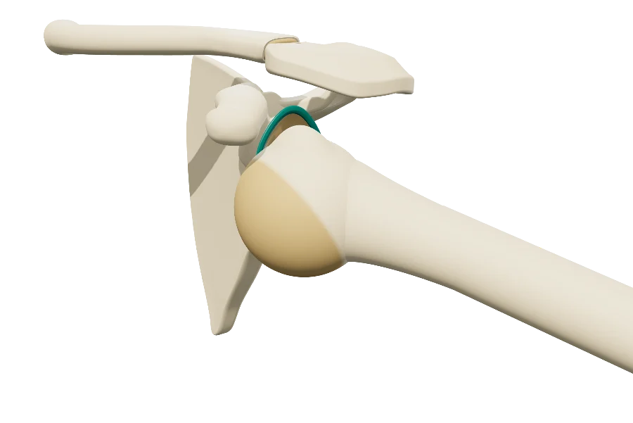
The infraspinatus, in 3D, for your patient
Open the rotator cuff model in your browser, no sign-up needed.
A colleague preparing a lecture on the cuff?