- EMET Education
- 3D classifications
- Patte classification
Classification · Rotator cuff tear
Patte Classification: 3D Visualization
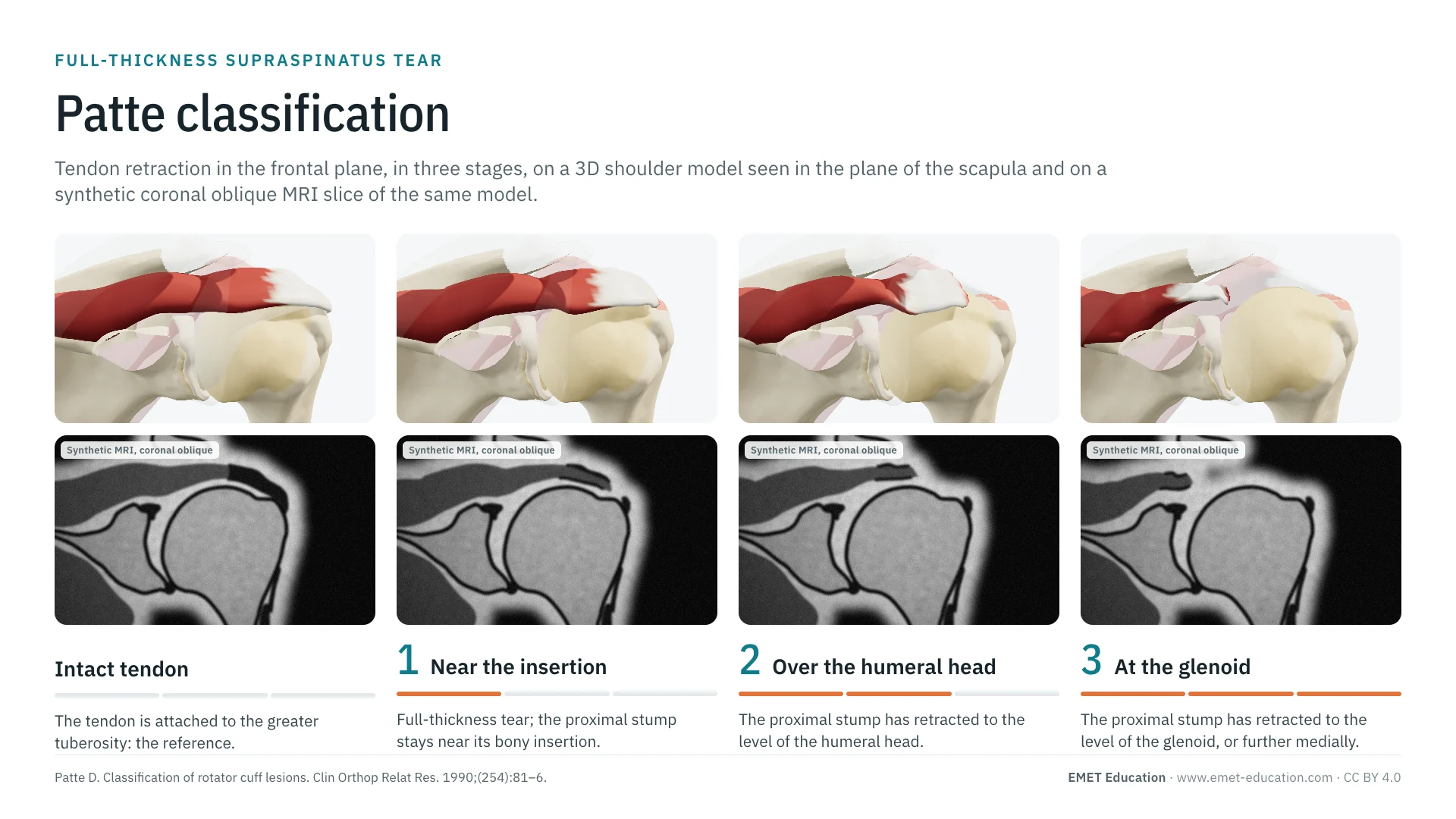
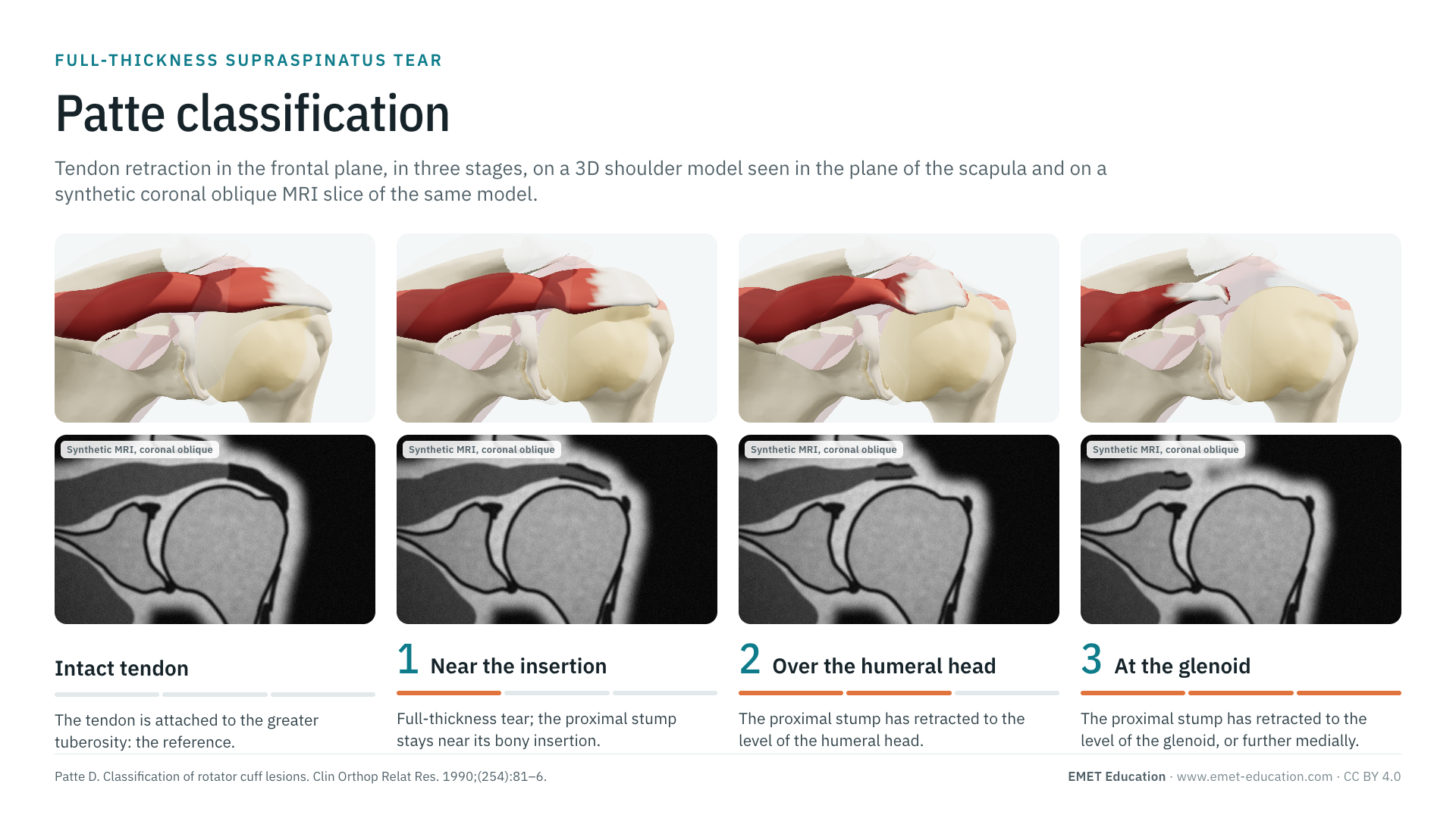
Retraction of the torn supraspinatus in the frontal plane, from a stump still near its insertion to a stump retracted to the glenoid, shown on a 3D model of the shoulder and on synthetic MRI slices of the same model. With the original article, its modified versions, reliability data, prognostic value and free infographics for teaching and publication.
Designed by an orthopedic surgeon
The 3D model opens in your browser, at the stage shown, with no sign-up. Infographics are free to reuse under CC BY 4.0.
- 1990Published by Patte in Clinical Orthopaedics and Related Research
- 1–3Three stages, from a stump near its insertion to a stump at the level of the glenoid
- 872Citations of the original article (OpenAlex, October 7, 2026)
- κ 0.58Interobserver agreement on MRI (Lippe et al.), the best of the three cuff classifications tested
The intact tendon and the three stages
From stage 1 to stage 3, in 3D and in section
Each image pairs a rendering of the EMET Education rotator cuff model, set to that stage, with a synthetic MRI slice of the same model. The 3D view looks from the front in the plane of the scapula, slightly from above: clavicle, coracoid and acromion are transparent, the infraspinatus is faded, the subscapularis and the long head of the biceps are hidden. The slice is coronal oblique, parallel to the supraspinatus, like the one the stage is graded on.
-
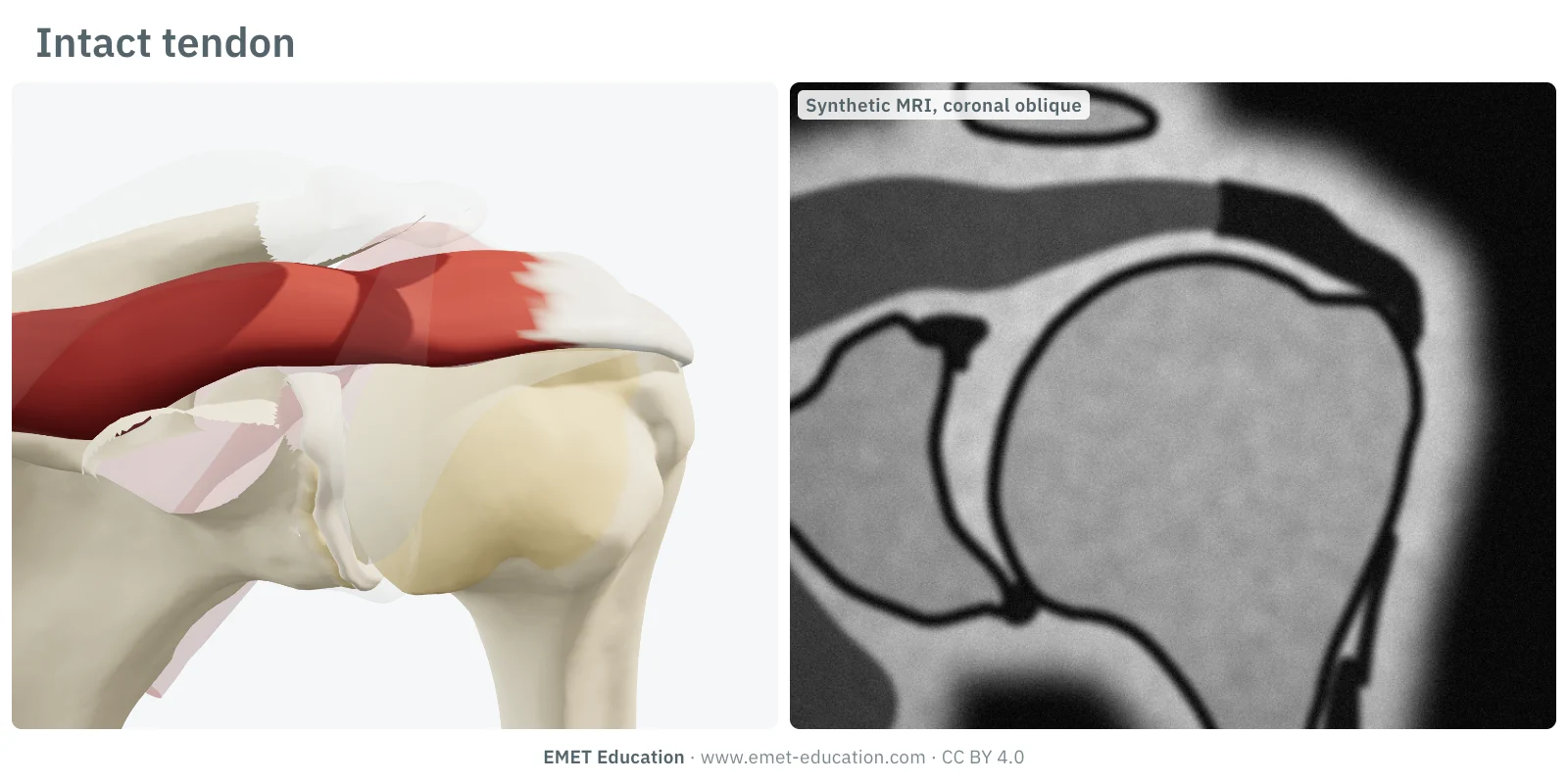
Intact tendon
- Criterion, frontal plane
- The tendon is attached to the greater tuberosity: this is the reference, with no stage.
- In the 3D model
- The supraspinatus runs over the top of the head and inserts on the superior facet of the greater tuberosity.
-
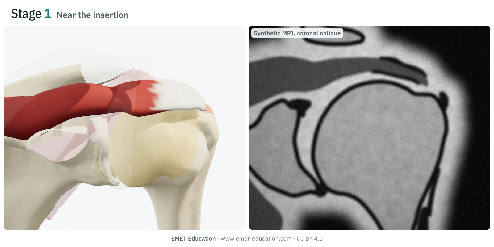
1Near the insertion
- Criterion, frontal plane
- Full-thickness tear whose proximal stump stays close to its bony insertion.
- In the 3D model
- The tendon has come off its footprint, now bare, but has stayed in place: no retraction.
-
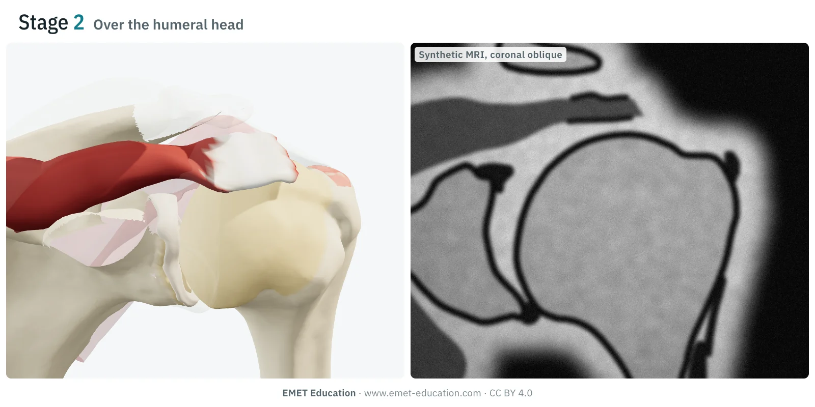
2Over the humeral head
- Criterion, frontal plane
- The proximal stump has retracted to the level of the humeral head.
- In the 3D model
- The tendon end stops slightly lateral to the top of the head: the footprint and the lateral dome of the head are uncovered.
-
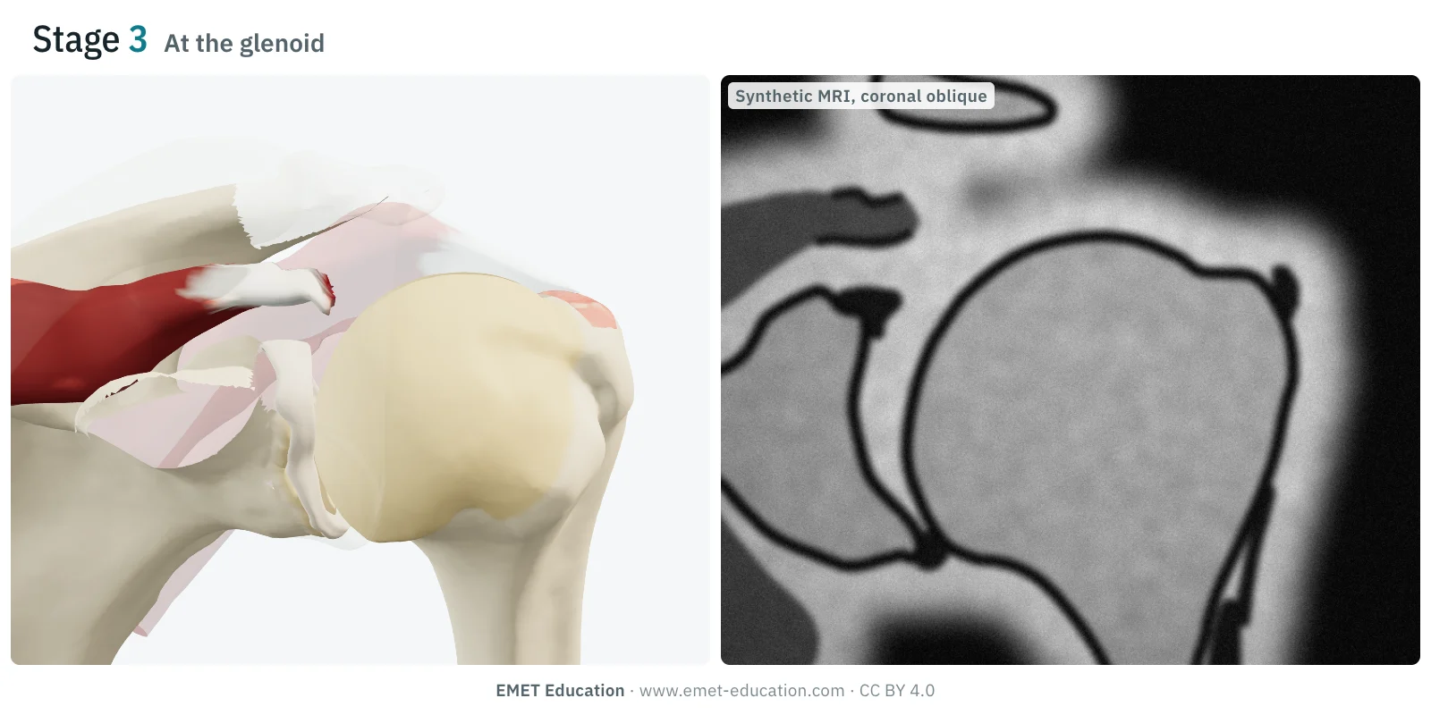
3At the glenoid
- Criterion, frontal plane
- The proximal stump has retracted to the level of the glenoid, or further medially.
- In the 3D model
- The tendon has retracted to the glenohumeral joint line and the muscle has bunched up: the whole top of the head is uncovered.
Schematic rendering, for teaching: the model illustrates each stage on a single shoulder; the slices are synthetic, not patient examinations. Criteria reworded from Patte (1990); no figure from the article is reproduced.
The original article
Patte, 1990
Daniel Patte (Les Fontaines clinic, Melun, France) published Classification of rotator cuff lesions in Clinical Orthopaedics and Related Research in 1990 (254:81–6, doi:10.1097/00003086-199005000-00012). His starting point: without a shared classification, series of rotator cuff repairs could not be compared with one another.
He proposed describing each tear by several criteria:
- the extent of the tear;
- its topography in the sagittal plane, that is, the tendons involved;
- its topography in the frontal plane: the retraction of the stump, in three stages, the subject of this page;
- the trophic quality of the muscles, and the state of the long head of the biceps.
Of these criteria, retraction in the frontal plane is the one that remained in everyday use, as the "Patte stage". It is now graded on the coronal oblique slice of MRI or CT arthrography. As of October 7, 2026, OpenAlex counts 872 citations of the original article. Muscle quality is now described mainly by fatty infiltration (Goutallier, 1994) and atrophy.
Reliability
How reproducible is the stage?
Moderately for the original version, better for the modified versions. In the study by Lippe et al., the Patte stage was nevertheless the most reproducible of the three cuff classifications tested on the same MRI studies.
| Study | Measure | Value |
|---|---|---|
| Lippe et al., 2012 | Interobserver kappa, Patte stage on T2 coronal images, three shoulder surgeons, 31 patients | 0.58 |
| Lippe et al., 2012 | Same study, for comparison: supraspinatus Goutallier grade in two classes (0 to 2, 3 and 4) | 0.53 |
| Guo et al., 2020 | Modified Patte, graded on two coronal slices: intra- and interobserver reliability (ICC) | excellent |
| Takeda et al., 2024 | Five-stage modified Patte: mean intraobserver kappa | 0.875 |
| Takeda et al., 2024 | Same classification: interobserver kappa | 0.797 |
Readers, sequences and populations differ between studies: the modified versions have not been compared with the original on the same images by the same readers. In the study by Lippe et al., supraspinatus and infraspinatus atrophy reached only 28% agreement.
Variants and related classifications
What Patte grades, and what it does not
The Patte stage locates the stump in the frontal plane. It says nothing about the anteroposterior extent of the tear, muscle quality or the other tendons: an MRI report completes it.
Two modified versions, and the classifications most often reported alongside it:
| Classification | What it grades | Typical use |
|---|---|---|
| Guo (2020) | The Patte stage graded on two coronal slices instead of one, in large and massive tears. | Predicting irreparability and retear: modified stage III had a specificity of 93.6% and 98.8%, versus 76.2% and 84.2% for original stage 3. |
| Takeda (2024) | The original stages combined with two cut-offs measured on the humeral head (its medial third, its medial fifth): five stages, I to V. | Predicting reparability (area under the curve 0.897) and healing (0.768) after arthroscopic supraspinatus repair. |
| Goutallier (1994) | Fatty infiltration of each muscle, from 0 (none) to 4 (more fat than muscle), described on CT then transposed to MRI (Fuchs, 1999). | With retraction, the main criterion for reparability and prognosis. |
| Atrophy: tangent sign (Zanetti, 1998), Warner (2001) | Muscle volume on the sagittal oblique slice: a healthy supraspinatus crosses the line tangent to the superior borders of the scapular spine and the coracoid. | A simple sign of a chronic tear, associated with irreparability (odds ratio 11.1 for the tangent sign in Hsu et al.). |
| Lafosse (2007) | Subscapularis lesions, from a partial lesion of its upper third to complete tendon rupture. | The anterior side of the cuff, which the Patte stage does not describe; a gauge in the same 3D model. |
Use
In research and in clinical practice
In research
The Patte stage describes retraction in surgical series and is used to predict reparability. In the meta-analysis by Hsu et al. (2024; 18 studies, 2,700 patients), stage 3 was associated with irreparability with an odds ratio of 8.0 (95% CI 4.3 to 14.9), alongside tear size, fatty infiltration, the tangent sign and superior migration of the head. In massive tears alone, Kim et al. (2017, 105 patients) did not find it predictive on its own; combined with infraspinatus fatty infiltration (Goutallier below 3 and Patte 2 at most), it gave the best prediction of reparability (area under the curve 0.874).
In clinical practice
The stage helps set the indication and inform the patient. A stump still near its insertion is brought back to bone without difficulty; a stump retracted to the glenoid raises the prospect of an incomplete or impossible repair, and of a retear. But the stage alone does not decide: age, duration of symptoms, muscle quality (fatty infiltration, atrophy), the acromiohumeral interval and function weigh as much.
Reparability is confirmed in the operating room, once the tendon is released: the imaging stage prepares the procedure, it does not replace it.
Grading pitfalls
Five pitfalls when grading a tear
- Grade on the right slice. The coronal oblique slice, parallel to the supraspinatus and perpendicular to the glenoid, on T2 or fat-saturated proton density. A strictly coronal slice cuts the tendon obliquely.
- Do not rely on a single slice in a large tear. Retraction varies from front to back. Guo et al. proposed grading two coronal slices, with better specificity for irreparability.
- Locate the stump, not the musculotendinous junction. The stage is read on the end of the torn tendon, relative to the insertion, the head and the glenoid.
- Keep the stage for full-thickness tears. A partial-thickness tear has no retracted stump: the Patte stage does not apply.
- There is no stage 4. Stage 3 covers a stump at the level of the glenoid or further medially. Versions with more than three stages, such as that of Takeda et al. (I to V), are different classifications: name the one you use.
Self-test
Which stage?
Four synthetic coronal oblique MRI slices of the same model, in random order: an intact tendon and the three stages. Find the stage of each one; the answer comes with the criterion.
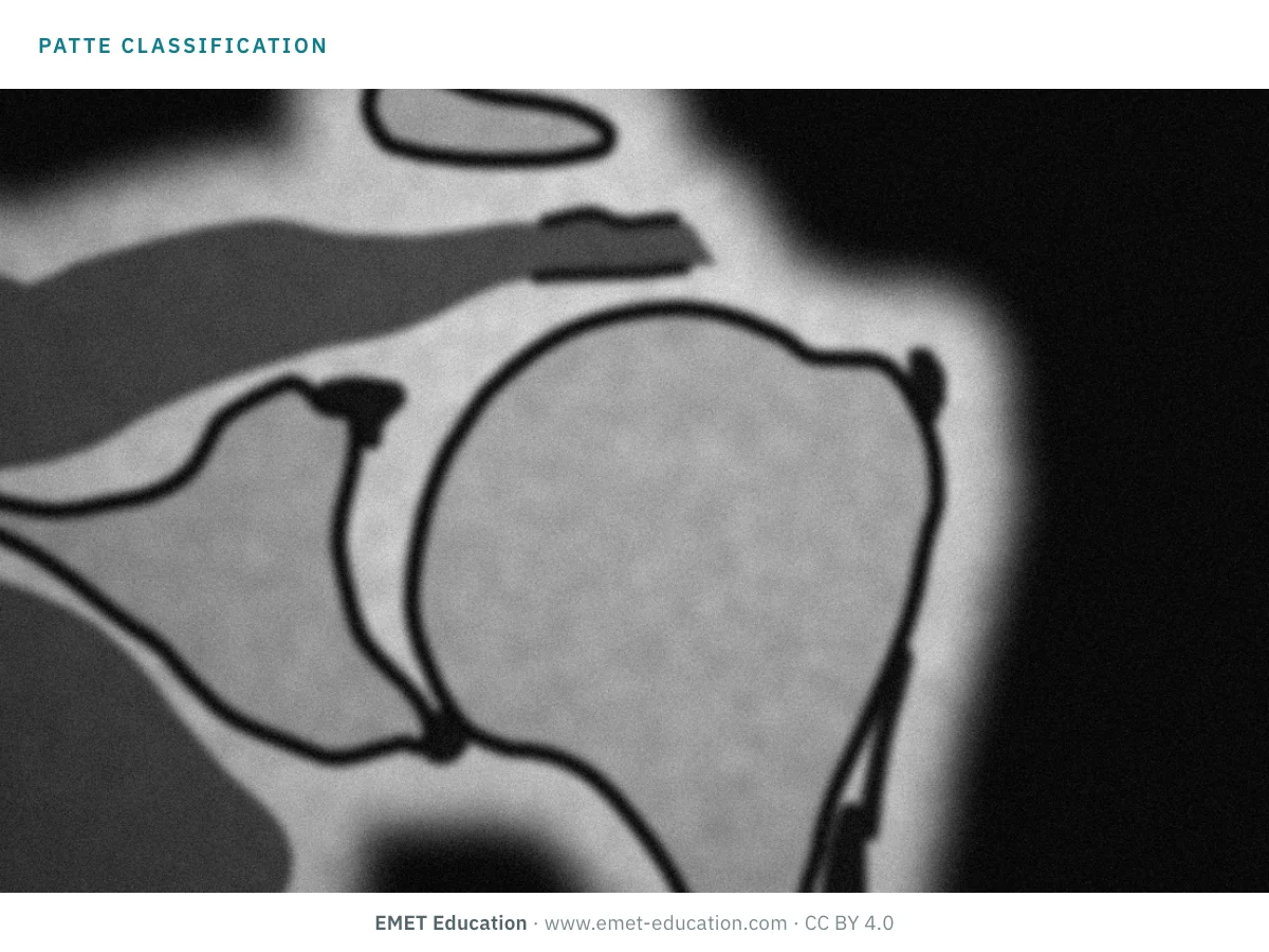
Which Patte stage is this?
Download and cite
Free infographics, for teaching and publication
Use them in a course, a presentation, a social media post or a scientific article, under the Creative Commons Attribution 4.0 license: free reuse, including commercial, provided EMET Education is credited with a link to this page.
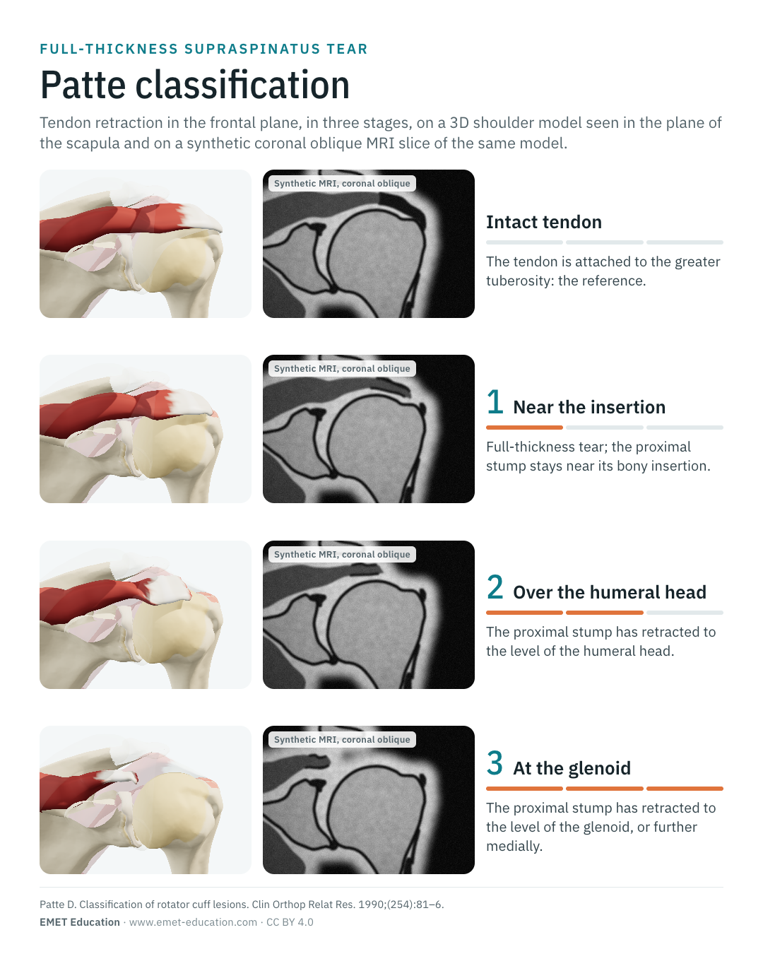
Cite this figure
Ready to paste. For a journal figure, add the original reference (Patte, 1990).
EMET Education. Patte classification of supraspinatus tendon retraction: 3D visualization [Internet]. 2026 [cited]. Available from: https://www.emet-education.com/en/classifications/patte/. CC BY 4.0.EMET Education. (2026). Patte classification of supraspinatus tendon retraction: 3D visualization [Infographic]. https://www.emet-education.com/en/classifications/patte/ (CC BY 4.0)<a href="https://www.emet-education.com/en/classifications/patte/"><img src="https://www.emet-education.com/en/classifications/patte/img/infographic.webp" alt="Patte classification in 3D" width="960"></a><br>Figure: <a href="https://www.emet-education.com/en/classifications/patte/">EMET Education</a>, CC BY 4.0Questions
Frequently asked questions
What are the stages of the Patte classification?
They describe the position of the proximal stump of a full-thickness tear in the frontal plane: at stage 1, it stays near its bony insertion; at stage 2, it has retracted to the level of the humeral head; at stage 3, to the level of the glenoid or further medially.
Is there a Patte stage 4?
No. The original classification has three stages in the frontal plane, and stage 3 already covers a stump at the level of the glenoid or further medially. The modified version by Takeda et al. (2024) has five stages, I to V, by adding cut-offs measured on the humeral head: it is a different classification, to be named as such.
Which MRI slice is the Patte stage graded on?
On the coronal oblique slice, parallel to the supraspinatus tendon, on T2 or fat-saturated proton density; coronal oblique CT arthrography works too. In a large tear, look at several slices: retraction varies from front to back.
Does stage 3 mean the tear is irreparable?
No, but the risk rises: in the meta-analysis by Hsu et al., stage 3 was associated with irreparability with an odds ratio of 8.0. The decision combines the stage, muscle fatty infiltration and atrophy, the acromiohumeral interval, age and function; reparability is confirmed in the operating room.
How does it differ from the Goutallier classification?
Patte describes the tendon: where the torn stump lies. Goutallier describes the muscle: how much fat has replaced the fibers, from 0 to 4. The two complement each other and are reported together in an MRI report of a rotator cuff tear.
Is the Patte stage reproducible?
Moderately: interobserver kappa of 0.58 between three shoulder surgeons (Lippe et al.), which made it the most reproducible of the three cuff classifications tested. The modified versions report better: interobserver kappa of 0.797 for that of Takeda et al..
Can I reuse these images in a course or an article?
Yes. The images and infographics on this page are under the Creative Commons Attribution 4.0 license: free reuse, including commercial, with credit to EMET Education and a link to this page. The "Cite this figure" box gives ready-to-paste citations.
How were the images made?
The 3D images are renderings of the EMET Education rotator cuff model, set to each stage. The MRI slices are synthetic: EMET's virtual imaging module cuts the geometry of the same model in the coronal oblique plane and gives it the look of a T2-weighted sequence. They are not patient examinations, and nothing is measured on them.
Sources
Author and references
- Patte D. Classification of rotator cuff lesions. Clin Orthop Relat Res. 1990;(254):81–6. doi:10.1097/00003086-199005000-00012
- Lippe J, Spang JT, Leger RR, Arciero RA, Mazzocca AD, Shea KP. Inter-rater agreement of the Goutallier, Patte, and Warner classification scores using preoperative magnetic resonance imaging in patients with rotator cuff tears. Arthroscopy. 2012;28(2):154–9. doi:10.1016/j.arthro.2011.07.016
- Guo S, Zhu Y, Song G, Jiang C. Assessment of tendon retraction in large to massive rotator cuff tears: a modified Patte classification based on 2 coronal sections on preoperative magnetic resonance imaging with higher specificity on predicting reparability. Arthroscopy. 2020;36(11):2822–30. doi:10.1016/j.arthro.2020.06.023
- Takeda Y, Fujii K, Suzue N, Kawasaki Y, Sumitomo J, Nishidono K, et al. A modified Patte classification system for rotator cuff tendon retraction to predict reparability and tendon healing in arthroscopic rotator cuff repair. Knee Surg Sports Traumatol Arthrosc. 2024;32(6):1579–90. doi:10.1002/ksa.12162
- Hsu KL, Kuan FC, Velasquez Garcia A, Hong CK, Chen Y, Shih CA, et al. Factors associated with reparability of rotator cuff tears: a systematic review and meta-analysis. J Shoulder Elbow Surg. 2024;33(9):e465–77. doi:10.1016/j.jse.2024.03.014
- Kim JY, Park JS, Rhee YG. Can preoperative magnetic resonance imaging predict the reparability of massive rotator cuff tears? Am J Sports Med. 2017;45(7):1654–63. doi:10.1177/0363546517694160
- Goutallier D, Postel JM, Bernageau J, Lavau L, Voisin MC. Fatty muscle degeneration in cuff ruptures. Pre- and postoperative evaluation by CT scan. Clin Orthop Relat Res. 1994;(304):78–83. PMID 8020238
- Fuchs B, Weishaupt D, Zanetti M, Hodler J, Gerber C. Fatty degeneration of the muscles of the rotator cuff: assessment by computed tomography versus magnetic resonance imaging. J Shoulder Elbow Surg. 1999;8(6):599–605. doi:10.1016/s1058-2746(99)90097-6
- Zanetti M, Gerber C, Hodler J. Quantitative assessment of the muscles of the rotator cuff with magnetic resonance imaging. Invest Radiol. 1998;33(3):163–70. doi:10.1097/00004424-199803000-00006
- Warner JJ, Higgins L, Parsons IM, Dowdy P. Diagnosis and treatment of anterosuperior rotator cuff tears. J Shoulder Elbow Surg. 2001;10(1):37–46. doi:10.1067/mse.2001.112022
- Lafosse L, Jost B, Reiland Y, Audebert S, Toussaint B, Gobezie R. Structural integrity and clinical outcomes after arthroscopic repair of isolated subscapularis tears. J Bone Joint Surg Am. 2007;89(6):1184–93. doi:10.2106/JBJS.F.00007
3D models
Show the stage to your patient
The images on this page come from an interactive model. In clinic, set the stage from your patient's MRI, then show the repair discussed.
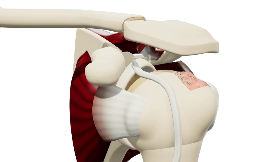 ShoulderRotator cuffFull-thickness (Patte stages) and partial-thickness tears, subscapularis (Lafosse), long head of the biceps, arthroscopic repair.
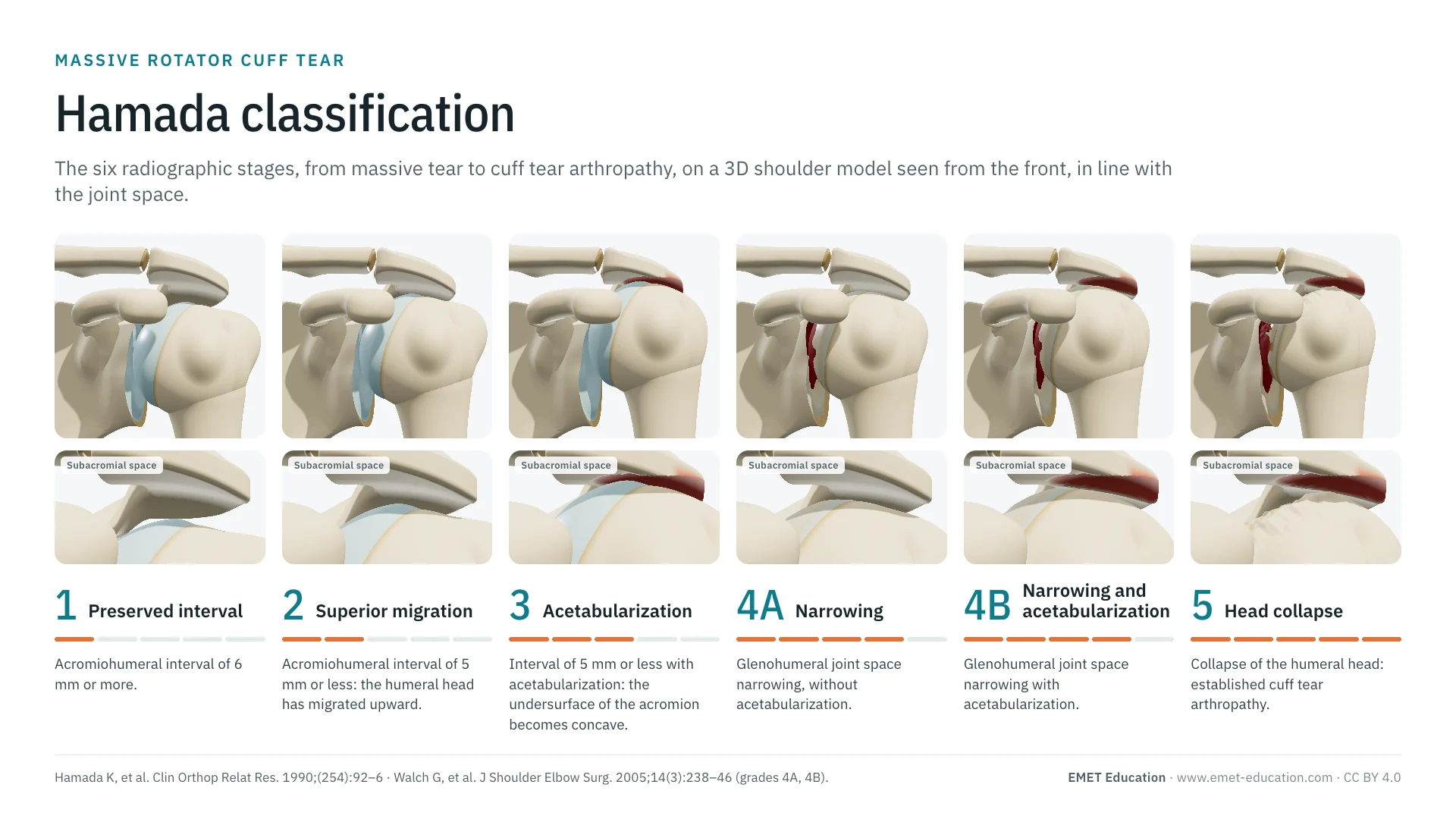 ClassificationsHamada classificationMassive tears: from superior migration of the head to arthropathy.
 ClassificationsAll 3D classificationsArranged by joint.
Explain the tear in 3D
The rotator cuff model opens in your browser, at the stage you choose, with no sign-up.
Share this page with a colleague or a resident: