- EMET Education
- 3D Anatomy
- Elbow
- Ulnar nerve at the elbow
Anatomy · Elbow · Nerves
The Ulnar Nerve at the Elbow in 3D
The ulnar nerve in 3D, from the skin to the nerve, then on labeled MRI slices. With the three compression sites, from arm to forearm, its branches, what flexion does to it, and the measurements used to examine it.
Designed by an orthopedic surgeon
Latin nervus ulnaris · French nerf ulnaire, nerf cubital
- 6.6 mm²Cross-sectional area of the healthy nerve at the medial epicondyle, on ultrasound (meta-analysis)
- 10 mm²Area cutoff favoring cubital tunnel syndrome: sensitivity 0.85, specificity 0.91
- −30 to −41%Cubital tunnel area from extension to 135° of flexion (20 specimens)
- 3 (1–6)Motor branches to the flexor carpi ulnaris, on average (39 elbows)
At a glance
Origin, course, relations
| Origin | Medial cord of the brachial plexus (roots C8 and T1, sometimes C7). |
|---|---|
| Course | It descends along the medial arm, pierces the medial intermuscular septum at mid-arm, passes behind the medial epicondyle in the retrocondylar groove, then between the two heads of the flexor carpi ulnaris (FCU). Its course is tortuous in extension, linear in flexion. |
| Cubital tunnel | Three parts: the retrocondylar groove, partly covered by the cubital tunnel retinaculum (Osborne's ligament), the humeroulnar arcade of the FCU and the deep flexor-pronator aponeurosis (Assmus). The nerve runs 3.8 cm within it (Karataş); the roof is tri-laminar, 523 ± 235 µm (Macchi). |
| Branches | At the elbow: on average one capsular branch, arising 7 mm above the medial epicondyle, and three motor branches to the FCU (1–6); variation is significant. Branches of the medial antebrachial cutaneous nerve cross it 2.9 cm below the epicondyle. |
| Caliber | Multifascicular trunk: 5.7 ± 1.5 fascicles 1 cm above the epicondyle, area 6.6 mm² at the medial epicondyle in healthy subjects (5.9 mm² at the elbow in the meta-analysis of 4,186 volunteers). |
| Mobility | It lengthens in flexion: 5.1 mm of excursion between 10° and 90° of elbow flexion, 4.9 mm for shoulder abduction from 30° to 110°. It shifts anteriorly in the tunnel and flattens against the medial epicondyle. |
| Relations | It passes behind the medial epicondyle, over the posterior band of the ulnar collateral ligament that forms the floor of the tunnel, against the olecranon, under the roof; then between the humeral and ulnar heads of the FCU, beneath their common aponeurosis with the flexor digitorum superficialis. |
Layer by layer
From the skin to the nerve
The same view, from behind and medial to the elbow in extension, removing one layer per image.
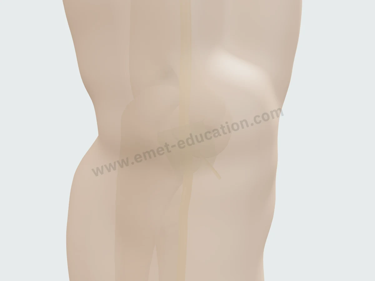
The skinThe nerve can be guessed under the skin, behind the bump of the medial epicondyle.
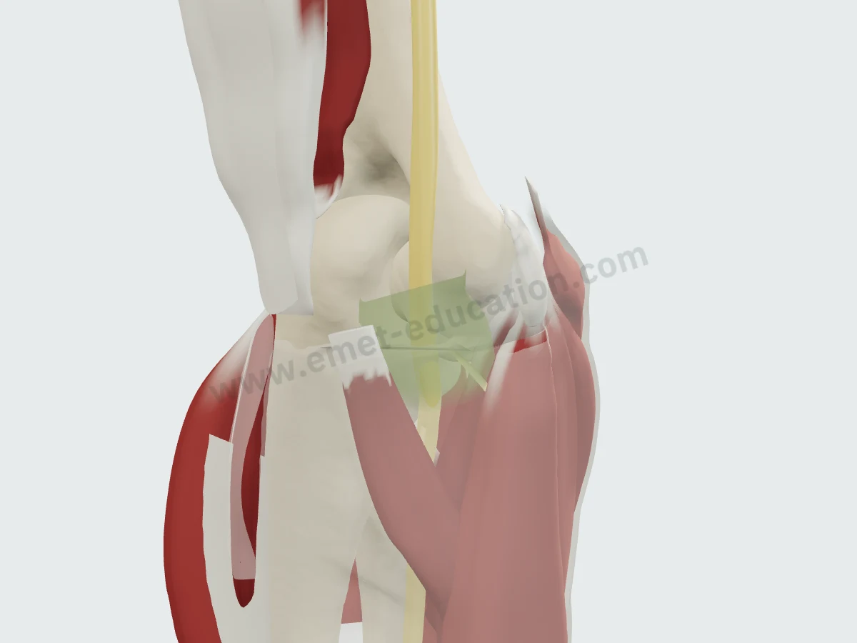
Muscles and fasciaSkin removed: the triceps behind, the flexor-pronators anteromedially, the aponeurosis that joins them.
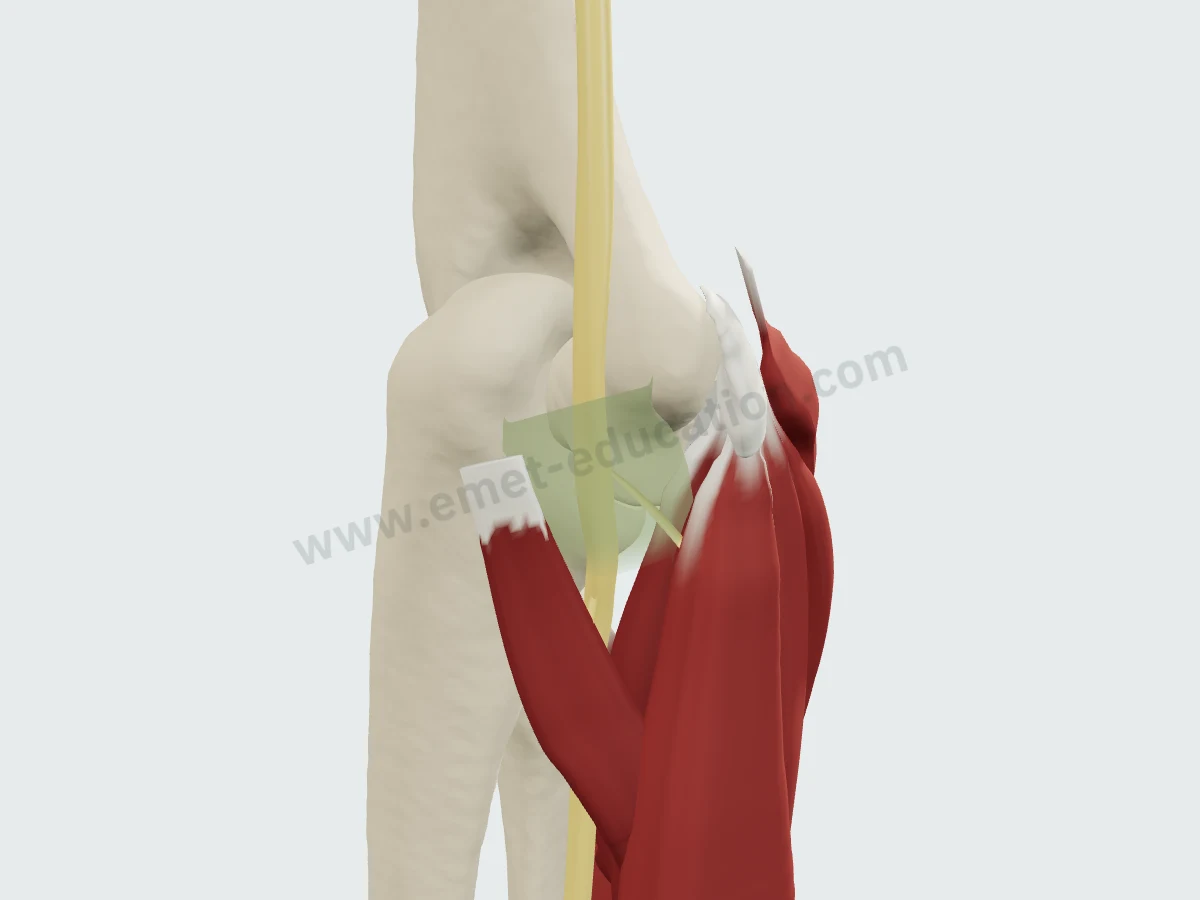
The flexorsTriceps, extensors and fascia removed: the nerve between the two heads of the FCU, with the common tendon on the epicondyle.
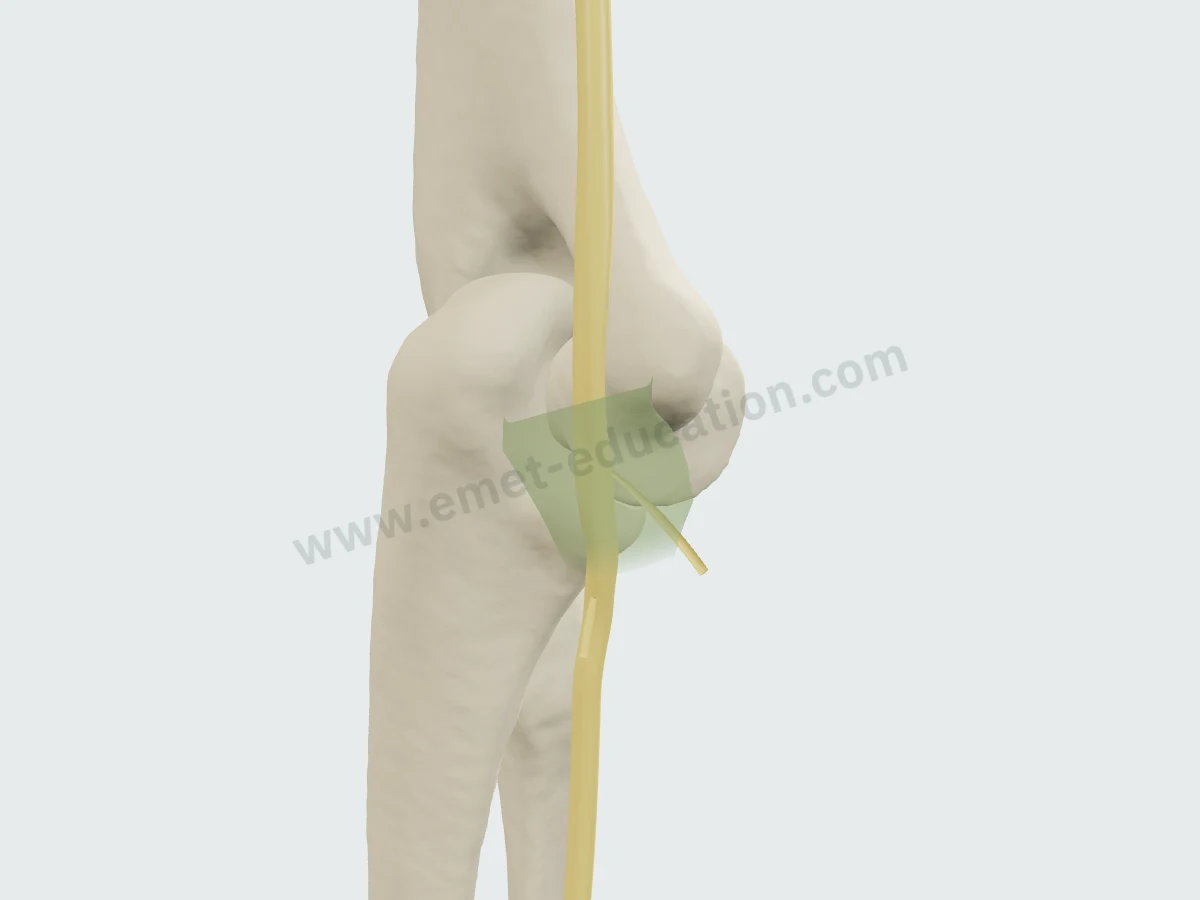
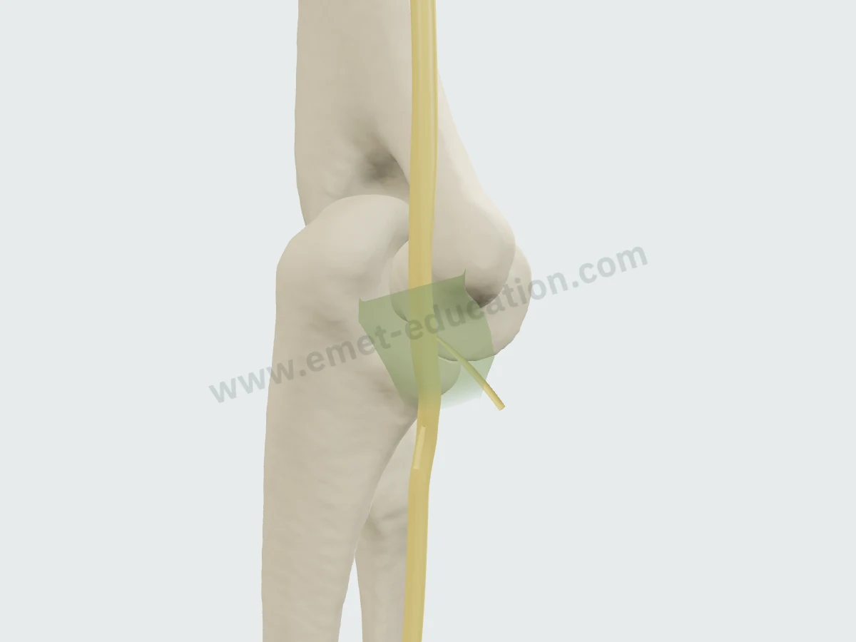
The nerve and its roofBone, nerve and tunnel roof: the retinaculum, continued by the FCU arcade, stretched between the epicondyle and the olecranon.
Compression
The three sites, from top to bottom
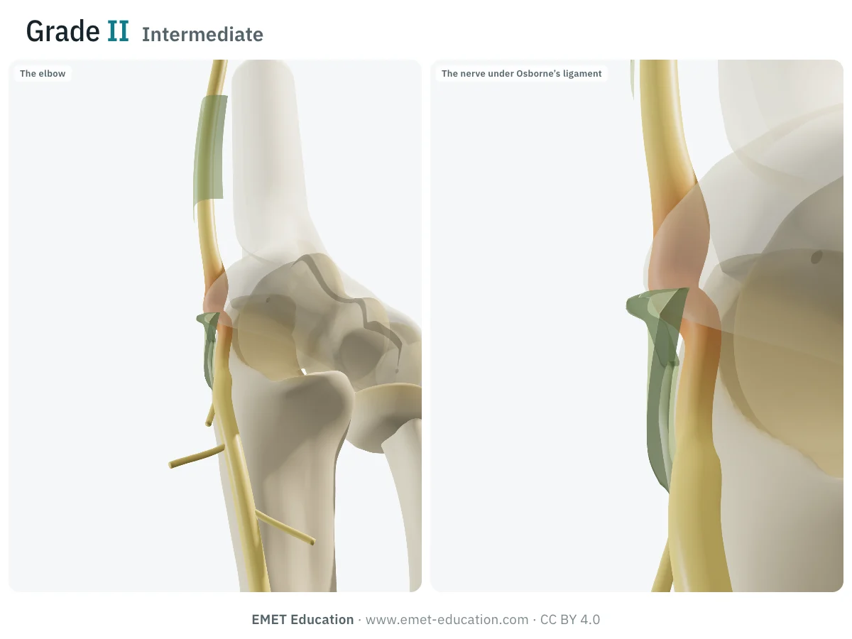
Compression occurs in the cubital tunnel, which includes the groove, the retinaculum, the humeroulnar arcade of the FCU and the deep flexor-pronator aponeurosis (Assmus); the arcade of Struthers, in the arm, remains debated.
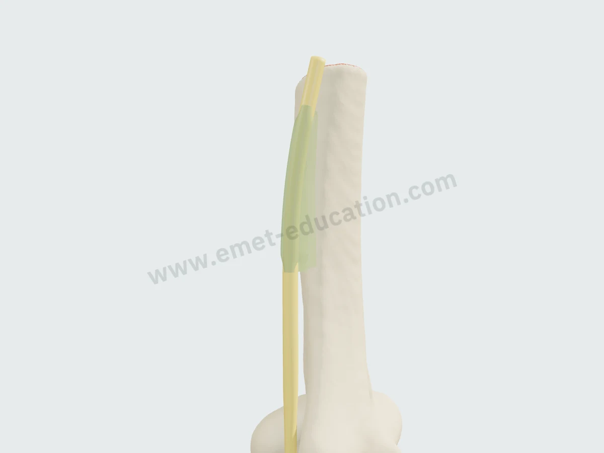
The arcade of StruthersIn the arm, 3.9 to 9.6 cm above the epicondyle depending on the specimen. Absent in 10 arms (Bartels), present on 86.7% of 30 sides (Tubbs), in 40 of 40 arms (Caetano): its existence depends on the definition.
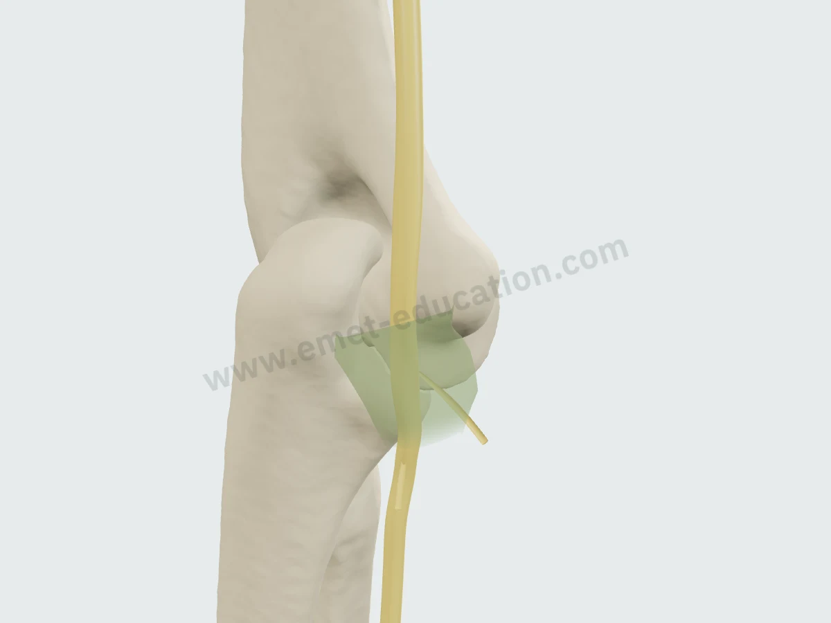
The tunnel retinaculumA band about 4 mm wide from the medial epicondyle to the olecranon (O'Driscoll); absent or replaced by the anconeus epitrochlearis in 4 of 27 elbows; Osborne's ligament is missing on 18.3% of 60 sides (Persons). Its tension in flexion can compress the nerve.
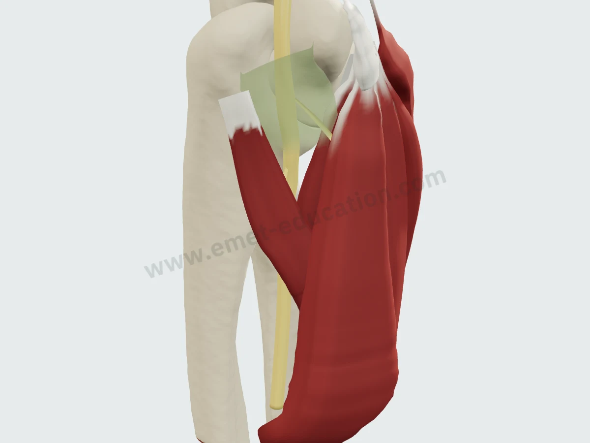
The FCU arcade and the aponeurosisThe nerve passes between the two heads of the FCU then begins to traverse their common aponeurosis with the flexor superficialis 3.9 cm below the epicondyle (Mahan).
Imaging
The ulnar nerve on MRI and CT
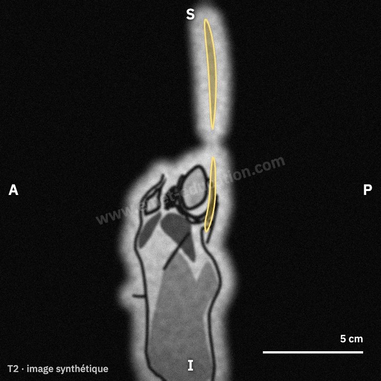
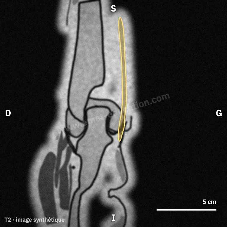
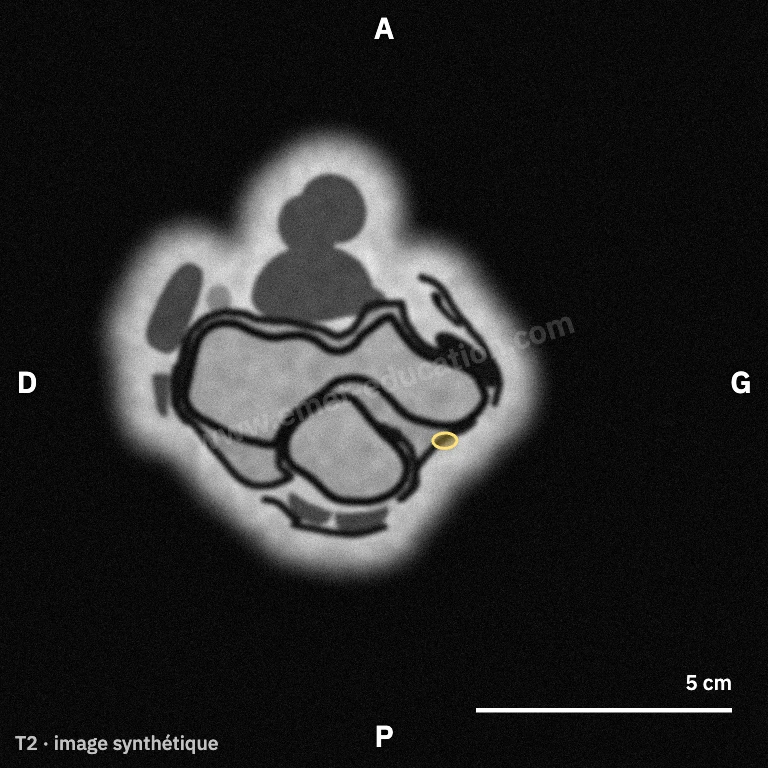
Synthetic images. These slices do not come from a patient: they are computed from the 3D model, by cutting each structure and giving it the signal it would have in the chosen sequence, on MRI or CT. They are for learning where the nerve lies on a slice; nothing is measured on them.
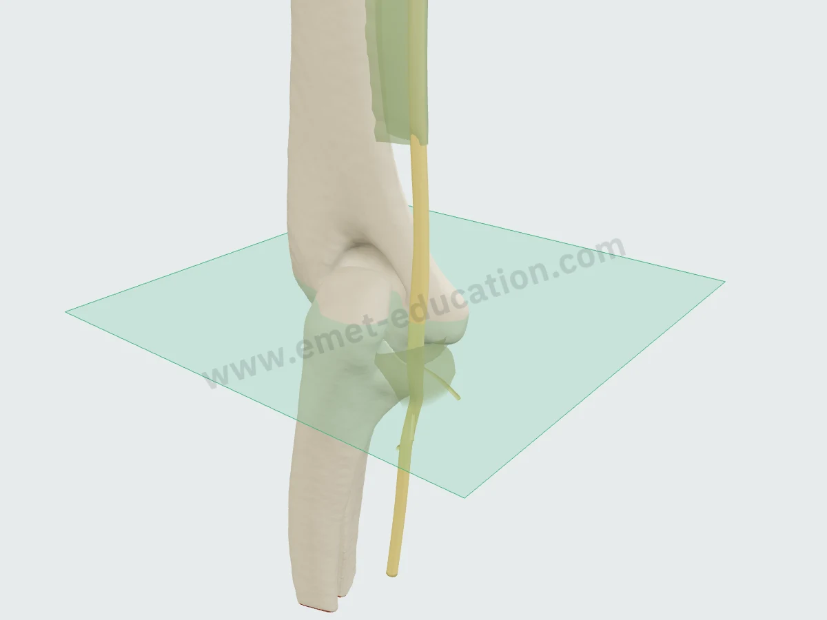
Where the axial view cuts
The axial plane (green) on the 3D model: horizontal, at the level of the medial epicondyle. Hover over a slice and click “Scroll” to page through it as on a workstation: upward, the nerve leaves the groove to run along the humerus; downward, it enters between the heads of the FCU.
In numbers
The caliber, the arcade and flexion, measured
Published mean values, with sample size and source under each figure.
| Value | Reference | Year | Sample | Note |
|---|---|---|---|---|
| 5.2 mm² | Fisse | 2021 | 1983 sites | Forearm |
| 5.5 mm² | Chang | 2017 | – | Arm, above the elbow |
| 5.9 mm² | Fisse | 2021 | 2551 sites | Elbow |
| 6 mm² | Macchi | 2014 | 9 specimens | Under the microscope, within the tunnel (± 1.5) |
| 6.54 mm² | Bedewi | 2017 | 50 subjects | 1 cm above the epicondyle (± 1.67), young men |
| 6.6 mm² | Chang | 2017 | – | At the medial epicondyle |
| 7.3 mm² | Chang | 2017 | – | At the cubital tunnel outlet |
| 7.4 mm² | Chang | 2017 | – | At the cubital tunnel inlet |
| Value | Reference | Year | Sample | Note |
|---|---|---|---|---|
| 3.9 cm | von Schroeder | 2003 | 11 specimens | Distal opening of the fibrous canal |
| 6.8 cm | Tubbs | 2011 | 30 sides | Distal end of the arcade (mean length 4.3 cm) |
| 9.6 cm | von Schroeder | 2003 | 11 specimens | Proximal opening of the fibrous canal |
| Value | Reference | Year | Sample | Note |
|---|---|---|---|---|
| 30 % | Gelberman | 1998 | 20 specimens | Tunnel, at the medial epicondyle |
| 33 % | Gelberman | 1998 | 20 specimens | Nerve, at the medial epicondyle |
| 34 % | Gelberman | 1998 | 20 specimens | Nerve, beneath the FCU |
| 39 % | Gelberman | 1998 | 20 specimens | Tunnel, beneath the aponeurosis |
| 41 % | Gelberman | 1998 | 20 specimens | Tunnel, beneath the FCU |
| 50 % | Gelberman | 1998 | 20 specimens | Nerve, beneath the aponeurosis |
Published mean values, from smallest to largest. Sample size is as each study reports it; populations, measurement levels and protocols differ from one study to the next.
Cross-sectional areas to scale, as discs of equal area: the real shape is oval. A 6.6 mm² nerve has an equivalent diameter of about 2.9 mm.
- 5.1 mmNerve excursion at the elbow for flexion from 10° to 90°; 4.9 mm for shoulder abduction from 30° to 110°10 arms · Wright 2001
- + 6.0 mm²Difference in area between compressed and healthy nerves at the medial epicondyle (95% CI 4.5–7.4): the level where the difference is largest14 trials · Chang 2017
- 3 (1–6)Motor branches to the FCU; on average one capsular branch (0–3), arising 7 mm above the epicondyle39 elbows · Gonzalez 2001
- 3.9 cmBelow the medial epicondyle, entry of the nerve beneath the common aponeurosis of the FCU and flexor superficialis (± 0.7): it kinks there after transposition if it is not released26 specimens · Mahan 2015
- 11 of 80Tunnels with no visible retinaculum, all with unstable nerves; the other 69, with stable nerves, had a retinaculum tightened in flexion40 healthy volunteers · Michelin 2017
Areas are measured at different levels across studies (arm, tunnel inlet, epicondyle, outlet), on ultrasound or under the microscope: they cannot be compared directly. The nerve is unstable at 30° of flexion in 51% of 153 healthy subjects (Endo).
History
How its description has changed since 1991
Classic anatomy places the ulnar nerve in the retrocondylar groove. Studies published since have described its roof, its behavior in flexion, its branches and the variants of its compression sites.
- 1991
A roof for the tunnel
The cubital tunnel retinaculum, 4 mm wide, in four types: its absence allows nerve displacement, excess tension causes dynamic compression, replacement by the anconeus epitrochlearis causes static compression. O'Driscoll
- 1998
What flexion does to the tunnel
From extension to 135° of flexion, the tunnel loses 30 to 41% of its area; the nerve, tortuous in extension, becomes linear, shifts anteriorly and flattens against the epicondyle. Gelberman, Patel
- 2001
Variable branches
On average one capsular branch and three motor branches to the FCU, ranging from 1 to 6: to be identified before a transposition. Gonzalez
- 2003
A disputed arcade
The arcade of Struthers, denied in 10 arms, then described as a 5.7 cm fibrous canal in 11 of 11 specimens, then as a thickening of the brachial fascia on 86.7% of 30 sides. Bartels, von Schroeder, Tubbs
- 2017
A roof that holds the nerve
On ultrasound, the retinaculum is absent in the 11 tunnels with unstable nerves and present, taut in flexion, in the 69 tunnels with stable nerves. Michelin
- 2026
Osborne, not a ligament
Five types on 60 sides: Osborne's ligament is absent in 18.3%, wide and thin in 51%, narrow and proximal in 39%, coexisting with the anconeus epitrochlearis in 6%. Persons
In practice
What anatomy changes in practice
- Examining it. The elbow flexion test combined with pressure on the nerve has a sensitivity of 0.91, versus 0.32 for flexion alone and 0.70 for the Tinel sign (Novak). An elbow lesion causes paresthesia in 98% of cases and palmar sensory loss in 96%, versus 60% and 30% at the wrist (Omejec).
- Measuring it. At the medial epicondyle, the area is 6.6 mm² in healthy subjects; a 10 mm² cutoff gives a sensitivity of 0.85 and a specificity of 0.91 (Chang). The level matters: the difference from healthy subjects is largest there, and normal values vary with the site (Fisse).
- Understanding flexion. The tunnel loses 30 to 41% of its area and the nerve 33 to 50% between extension and 135° (Gelberman); nerve flattening increases from 60° and the trochlea protrudes into the tunnel (Hasegawa). The nerve must lengthen by 5.1 mm between 10° and 90° (Wright).
- Releasing or transposing. Releasing the common aponeurosis of the FCU and flexor superficialis, where the nerve enters 3.9 cm below the epicondyle, prevents kinking after transposition (Mahan); identify the motor branches to the FCU, from 1 to 6 (Gonzalez), and the medial antebrachial cutaneous branches crossing it at 2.9 cm (Mirza).
- Looking for a variant. The retinaculum is absent in unstable nerves (Michelin), and the nerve is unstable at 30° in 51% of healthy subjects (Endo): instability is not always pathological. The anconeus epitrochlearis exists on 10% of 324 sides (Banerjee), and the arcade of Struthers is sought 2.5–7.0 cm from the epicondyle (Caetano).
Your turn
Where is the ulnar nerve?
Six unlabeled synthetic slices, in random order: click the ulnar nerve. The answer shows its outline.
Click the ulnar nerve.
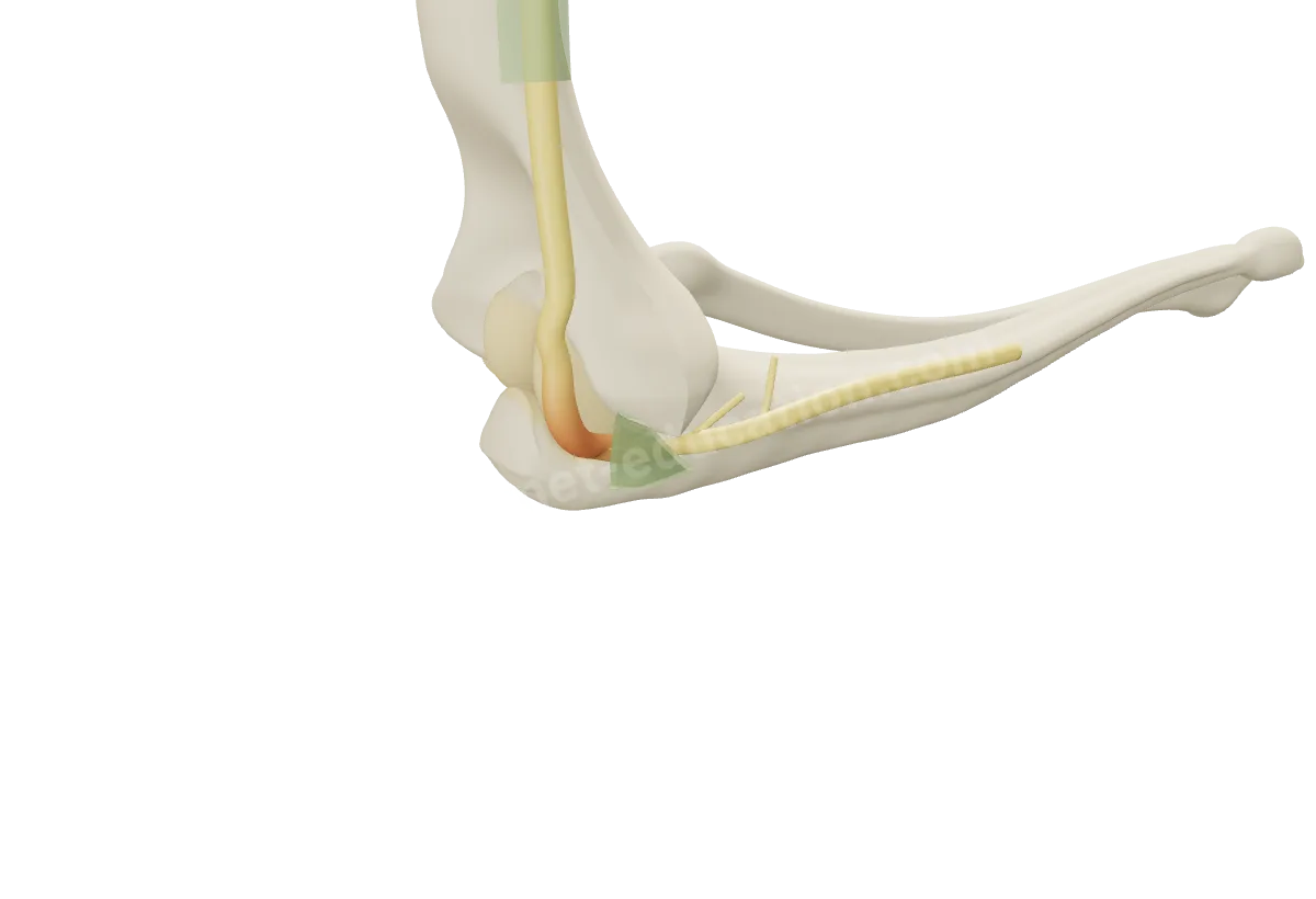
Classifications
Related classifications
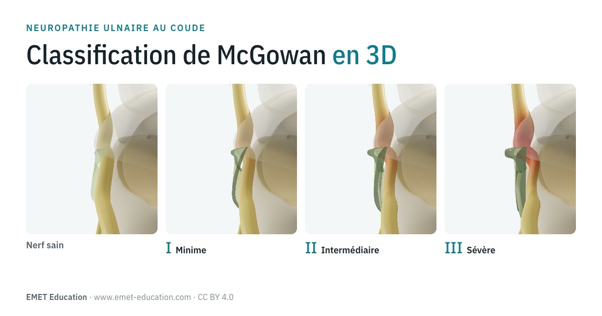 McGowanClassification of ulnar nerve compression at the elbow
- SoonO'DriscollCubital tunnel retinaculum: types 0, Ia, Ib and II (1991)
Neighbors
Its neighbors
- SoonTriceps brachiiPosterior
- SoonCollateral ligamentsDeep
- SoonMedial epicondyleAnterior
- SoonFlexor carpi ulnarisSuperficial
Questions
Frequently asked questions
Where does the ulnar nerve pass at the elbow?
Behind the medial epicondyle, in the retrocondylar groove, beneath the roof of the cubital tunnel, then between the two heads of the flexor carpi ulnaris (Gray's Anatomy). It runs 3.8 cm within the tunnel (Karataş).
Where can the ulnar nerve be compressed at the elbow?
In the cubital tunnel, which includes the groove, the retinaculum (Osborne's ligament), the humeroulnar arcade of the FCU and the deep flexor-pronator aponeurosis (Assmus); higher up, the arcade of Struthers, whose existence is debated (Bartels, Tubbs).
What is the normal area of the ulnar nerve?
On ultrasound, 6.6 mm² at the medial epicondyle, 7.4 mm² at the tunnel inlet and 5.5 mm² at the forearm in healthy subjects (Chang); 5.9 mm² at the elbow in a meta-analysis of 4,186 volunteers (Fisse). A 10 mm² cutoff favors cubital tunnel syndrome.
Why can elbow flexion compress the nerve?
Because the tunnel loses 30 to 41% of its area between extension and 135° and the nerve, which must lengthen by 5.1 mm, flattens there against the epicondyle (Gelberman, Patel, Wright). A retinaculum tight between 90° and 120° (type Ib) can compress the nerve dynamically (O'Driscoll).
Is an unstable ulnar nerve always pathological?
No: at 30° of flexion, 51% of 153 healthy subjects had instability on at least one side (Endo). The retinaculum is missing in unstable nerves (Michelin).
Are the MRI images on this page real?
No: they are synthetic, computed from the 3D model. They show where the nerve lies on a slice; they do not replace an actual exam.
Sources
Author and references
- Standring S, ed. Gray's Anatomy: The Anatomical Basis of Clinical Practice. 42nd ed. Elsevier; 2020.
- O'Driscoll SW, Horii E, Carmichael SW, Morrey BF. The cubital tunnel and ulnar neuropathy. J Bone Joint Surg Br. 1991;73(4):613-7. doi:10.1302/0301-620X.73B4.2071645
- Persons EM, Hines BL, Herrera M, Lesser ER, Anbalagan M, Dave M, et al. Osborne's ligament: Anatomical study with application to better understanding ulnar nerve compression at the elbow. Neurosurg Rev. 2026;49(1). doi:10.1007/s10143-026-04307-9
- Michelin P, Leleup G, Ould-Slimane M, Merlet MC, Dubourg B, Duparc F. Ultrasound biomechanical anatomy of the soft structures in relation to the ulnar nerve in the cubital tunnel of the elbow. Surg Radiol Anat. 2017;39(11):1215-1221. doi:10.1007/s00276-017-1879-y
- Assmus H, Antoniadis G, Bischoff C, Hoffmann R, Martini AK, Preissler P, et al. Cubital tunnel syndrome - a review and management guidelines. Cent Eur Neurosurg. 2011;72(2):90-8. doi:10.1055/s-0031-1271800
- Karatas A, Apaydin N, Uz A, Tubbs R, Loukas M, Gezen F. Regional anatomic structures of the elbow that may potentially compress the ulnar nerve. J Shoulder Elbow Surg. 2009;18(4):627-31. doi:10.1016/j.jse.2009.03.004
- Macchi V, Tiengo C, Porzionato A, Stecco C, Sarasin G, Tubbs S, et al. The cubital tunnel: a radiologic and histotopographic study. J Anat. 2014;225(2):262-9. doi:10.1111/joa.12206
- Bedewi MA, Yousef AMM, Abd-Elghany AA, El-Sharkawy MS, Awad EM. Estimation of ultrasound reference values for the ulnar nerve fascicular number and cross-sectional area in young males: A cross-sectional study. Medicine (Baltimore). 2017;96(10):e6204. doi:10.1097/MD.0000000000006204
- Chang KV, Wu WT, Han DS, Özçakar L. Ulnar Nerve Cross-Sectional Area for the Diagnosis of Cubital Tunnel Syndrome: A Meta-Analysis of Ultrasonographic Measurements. Arch Phys Med Rehabil. 2017;99(4):743-757. doi:10.1016/j.apmr.2017.08.467
- Fisse AL, Katsanos AH, Gold R, Pitarokoili K, Krogias C. Cross-sectional area reference values for peripheral nerve ultrasound in adults: a systematic review and meta-analysis-Part I: Upper extremity nerves. Eur J Neurol. 2021;28(5):1684-1691. doi:10.1111/ene.14759
- Gelberman RH, Yamaguchi K, Hollstien SB, Winn SS, Heidenreich FP, Bindra RR, et al. Changes in interstitial pressure and cross-sectional area of the cubital tunnel and of the ulnar nerve with flexion of the elbow. An experimental study in human cadavera. J Bone Joint Surg Am. 1998;80(4):492-501. doi:10.2106/00004623-199804000-00005
- Patel VV, Heidenreich FP, Bindra RR, Yamaguchi K, Gelberman RH. Morphologic changes in the ulnar nerve at the elbow with flexion and extension: a magnetic resonance imaging study with 3-dimensional reconstruction. J Shoulder Elbow Surg. 1998;7(4):368-74. doi:10.1016/s1058-2746(98)90025-8
- Hasegawa H, Omokawa S, Nakanishi Y, Nakano K, Nagashima M, Sananpanich K, et al. Dynamic analysis of the ulnar nerve and cubital tunnel morphology using ultrasonography: a cadaveric study. J Shoulder Elbow Surg. 2022;31(11):2322-2327. doi:10.1016/j.jse.2022.05.026
- Wright TW, Glowczewskie F, Cowin D, Wheeler DL. Ulnar nerve excursion and strain at the elbow and wrist associated with upper extremity motion. J Hand Surg Am. 2001;26(4):655-62. doi:10.1053/jhsu.2001.26140
- Gonzalez MH, Lotfi P, Bendre A, Mandelbroyt Y, Lieska N. The ulnar nerve at the elbow and its local branching: an anatomic study. J Hand Surg Br. 2001;26(2):142-4. doi:10.1054/jhsb.2000.0532
- Mahan MA, Gasco J, Mokhtee DB, Brown JM. Anatomical considerations of fascial release in ulnar nerve transposition: a concept revisited. J Neurosurg. 2015;123(5):1216-22. doi:10.3171/2014.10.JNS141379
- Mirza A, Mirza JB, Lee BK, Adhya S, Litwa J, Lorenzana DJ. An anatomical basis for endoscopic cubital tunnel release and associated clinical outcomes. J Hand Surg Am. 2014;39(7):1363-9. doi:10.1016/j.jhsa.2014.04.030
- Bartels RH, Grotenhuis JA, Kauer JM. The arcade of Struthers: an anatomical study. Acta Neurochir (Wien). 2003;145(4):295-300; discussion 300. doi:10.1007/s00701-003-0006-5
- von Schroeder HP, Scheker LR. Redefining the "Arcade of Struthers". J Hand Surg Am. 2003;28(6):1018-21. doi:10.1016/s0363-5023(03)00421-0
- Tubbs RS, Deep A, Shoja MM, Mortazavi MM, Loukas M, Cohen-Gadol AA. The arcade of Struthers: An anatomical study with potential neurosurgical significance. Surg Neurol Int. 2011;2:184. doi:10.4103/2152-7806.91139
- Caetano EB, Sabongi Neto JJ, Vieira LA, Caetano MF. The arcade of Struthers: an anatomical study and clinical implications. Rev Bras Ortop. 2017;52(3):331-336. doi:10.1016/j.rboe.2016.07.006
- Endo F, Tajika T, Kuboi T, Shinagawa S, Tsukui T, Nakajima T, et al. The ultrasonographic assessment of the morphologic changes in the ulnar nerve at the cubital tunnel in Japanese volunteers: relationship between dynamic ulnar nerve instability and clinical symptoms. JSES Int. 2021;5(5):942-947. doi:10.1016/j.jseint.2021.05.005
- Banerjee S, Nguyen K, Bishop JL, Pujol JE, Rosbrugh JE, Yurasakpong L, et al. New Insights Into the Anconeus Epitrochlearis Muscle and Its Relationship to the Ulnar Nerve: Anatomical Study. Clin Anat. 2026. doi:10.1002/ca.70123
- Omejec G, Podnar S. Differentiation of ulnar neuropathy at the wrist due to ganglion cyst from ulnar neuropathy at the elbow. Neurophysiol Clin. 2020;50(5):345-351. doi:10.1016/j.neucli.2020.08.004
- Novak CB, Lee GW, Mackinnon SE, Lay L. Provocative testing for cubital tunnel syndrome. J Hand Surg Am. 1994;19(5):817-20. doi:10.1016/0363-5023(94)90193-7
In clinic
Show the cubital tunnel to your patient
The images on this page come from an interactive model. In clinic, show the nerve compression, its signs and the surgical options.
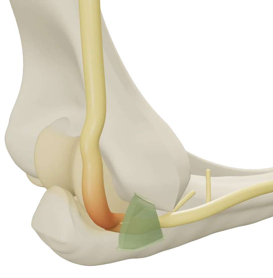
The ulnar nerve, in 3D, for your patient
Open the cubital tunnel model in your browser, no sign-up needed.
A colleague preparing a lecture on the elbow?