- EMET Education
- 3D classifications
- Neer classification (proximal humerus)
Classification · Proximal humerus fracture
Neer Classification of Proximal Humerus Fractures in 3D
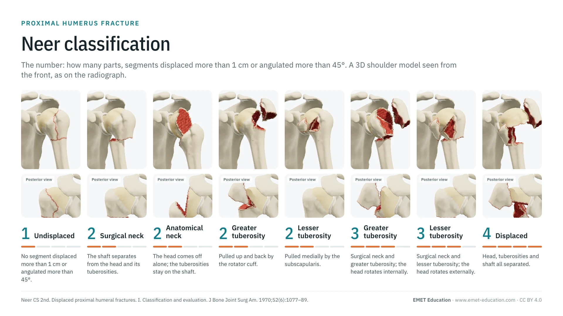
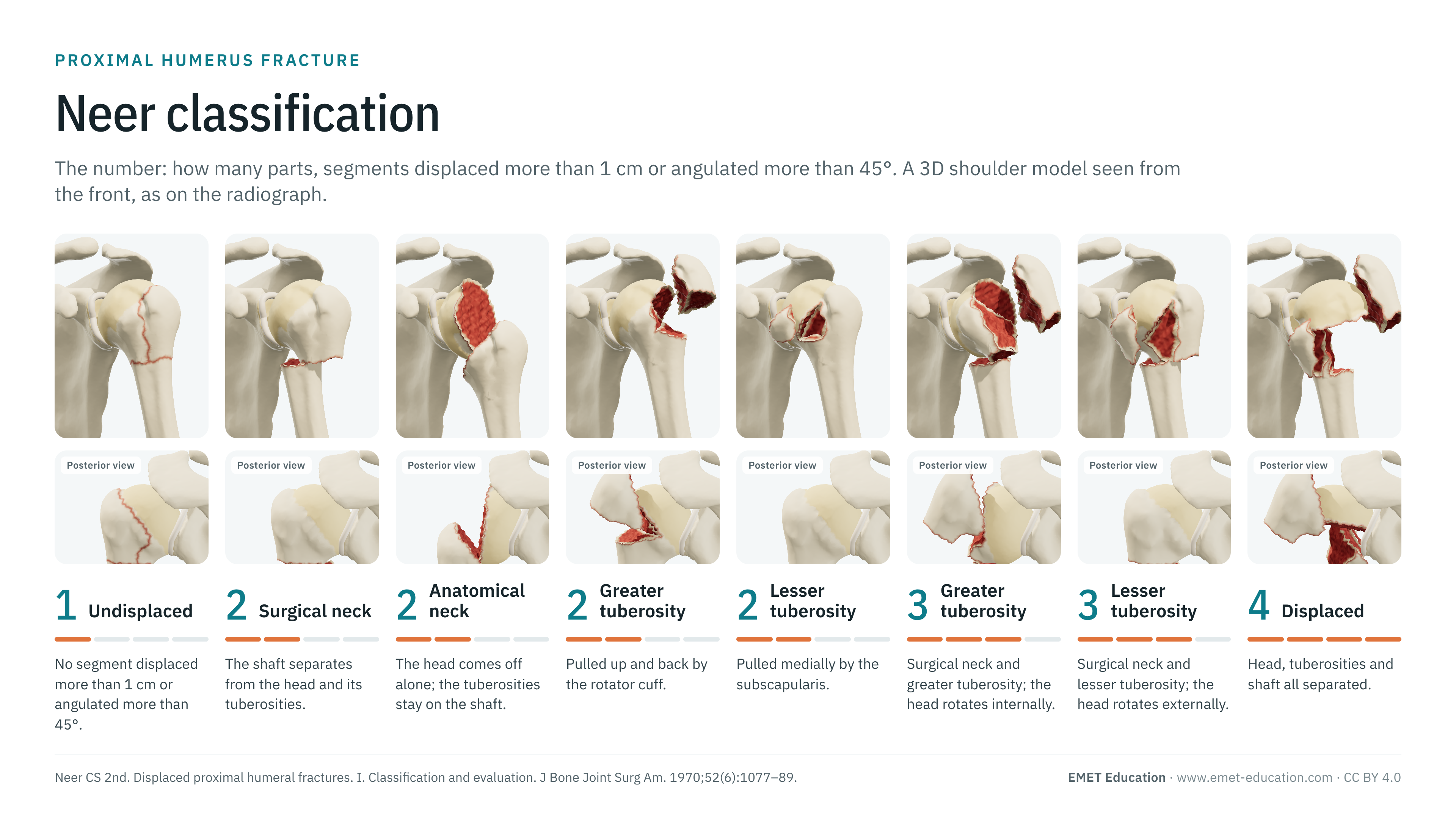
The categories of the Neer classification, by the number of displaced segments, on the proximal humerus fracture 3D model, seen from the front as on the radiograph. With the origin of the classification, reliability data, the link with AO/OTA, grading pitfalls and free infographics for teaching and publication.
Designed by an orthopedic surgeon
The 3D model opens in your browser, at the category shown, with no sign-up. Infographics are free to reuse under CC BY 4.0.
- 1970Published by Neer, in the Journal of Bone and Joint Surgery
- 1–4Parts: the segments displaced more than 1 cm or angulated more than 45°
- ≈ 1,950Citations of the 1970 article (OpenAlex, October 2026)
- κ 0.17–0.52Mean interobserver agreement on radiographs, in 11 studies (systematic review)
The categories
From one to four parts, in 3D
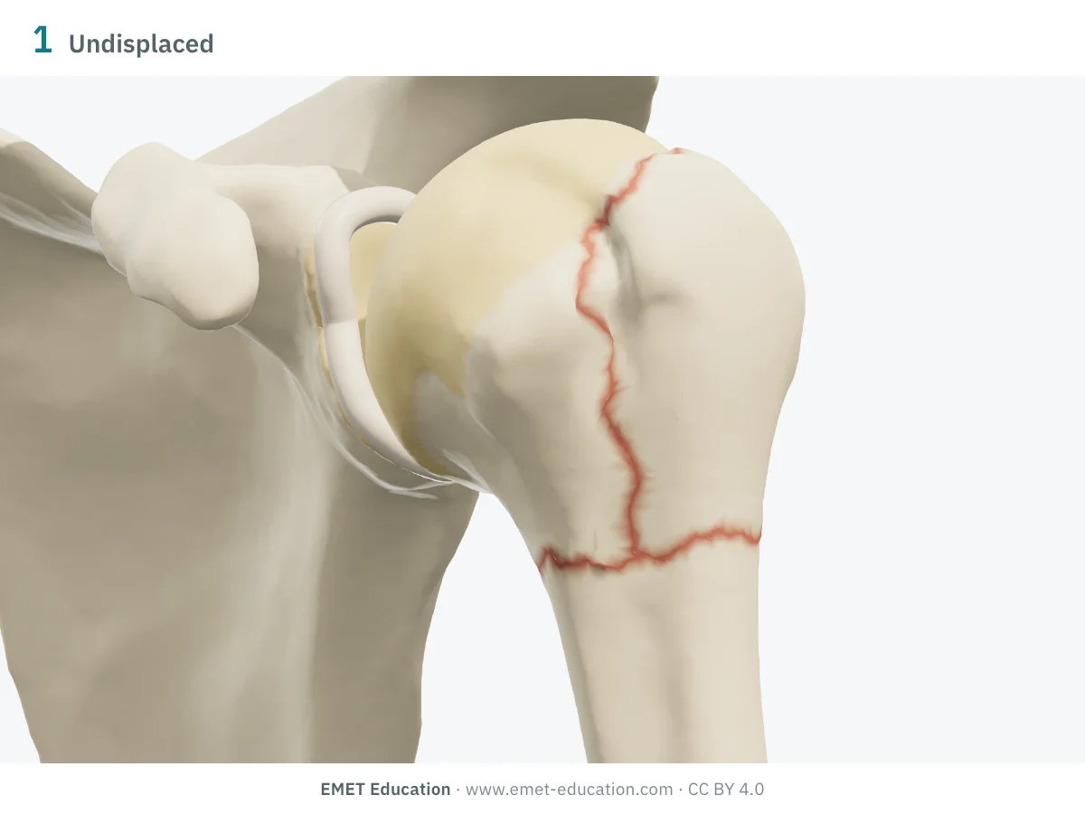
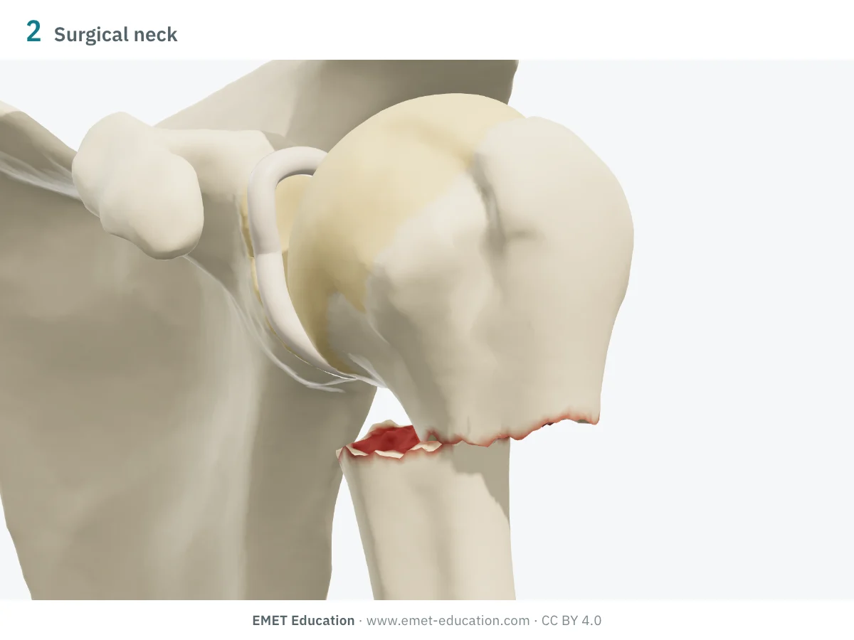
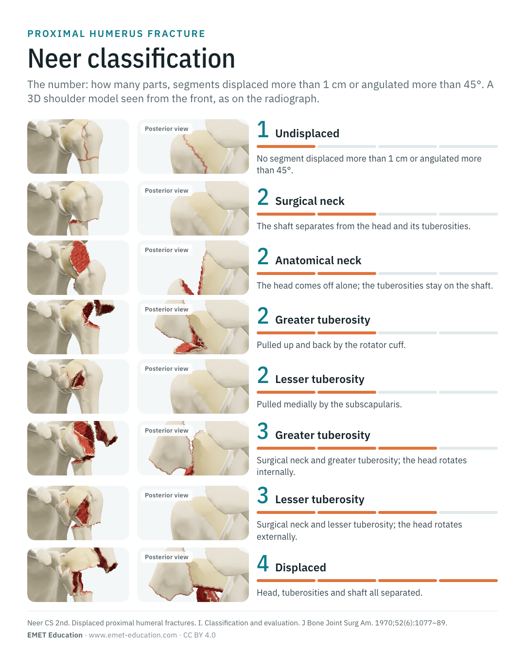
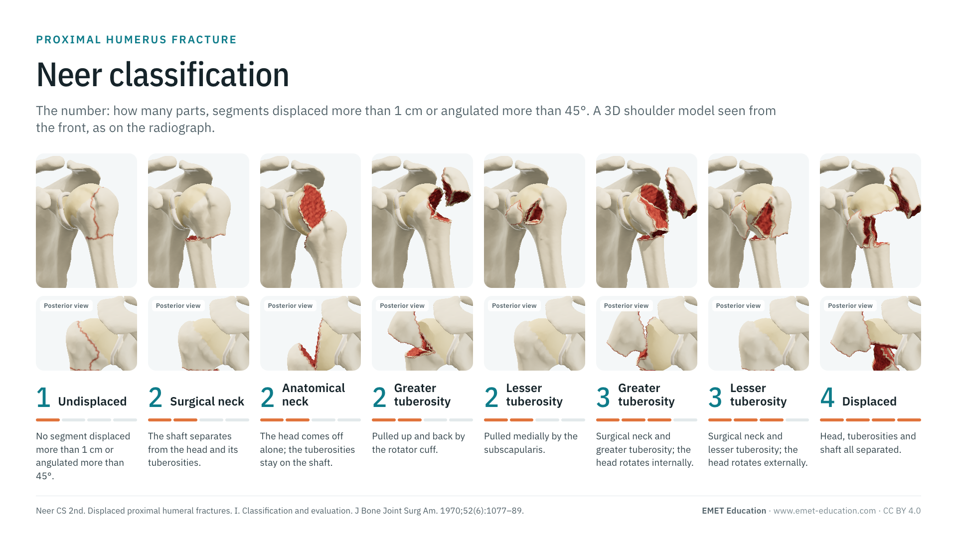
Each image is a rendering of the EMET Education proximal humerus fracture model at that category, left shoulder seen from the front, with a narrow field to approach the projection of an AP radiograph. The four segments are Neer's: the head (at the anatomical neck), the greater tuberosity, the lesser tuberosity and the shaft (at the surgical neck). A displaced part is displaced beyond Neer's thresholds; a red rim outlines every fracture line, even an undisplaced one. Under each category, its AO/OTA 2018 code.
-
1part · minimal displacement
- Criteria
- One or more fracture lines, but no segment displaced more than 1 cm or angulated more than 45°: the fracture counts as a single part, whatever the number of lines.
- AO/OTA 2018
- The code of its lines, with modifier [1] (undisplaced)
- In the 3D model
- Surgical neck and greater tuberosity lines, without displacement: the fragments stay in place, only the red rims outline them.
-
2parts · surgical neck
- Criteria
- The shaft is displaced relative to the head, which keeps both tuberosities.
- AO/OTA 2018
- 11A2.1 (simple), 11A2.2 (wedge) or 11A2.3 (multifragmentary)
- In the 3D model
- Shaft translated 15 mm medially, toward the scapula, under the pull of the pectoralis major and latissimus dorsi; the head stays in the glenoid.
-
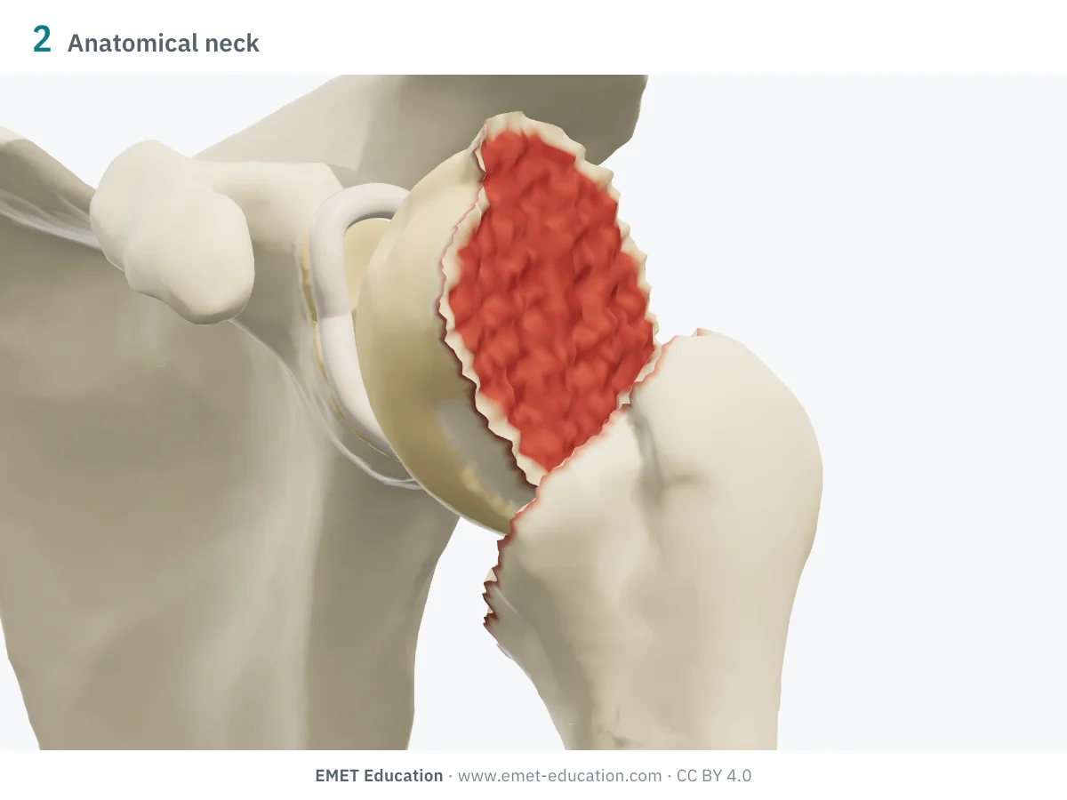
2parts · anatomical neck
- Criteria
- The articular head comes off alone, at the anatomical neck; the tuberosities stay on the shaft. Rare, it deprives the head of most of its attachments: the risk of ischaemia is high (Hertel).
- AO/OTA 2018
- 11C1.3, isolated anatomical neck
- In the 3D model
- The head tilts 45° into varus about its centre, staying in the glenoid; the block of tuberosities and shaft does not move.
-
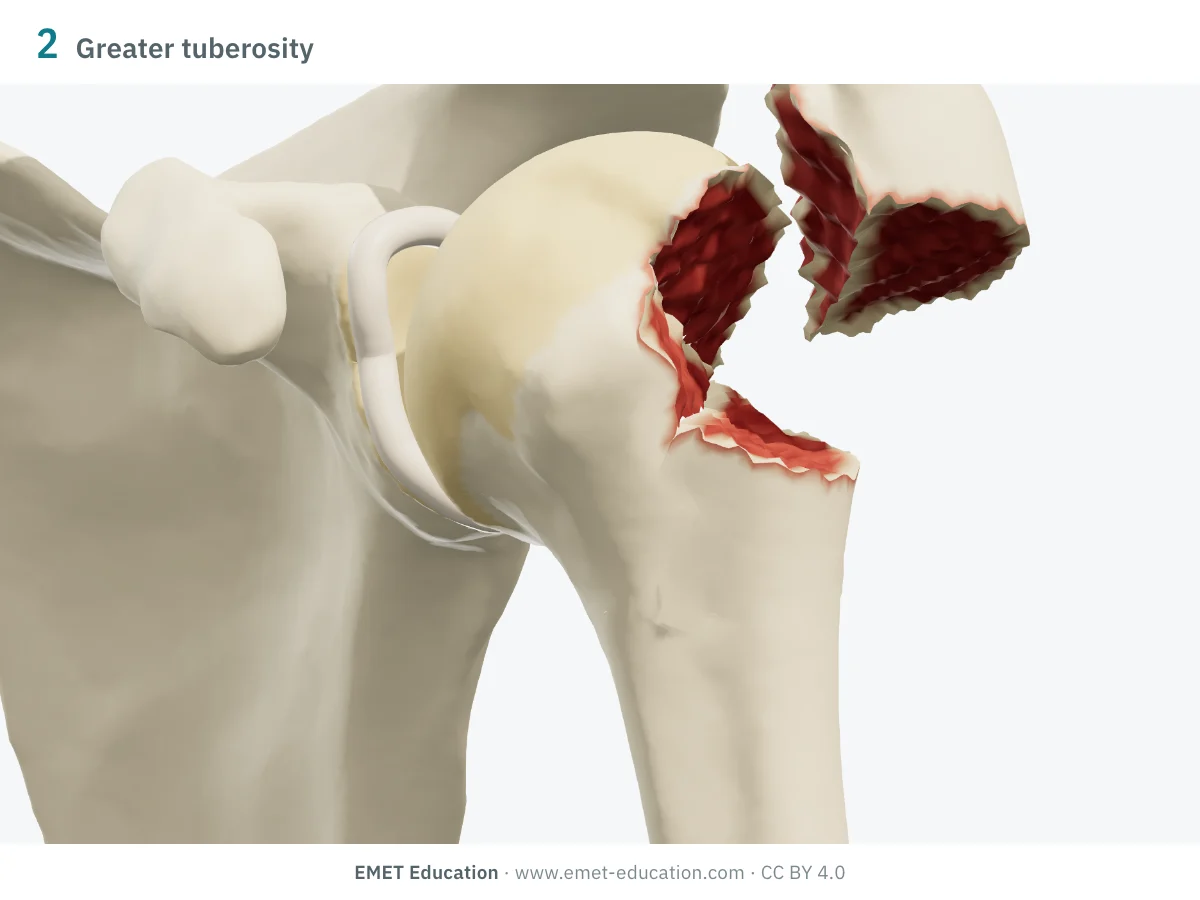
2parts · greater tuberosity
- Criteria
- The displaced greater tuberosity is pulled up and back by the supraspinatus, infraspinatus and teres minor; the rest of the humerus is in one piece.
- AO/OTA 2018
- 11A1.1
- In the 3D model
- Greater tuberosity displaced 15 mm, tilted 20° backward and 30° upward: its outer surface faces the acromion. The posterior view shows how far it moves back.
-
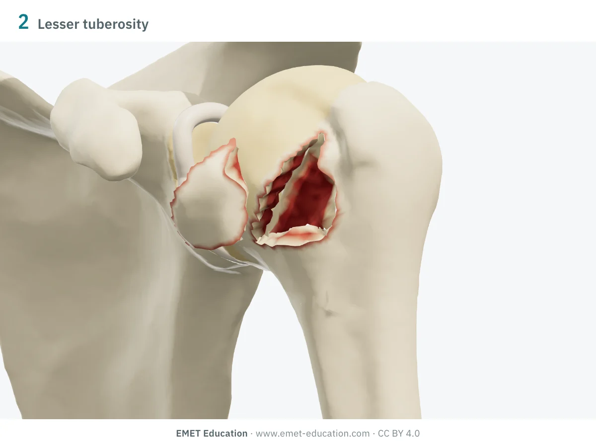
2parts · lesser tuberosity
- Criteria
- The displaced lesser tuberosity is pulled medially by the subscapularis. Rare in isolation, it should prompt a search for a posterior dislocation.
- AO/OTA 2018
- 11A1.2
- In the 3D model
- Lesser tuberosity displaced 15 mm medially, internally rotated 15°.
-
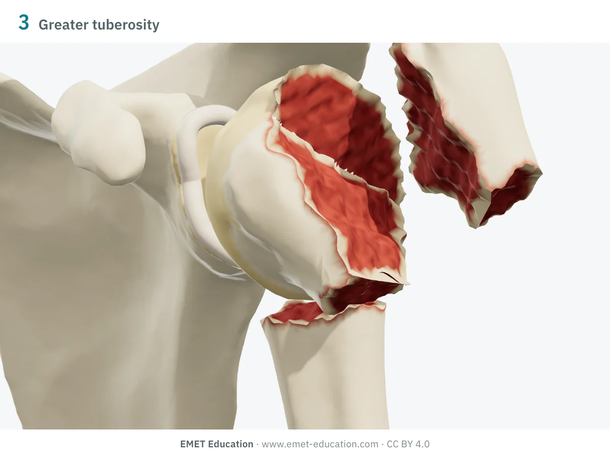
3parts · neck and greater tuberosity
- Criteria
- Surgical neck and greater tuberosity displaced. The head, still attached to the lesser tuberosity and held by the subscapularis, rotates internally.
- AO/OTA 2018
- 11B1.1, neck wedge intact (u) or fragmentary (v)
- In the 3D model
- Greater tuberosity as in the two-part fracture; shaft translated 10 mm; head tilted 30° into varus and rotated internally with the lesser tuberosity.
-
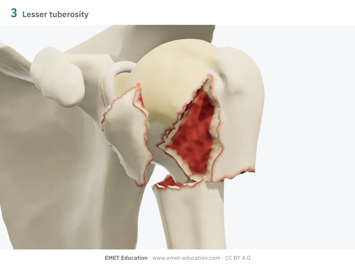
3parts · neck and lesser tuberosity
- Criteria
- Surgical neck and lesser tuberosity displaced. The head, still attached to the greater tuberosity and held by the posterosuperior cuff, rotates externally.
- AO/OTA 2018
- 11B1.2, same qualifications
- In the 3D model
- Lesser tuberosity displaced 15 mm medially; shaft translated 10 mm; the head rotates externally with the greater tuberosity.
-
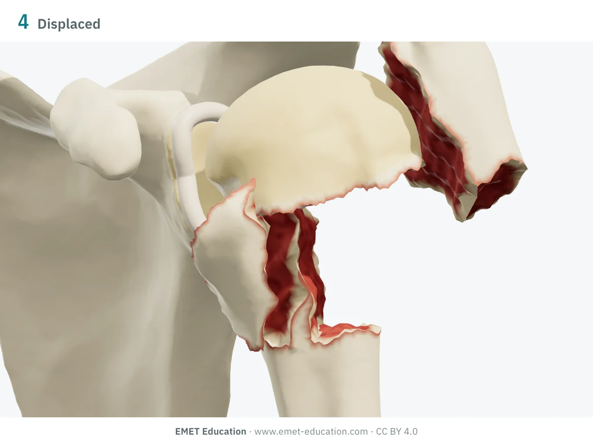
4parts
- Criteria
- The head, both tuberosities and the shaft are all separated and displaced. The head, cut off from its attachments, is exposed to necrosis.
- AO/OTA 2018
- 11C3.1, 11C3.2 or 11C3.3 depending on the articular surface and the shaft
- In the 3D model
- Head tilted 45° into valgus; greater tuberosity displaced 15 mm up and back; lesser tuberosity staying close to the shaft, translated 10 mm.
-
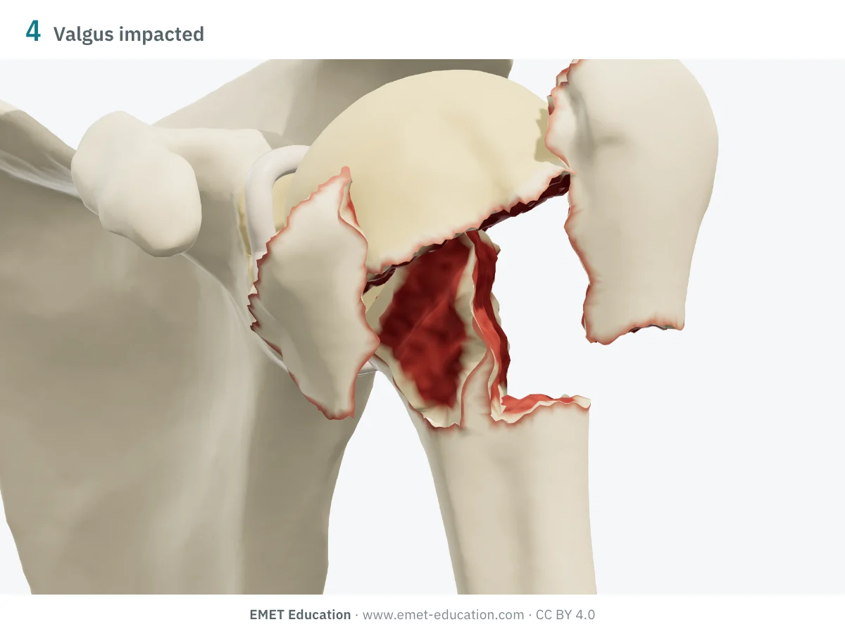
4parts · valgus impacted
- Criteria
- The head impacts into valgus on the metaphysis and splays the tuberosities; the medial periosteal hinge is preserved. Described by Jakob et al. in 1991, added by Neer in 2002: head necrosis is less frequent than in other four-part fractures.
- AO/OTA 2018
- 11C1.1, qualification p (both tuberosities)
- In the 3D model
- Head in 30° of valgus, shaft driven up beneath it (impaction); tuberosities splayed 10 mm each, without tilt.
-
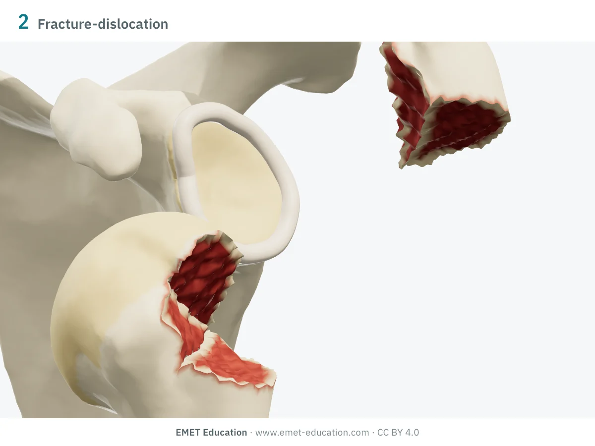
2parts · fracture-dislocation
- Criteria
- A two-, three- or four-part fracture in which the head has left the glenoid, anteriorly or posteriorly. Neer counts them as separate categories: here, a two-part greater tuberosity fracture with anterior dislocation.
- AO/OTA 2018
- 11A1.1[5a]: the fracture code and the anterior dislocation modifier
- In the 3D model
- The whole humerus comes out anteriorly, below the coracoid; the greater tuberosity, held by the cuff, stays up, close to its place.
-
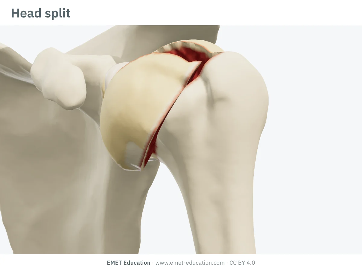
Head split
- Criteria
- Articular surface fracture: a line crosses the head and separates a cartilage-bearing fragment. Neer places these fractures, with impression fractures, in a separate group.
- AO/OTA 2018
- No code for an isolated head split; 11C3.2 or 11C3.3 if the metaphysis is fragmented
- In the 3D model
- A plane containing the axis of the head, 3 mm in front of its centre; the anterior fragment opens like a book by 10 mm, forward and medially.
-
Impression fracture of the articular surface
- Criteria
- The other articular surface fracture: an impacted notch in the head, left by the glenoid rim during a dislocation (Hill-Sachs lesion anteriorly, McLaughlin lesion posteriorly).
- AO/OTA 2018
- Modifier [3a] (articular impaction), added to the fracture code
- In the 3D model
- Not shown in this model: the notch is shown in the shoulder instability model.
Schematic rendering, for teaching: the model illustrates each category on a single shoulder, it does not reproduce a radiograph. Criteria reworded from Neer (1970), Neer (2002), Jakob et al. (1991) and Brorson et al. (2013); Neer's figure is not reproduced. The number and the bar show the number of parts.
The origin
Codman, 1934; Neer, 1970 and 2002
In 1970, Charles S. Neer II, in New York, published two consecutive articles in the Journal of Bone and Joint Surgery: the first proposed a classification of displaced fractures of the proximal humerus (part I), the second dealt with three- and four-part displacement (part II). He started from the four segments described by Codman (1934), and classified the fracture not by the number of fracture lines but by the number of displaced segments.
The classification comes down to a few rules:
- four segments: the head (articular surface, at the anatomical neck), the greater tuberosity, the lesser tuberosity and the shaft (at the surgical neck);
- a segment counts as a part if it is displaced more than 1 cm or angulated more than 45° (Brorson et al.); otherwise, even if a fracture line separates it, it does not count: the "one-part" fracture is a minimally displaced fracture, with any number of lines;
- the categories: one-part; two-part (surgical neck, anatomical neck, greater tuberosity, lesser tuberosity); three-part (surgical neck and one tuberosity); four-part;
- fracture-dislocations with two, three and four parts, anterior or posterior, and articular surface fractures (impression, head split) form separate categories: 16 in all (Brorson et al.);
- in 2002, Neer revisited his system ("purpose and reliable use") and added the four-part valgus impacted fracture of Jakob et al. (1991), until then found only in the AO classification.
Most of these fractures are not displaced. In the Edinburgh series of 1,027 proximal humerus fractures, half were minimally displaced, and nearly nine in ten fell into only three Neer categories (Court-Brown et al., 2001). The most common fracture there was valgus impacted, a pattern Neer had not yet included.
Reliability
How reproducible is the category?
Moderately at best, since the first studies. On radiographs, two readers agree only fairly or moderately; CT changes almost nothing. The systematic review by Brorson and Hróbjartsson found mean kappas of 0.17 to 0.52 across studies.
| Study | Measure | Value |
|---|---|---|
| Sidor et al., 1993 | Interobserver kappa at both readings, 5 readers, 50 fractures, radiographs (mean intraobserver 0.66) | 0.48 · 0.52 |
| Siebenrock and Gerber, 1993 | Inter- then intraobserver kappa, 5 shoulder surgeons, 95 fractures (AO: 0.53 · 0.58) | 0.40 · 0.60 |
| Bernstein et al., 1996 | Interobserver kappa, radiographs then radiographs and CT, 4 readers, 20 fractures | 0.52 · 0.50 |
| Sjödén et al., 1997 | Interobserver kappa, radiographs then CT, 10 readers, 26 fractures (AO: 0.31 · 0.30) | 0.42 · 0.45 |
| Brorson and Hróbjartsson, 2008 | Mean interobserver kappa, systematic review of 11 studies (88 readers, 468 cases) | 0.17–0.52 |
| Majed et al., 2011 | Interobserver kappa on 3D-printed models, 4 readers, 96 complex fractures (AO 0.11; Codman-Hertel 0.44) | 0.33 |
| Brorson et al., 2012 | Interobserver kappa at both readings, 5 shoulder surgeons, 193 fractures (treatment recommendation: 0.48 · 0.52) | 0.33 · 0.36 |
| Marongiu et al., 2020 | Mean inter- then intraobserver kappa, 3 readers, 116 fractures (AO/OTA 2018: 0.67 · 0.75) | 0.67 · 0.85 |
What improves agreement is mainly training: in the randomised trial by Brorson et al. (2002), kappa rose from 0.27 to 0.62 after training in the system (from 0.30 to 0.79 among specialists). Three-dimensional imaging gives inconsistent results: 3D-printed models raised kappa from 0.29 on radiographs to 0.47 in Bougher et al., but added nothing in Spek et al. for three- and four-part fractures.
Related classifications
What the other classifications add
Neer counts the displaced parts; it says nothing of the direction in which the head tilts (varus or valgus), the state of the medial hinge, or the length of calcar left on the head, which bear on head perfusion and on the choice of treatment. Other classifications describe them.
The most used:
| Classification | What it grades | Typical use |
|---|---|---|
| AO/OTA 2018 (11) | An alphanumeric code: types A (extra-articular, unifocal), B (bifocal) and C (articular or four-fragment), 13 subgroups; since 2018, the types are defined by Neer's two, three and four parts (Meinberg et al.). | Registries and research; reliability close to Neer's in Marongiu et al. |
| Jakob et al. (1991) | The four-part valgus impacted fracture: head driven into valgus, tuberosities splayed, medial hinge preserved. | Less head necrosis than other four-part fractures; 74% satisfactory results after closed or limited open reduction and minimal fixation. |
| Hertel et al. (2004) | A binary description of the fracture planes, in 12 patterns, and predictors of head ischaemia: metaphyseal extension under 8 mm, disrupted medial hinge. | Vascular prognosis: combined with an anatomical neck line, these criteria predicted ischaemia with a positive predictive value of up to 97%. |
| Resch et al. (2016) | On 3D CT, the position of the head: undisplaced (1), sagittal deformity (2), valgus (3), varus (4), fracture-dislocation (5), and the fractured tuberosities. | Interobserver kappa 0.70 and intraobserver 0.92 in the original study. |
Use
In research and in clinical practice
In research
Neer remains the common language of trials. The PROFHER trial included 250 adults with a displaced fracture involving the surgical neck (18 one-part, 128 two-part and 104 three- or four-part fractures on independent review): at two years, the Oxford Shoulder Score did not differ between surgery and nonoperative treatment (39.1 versus 38.3) (Rangan et al., 2015). The 2022 Cochrane review concluded, with high-certainty evidence, that there was no clinically important difference in function at one and two years between these two options for most displaced fractures (Handoll et al.).
In clinical practice
The category points the way, it does not decide. Surgeons agree more on treatment than on classification: in Brorson et al. (2012), a reader changed Neer category between readings in 36% of cases, but only a third of these changes altered the proposed treatment. Age, bone quality, functional demand and head perfusion weigh as much as the number of parts.
Two patterns call for a separate look. At the anatomical neck, the head loses its attachments: Hertel's criteria (short calcar, disrupted medial hinge) refine the risk of ischaemia. Valgus impacted, the four-part fracture keeps its medial hinge and undergoes necrosis less often than the others (Jakob et al.).
Grading pitfalls
Five pitfalls when classifying a proximal humerus fracture
- Count parts, not lines. A fracture whose lines separate all four segments without displacing any is still a one-part fracture. Only a segment displaced more than 1 cm or angulated more than 45° counts.
- At least two views. An AP in the plane of the scapula and an axillary view, without bony overlap (Brorson et al.). On the AP alone, a greater tuberosity pulled backward projects over the head and its displacement is underestimated: this is what the posterior view of the infographic shows.
- Displaced relative to what? Neer does not always say where to measure from. Measuring each segment relative to the head fragment raised interobserver agreement on radiographs from 0.40 to 0.52 in Matsumura et al..
- Recognise valgus impaction. The head is not rotated: the fracture can pass for a one-, two- or three-part fracture. It was the most common fracture in the Edinburgh series (Court-Brown et al.).
- Record what Neer leaves out. The direction of head tilt (varus or valgus), calcar length and the medial hinge (Hertel) change the prognosis of the head for the same number of parts; describe them alongside the category.
Self-test
Which category?
The same shoulder seen from the front at three-quarters, at the model's eleven categories, in random order. Find the category of each one; the answer comes with the criteria.
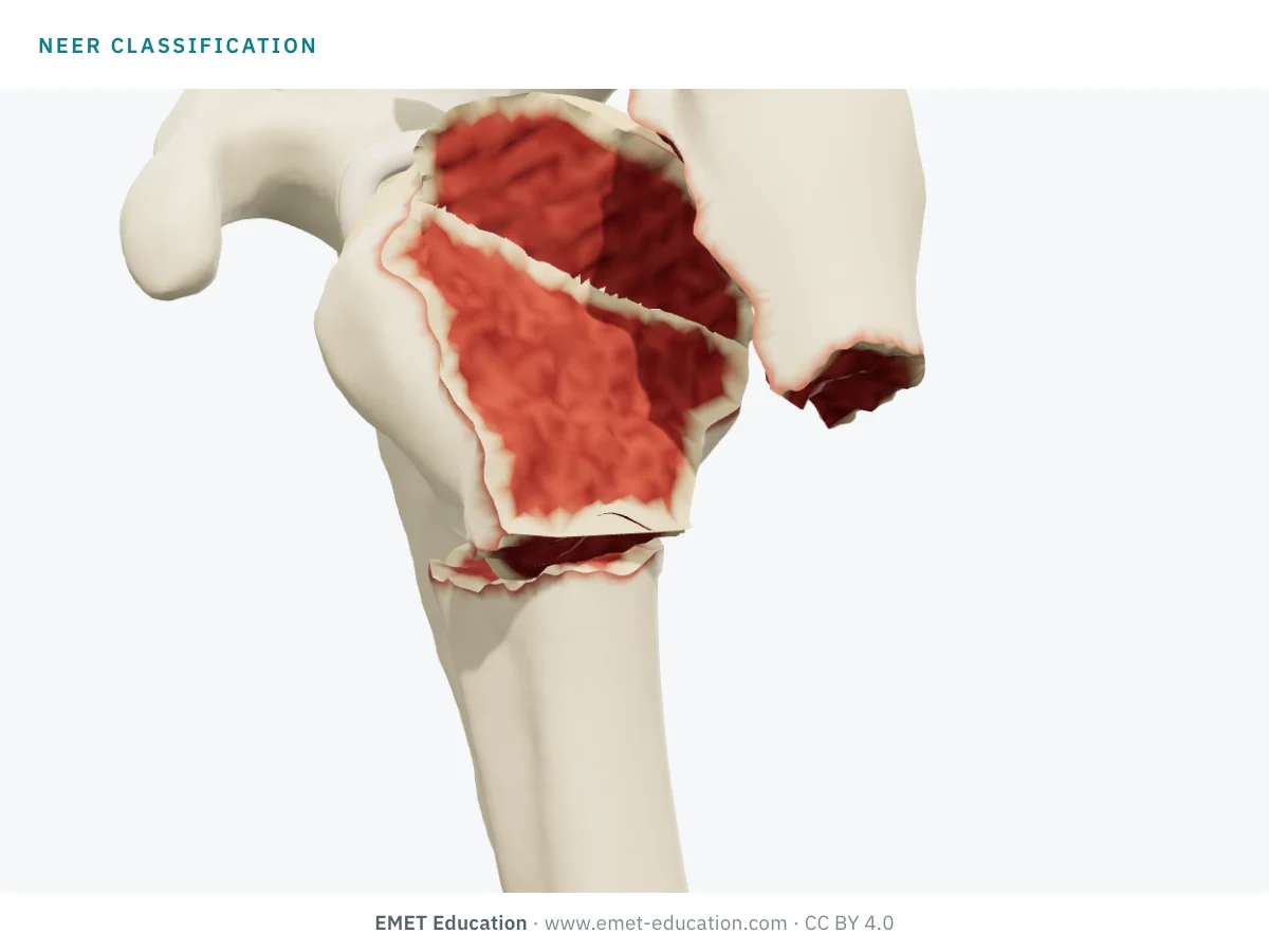
Which Neer category is this?
Download and cite
Free infographics, for teaching and publication
Use them in a course, a presentation, a social media post or a scientific article, under the Creative Commons Attribution 4.0 license: free reuse, including commercial, provided EMET Education is credited with a link to this page.
Cite this figure
Ready to paste. For a journal figure, add the original reference (Neer, 1970).
EMET Education. Neer classification of proximal humerus fractures: 3D visualization [Internet]. 2026 [cited]. Available from: https://www.emet-education.com/en/classifications/neer-proximal-humerus/. CC BY 4.0.EMET Education. (2026). Neer classification of proximal humerus fractures: 3D visualization [Infographic]. https://www.emet-education.com/en/classifications/neer-proximal-humerus/ (CC BY 4.0)<a href="https://www.emet-education.com/en/classifications/neer-proximal-humerus/"><img src="https://www.emet-education.com/en/classifications/neer-proximal-humerus/img/infographic.webp" alt="Neer classification of proximal humerus fractures in 3D" width="960"></a><br>Figure: <a href="https://www.emet-education.com/en/classifications/neer-proximal-humerus/">EMET Education</a>, CC BY 4.0Questions
Frequently asked questions
What is a "part" in the Neer classification?
One of the four segments of the proximal humerus (head, greater tuberosity, lesser tuberosity, shaft), when it is displaced more than 1 cm or angulated more than 45° relative to the others. A fragment separated by a fracture line but left in place does not count as a part.
Does a "one-part" fracture have only one fracture line?
No. It may have several lines, between all the segments; it is "one-part" because no segment is displaced beyond the thresholds. It is the most common pattern: about half of proximal humerus fractures.
How does it relate to the AO/OTA classification?
Since the 2018 revision, the AO/OTA types of the proximal humerus are defined by Neer's parts: type A for two-part fractures, B for three-part, C for articular or four-part fractures. The AO/OTA code adds the site of the line and, through its modifiers, dislocation or impaction. The page on the AO/OTA classification of the proximal humerus shows them in 3D, code by code.
Is it the same "Neer classification" as the clavicle one?
No. Neer also described a classification of distal clavicle fractures, by the position of the fracture line relative to the coracoclavicular ligaments, and the stages of subacromial impingement. This page covers fractures of the proximal humerus.
Does the number of parts decide on surgery?
Not on its own. For most displaced fractures in adults, trials have not shown better function after surgery than after nonoperative treatment. The decision takes into account age, bone quality, functional demand, dislocation and the risk of head necrosis, not just the category.
Can I reuse these images in a course or an article?
Yes. The images and infographics on this page are under the Creative Commons Attribution 4.0 license: free reuse, including commercial, with credit to EMET Education and a link to this page. The "Cite this figure" box gives ready-to-paste citations.
How were the 3D images made?
They are renderings of the EMET Education proximal humerus fracture model, set to each category: the fragments are cut from a humerus taken from CT, along the anatomical neck, the surgical neck and the bicipital groove, then displaced the way the muscles attached to them pull. It is a schematic model for teaching and patient explanation; it does not reproduce a radiograph.
Sources
Author and references
- Neer CS 2nd. Displaced proximal humeral fractures. I. Classification and evaluation. J Bone Joint Surg Am. 1970;52(6):1077–89. doi:10.2106/00004623-197052060-00001
- Neer CS 2nd. Displaced proximal humeral fractures. II. Treatment of three-part and four-part displacement. J Bone Joint Surg Am. 1970;52(6):1090–103. doi:10.2106/00004623-197052060-00002
- Neer CS 2nd. Four-segment classification of proximal humeral fractures: purpose and reliable use. J Shoulder Elbow Surg. 2002;11(4):389–400. doi:10.1067/mse.2002.124346
- Codman EA. The Shoulder: Rupture of the Supraspinatus Tendon and Other Lesions in or about the Subacromial Bursa. Boston: Thomas Todd; 1934.
- Brorson S, Eckardt H, Audigé L, Rolauffs B, Bahrs C. Translation between the Neer- and the AO/OTA-classification for proximal humeral fractures: do we need to be bilingual to interpret the scientific literature? BMC Res Notes. 2013;6:69. doi:10.1186/1756-0500-6-69
- Sidor ML, Zuckerman JD, Lyon T, Koval K, Cuomo F, Schoenberg N. The Neer classification system for proximal humeral fractures. An assessment of interobserver reliability and intraobserver reproducibility. J Bone Joint Surg Am. 1993;75(12):1745–50. doi:10.2106/00004623-199312000-00002
- Siebenrock KA, Gerber C. The reproducibility of classification of fractures of the proximal end of the humerus. J Bone Joint Surg Am. 1993;75(12):1751–5. doi:10.2106/00004623-199312000-00003
- Bernstein J, Adler LM, Blank JE, Dalsey RM, Williams GR, Iannotti JP. Evaluation of the Neer system of classification of proximal humeral fractures with computerized tomographic scans and plain radiographs. J Bone Joint Surg Am. 1996;78(9):1371–5. doi:10.2106/00004623-199609000-00012
- Sjödén GO, Movin T, Güntner P, et al. Poor reproducibility of classification of proximal humeral fractures. Additional CT of minor value. Acta Orthop Scand. 1997;68(3):239–42. doi:10.3109/17453679708996692
- Brorson S, Hróbjartsson A. Training improves agreement among doctors using the Neer system for proximal humeral fractures in a systematic review. J Clin Epidemiol. 2008;61(1):7–16. doi:10.1016/j.jclinepi.2007.04.014
- Brorson S, Bagger J, Sylvest A, Hróbjartsson A. Improved interobserver variation after training of doctors in the Neer system. A randomised trial. J Bone Joint Surg Br. 2002;84(7):950–4. doi:10.1302/0301-620x.84b7.13010
- Majed A, Macleod I, Bull AM, et al. Proximal humeral fracture classification systems revisited. J Shoulder Elbow Surg. 2011;20(7):1125–32. doi:10.1016/j.jse.2011.01.020
- Brorson S, Olsen BS, Frich LH, et al. Surgeons agree more on treatment recommendations than on classification of proximal humeral fractures. BMC Musculoskelet Disord. 2012;13:114. doi:10.1186/1471-2474-13-114
- Marongiu G, Leinardi L, Congia S, Frigau L, Mola F, Capone A. Reliability and reproducibility of the new AO/OTA 2018 classification system for proximal humeral fractures: a comparison of three different classification systems. J Orthop Traumatol. 2020;21(1):4. doi:10.1186/s10195-020-0543-1
- Matsumura N, Furuhata R, Seto T, et al. Reproducibility of the modified Neer classification defining displacement with respect to the humeral head fragment for proximal humeral fractures. J Orthop Surg Res. 2020;15(1):438. doi:10.1186/s13018-020-01966-2
- Court-Brown CM, Garg A, McQueen MM. The epidemiology of proximal humeral fractures. Acta Orthop Scand. 2001;72(4):365–71. doi:10.1080/000164701753542023
- Jakob RP, Miniaci A, Anson PS, Jaberg H, Osterwalder A, Ganz R. Four-part valgus impacted fractures of the proximal humerus. J Bone Joint Surg Br. 1991;73(2):295–8. doi:10.1302/0301-620X.73B2.2005159
- Hertel R, Hempfing A, Stiehler M, Leunig M. Predictors of humeral head ischemia after intracapsular fracture of the proximal humerus. J Shoulder Elbow Surg. 2004;13(4):427–33. doi:10.1016/j.jse.2004.01.034
- Rangan A, Handoll H, Brealey S, et al. Surgical vs nonsurgical treatment of adults with displaced fractures of the proximal humerus: the PROFHER randomized clinical trial. JAMA. 2015;313(10):1037–47. doi:10.1001/jama.2015.1629
- Handoll HH, Elliott J, Thillemann TM, Aluko P, Brorson S. Interventions for treating proximal humeral fractures in adults. Cochrane Database Syst Rev. 2022;6(6):CD000434. doi:10.1002/14651858.CD000434.pub5
- Meinberg EG, Agel J, Roberts CS, Karam MD, Kellam JF. Fracture and Dislocation Classification Compendium—2018. J Orthop Trauma. 2018;32 Suppl 1:S1–S170. doi:10.1097/BOT.0000000000001063
- Resch H, Tauber M, Neviaser RJ, et al. Classification of proximal humeral fractures based on a pathomorphologic analysis. J Shoulder Elbow Surg. 2016;25(3):455–62. doi:10.1016/j.jse.2015.08.006
- Spek RWA, Schoolmeesters BJA, Oosterhoff JHF, et al. 3D-printed handheld models do not improve recognition of specific characteristics and patterns of three-part and four-part proximal humerus fractures. Clin Orthop Relat Res. 2022;480(1):150–9. doi:10.1097/CORR.0000000000001921
- Bougher H, Buttner P, Smith J, et al. Interobserver and intraobserver agreement of three-dimensionally printed models for the classification of proximal humeral fractures. JSES Int. 2021;5(2):198–204. doi:10.1016/j.jseint.2020.10.019
- Iordens GI, Mahabier KC, Buisman FE, et al. The reliability and reproducibility of the Hertel classification for comminuted proximal humeral fractures compared with the Neer classification. J Orthop Sci. 2016;21(5):596–602. doi:10.1016/j.jos.2016.05.011
- Brorson S, Bagger J, Sylvest A, Hróbjartsson A. Diagnosing displaced four-part fractures of the proximal humerus: a review of observer studies. Int Orthop. 2009;33(2):323–7. doi:10.1007/s00264-008-0591-2
3D models
Show the fracture to your patient
The images on this page come from an interactive model, the proximal humerus fracture 3D model. In clinic, choose the category and set the displacement of each part from your patient's imaging: Neer is recalculated on its thresholds. Then show the proposed treatment: nonoperative care, nail, locking plate or reverse arthroplasty.
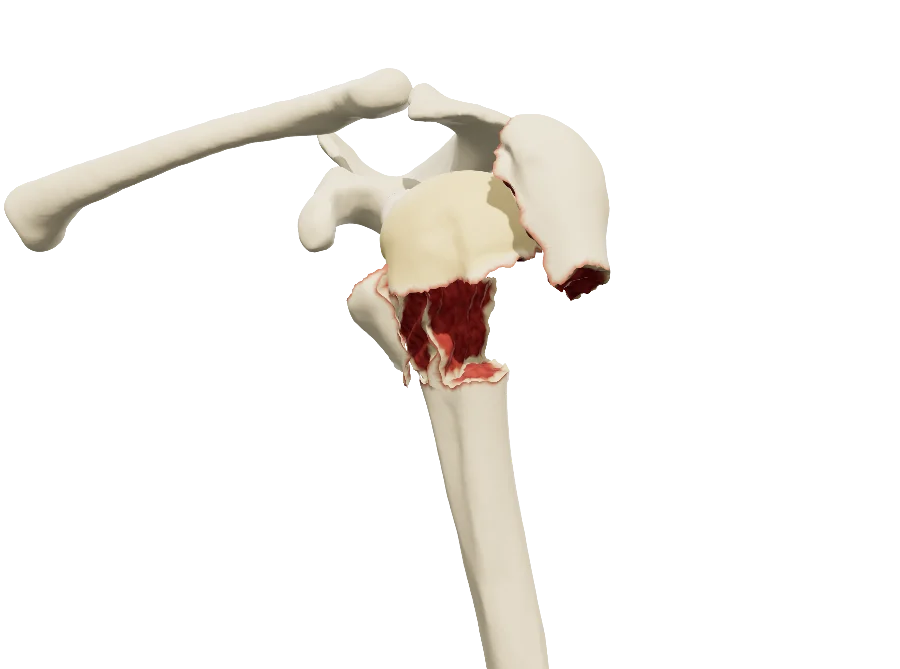 ShoulderProximal humerus fractureNeer and AO/OTA, displacement part by part, dislocation, healing, nail, plate and reverse arthroplasty.
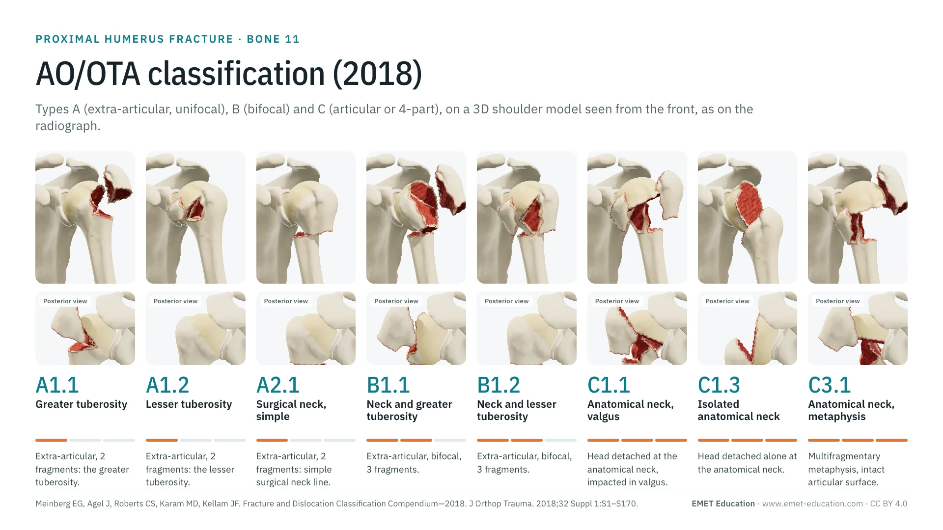 ClassificationsAO/OTA classification of the proximal humerusThe 2018 types A, B and C, code by code, in 3D.
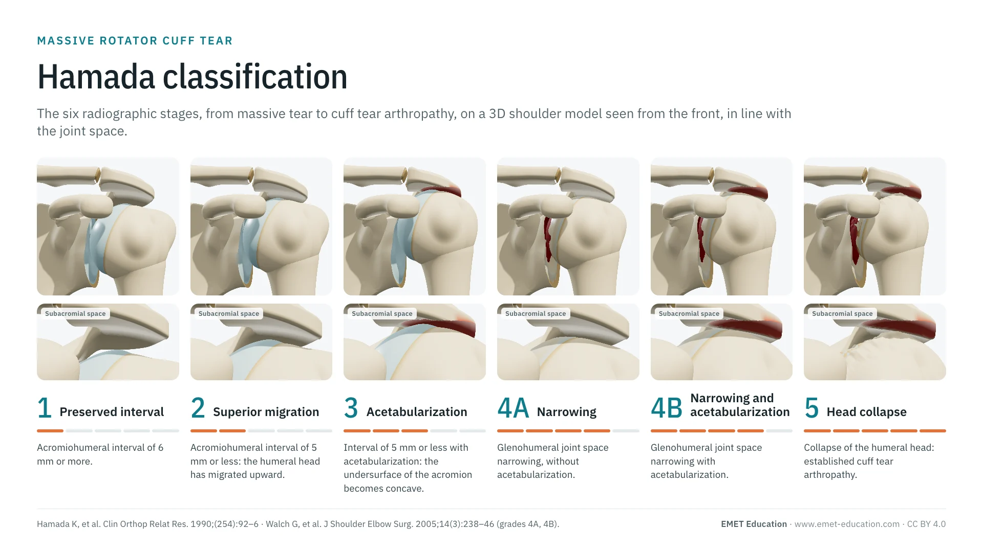 ClassificationsAll 3D classificationsArranged by joint.
Explain the fracture in 3D
The proximal humerus fracture model opens in your browser, at the category you choose, with no sign-up.
Share this page with a colleague or a resident: