- EMET Education
- 3D classifications
- Mason classification (radial head)
Classification · Radial head fracture
Mason Classification of Radial Head Fractures in 3D
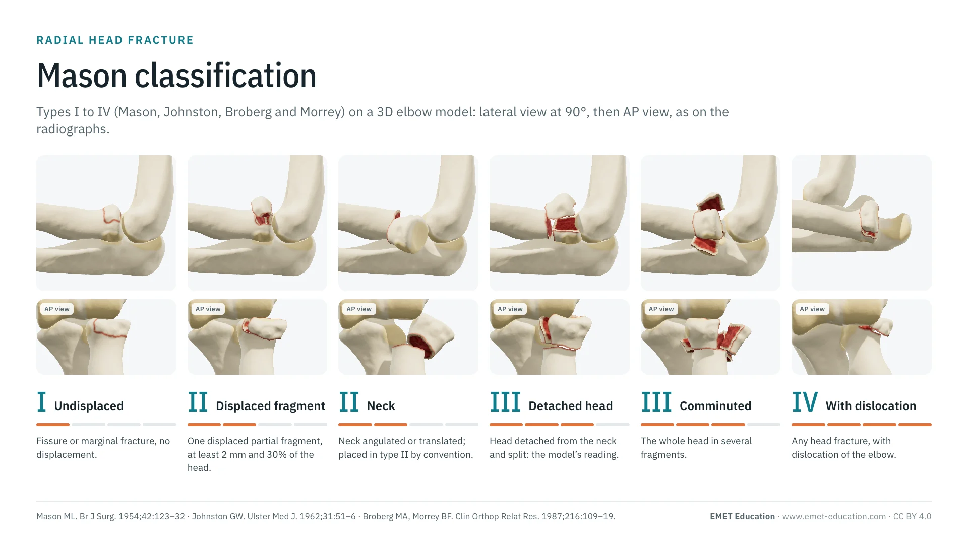
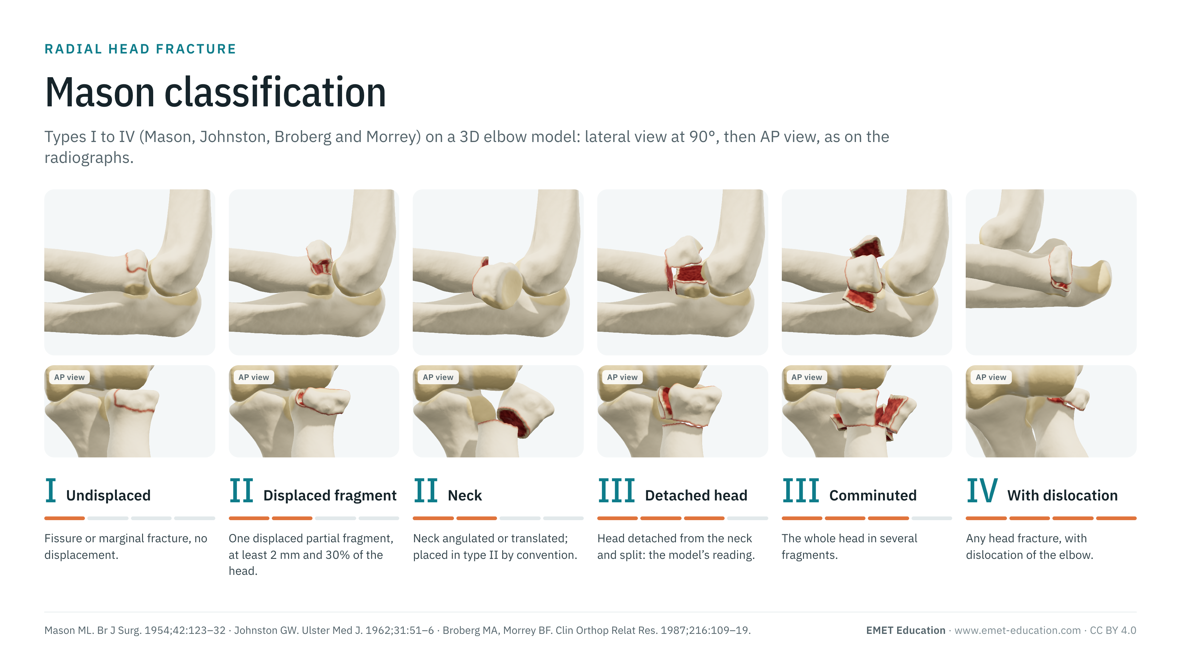
Mason types I to IV, with the modifications of Johnston and of Broberg and Morrey, on the radial head fracture 3D model, seen in lateral view at 90° and AP view as on elbow radiographs. With the 1954 paper, reliability data, related classifications (Hotchkiss, AO/OTA, the Mayo suffixes), the link with treatment, grading pitfalls and free infographics for teaching and publication.
Designed by an orthopedic surgeon
The 3D model opens in your browser, at the type shown, with no sign-up. Infographics are free to reuse under CC BY 4.0.
- 1954Mason's paper in the British Journal of Surgery: one hundred radial head fractures (Mason)
- 3 → 4Types in Mason's paper (1954), four since Johnston (1962): type IV is not Mason's (Johnston)
- 956Citations of Mason's paper (OpenAlex, October 31, 2026)
- κ 0.33–0.56Interobserver agreement on radiographs depending on the study (Ayyaswamy, Matsunaga, Calderazzi)
The types
Types I to IV, in 3D
Each image is a rendering of the EMET Education radial head fracture model at that type, left elbow seen in lateral view at 90° of flexion, the head beneath the capitellum and the anterior fragment above it, with a narrow field to approach the projection of a lateral radiograph; the infographic adds the AP view. The model draws seven cases covering the four types: type II has three (single fragment, multiple fragments, neck) and type III two (detached head, comminuted). The displacements shown are typical cases, adjustable in the model. Under each type, the closest AO/OTA 2018 code according to the model.
-
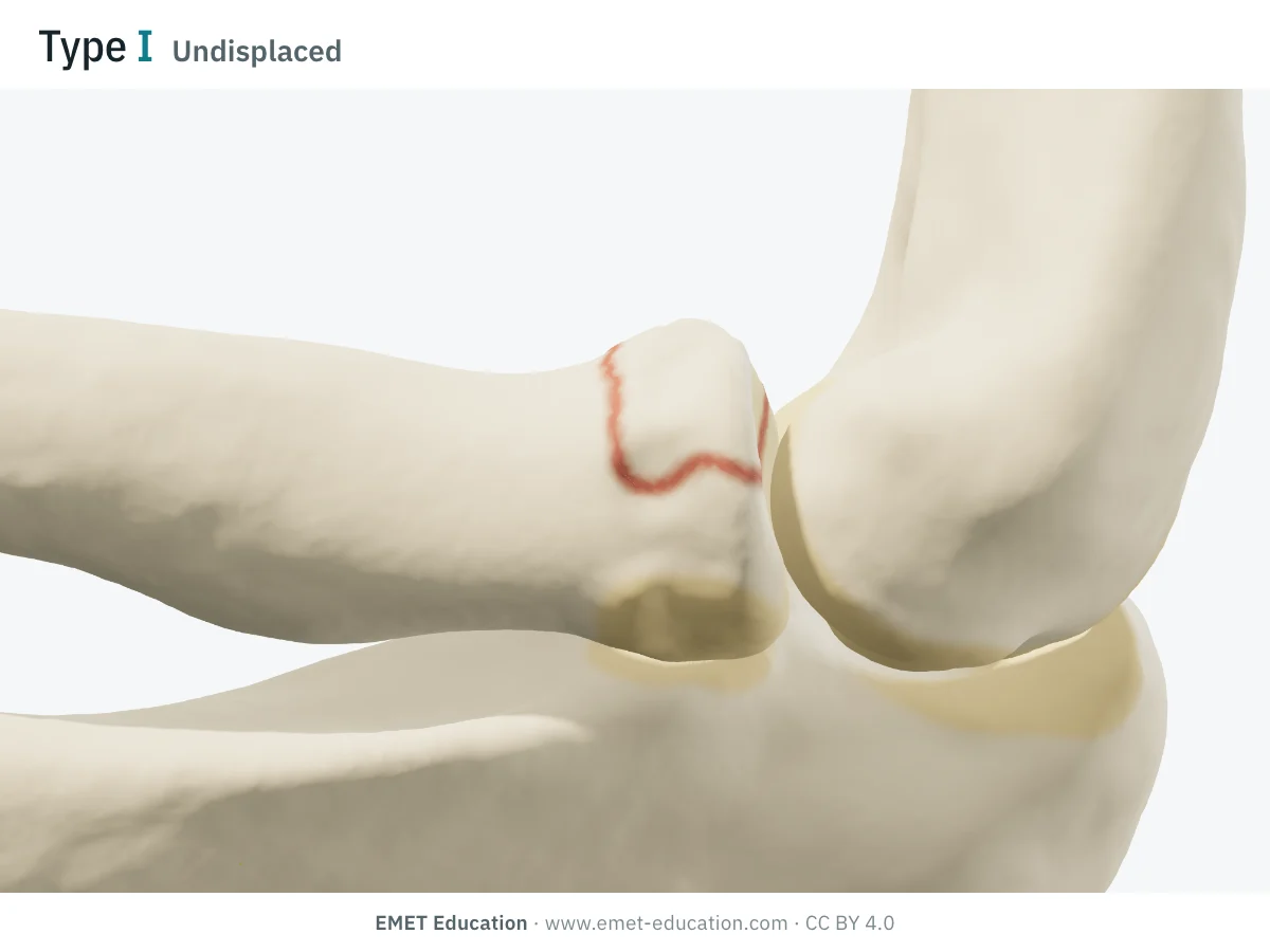
IUndisplaced
- Definition
- Fissure or marginal fracture of the head with no displacement: one line, no step-off (Iannuzzi and Leopold). Since Broberg and Morrey, a displaced partial fragment of less than 2 mm, or involving less than 30% of the articular surface, remains type I (van der Windt et al.).
- AO/OTA 2018
- 2R1B1 (partial articular, simple; AO Surgery Reference), with universal modifier [1] (undisplaced; compendium)
- In the 3D model
- A 120° anterolateral wedge of the head, fissured, with no displacement at all.
-
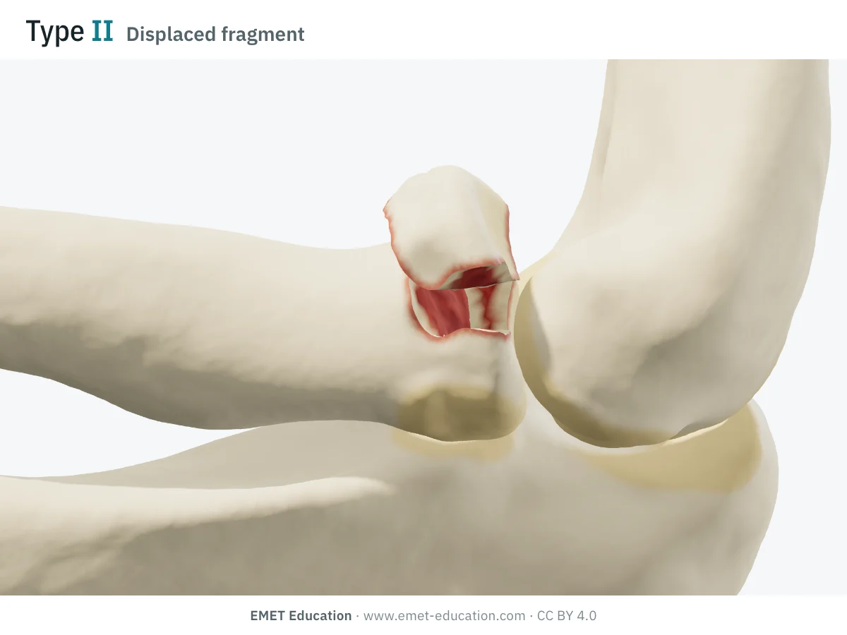
IIDisplaced partial fragment
- Definition
- Fracture of a marginal sector of the head, displaced: depression, tilt or step-off. The threshold attributed to Broberg and Morrey is at least 2 mm of displacement and at least 30% of the articular surface (van der Windt et al.). The fragment is almost always anterolateral, with the forearm in neutral rotation: 22 of 24 on CT (van Leeuwen et al.), 98% of 66 fractures (Mellema et al.).
- AO/OTA 2018
- 2R1B1 (partial articular, simple), universal modifier [2] (displaced)
- In the 3D model
- Anterolateral fragment of 135° (about 34% of the head), displaced 3 mm, tilted 20° and depressed 1 mm. The model reads type I again if the fragment is brought back under 2 mm or under 30% of the head.
-
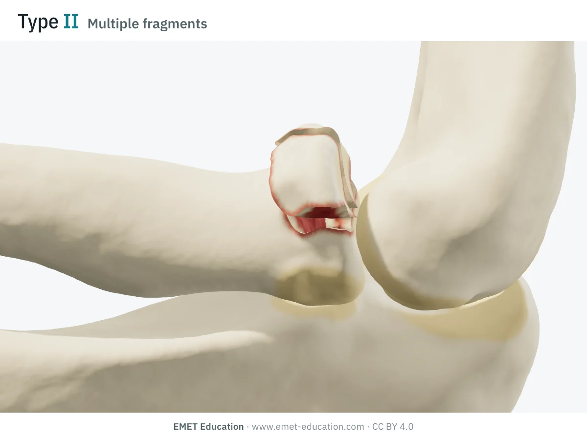
IIMultiple fragments
- Definition
- Displaced partial fracture whose sector is itself fragmented (2 or 3 pieces), the rest of the head remaining attached to the neck. Hotchkiss places these fractures in type II as long as they are reconstructable; Mason reserved type III for the whole head being comminuted (van der Windt et al.).
- AO/OTA 2018
- 2R1B3 (partial articular, multifragmentary)
- In the 3D model
- A 135° sector in 2 fragments (3 if chosen), displaced 3 mm, tilted 15° and depressed 1.5 mm.
-
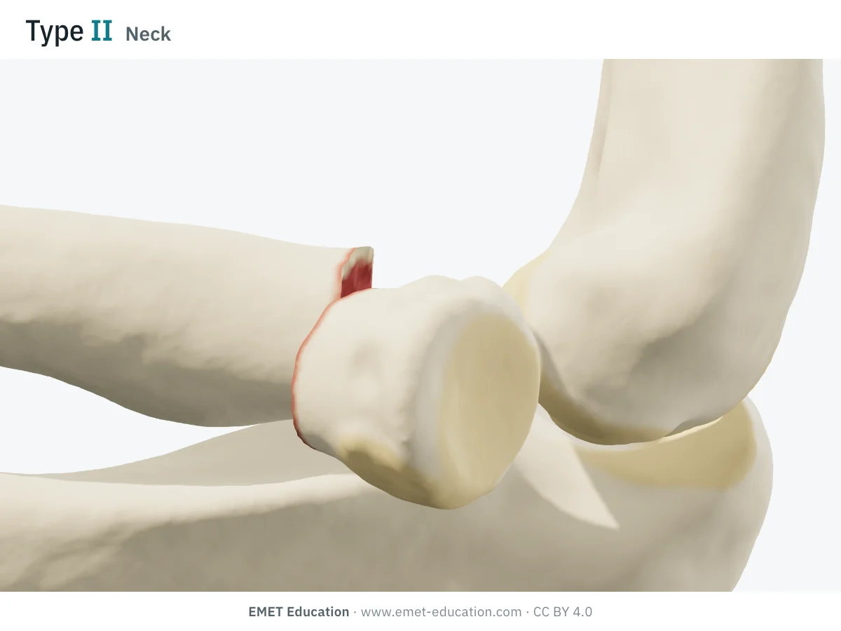
IIRadial neck
- Definition
- Fracture of the neck, the head angulated or translated on the neck. Mason described only the head: reviews place a displaced or angulated neck in type II (Mason–Hotchkiss), by convention (van der Windt et al.). No angulation threshold is established in adults; some authors tolerate up to 4 mm of displacement.
- AO/OTA 2018
- 2R1A2 (extra-articular, simple neck) (AO Surgery Reference)
- In the 3D model
- The head tilts 35° outward, translated 2 mm. The model reads type II from 30° or 2 mm: this is a choice of the model, not a published threshold.
-
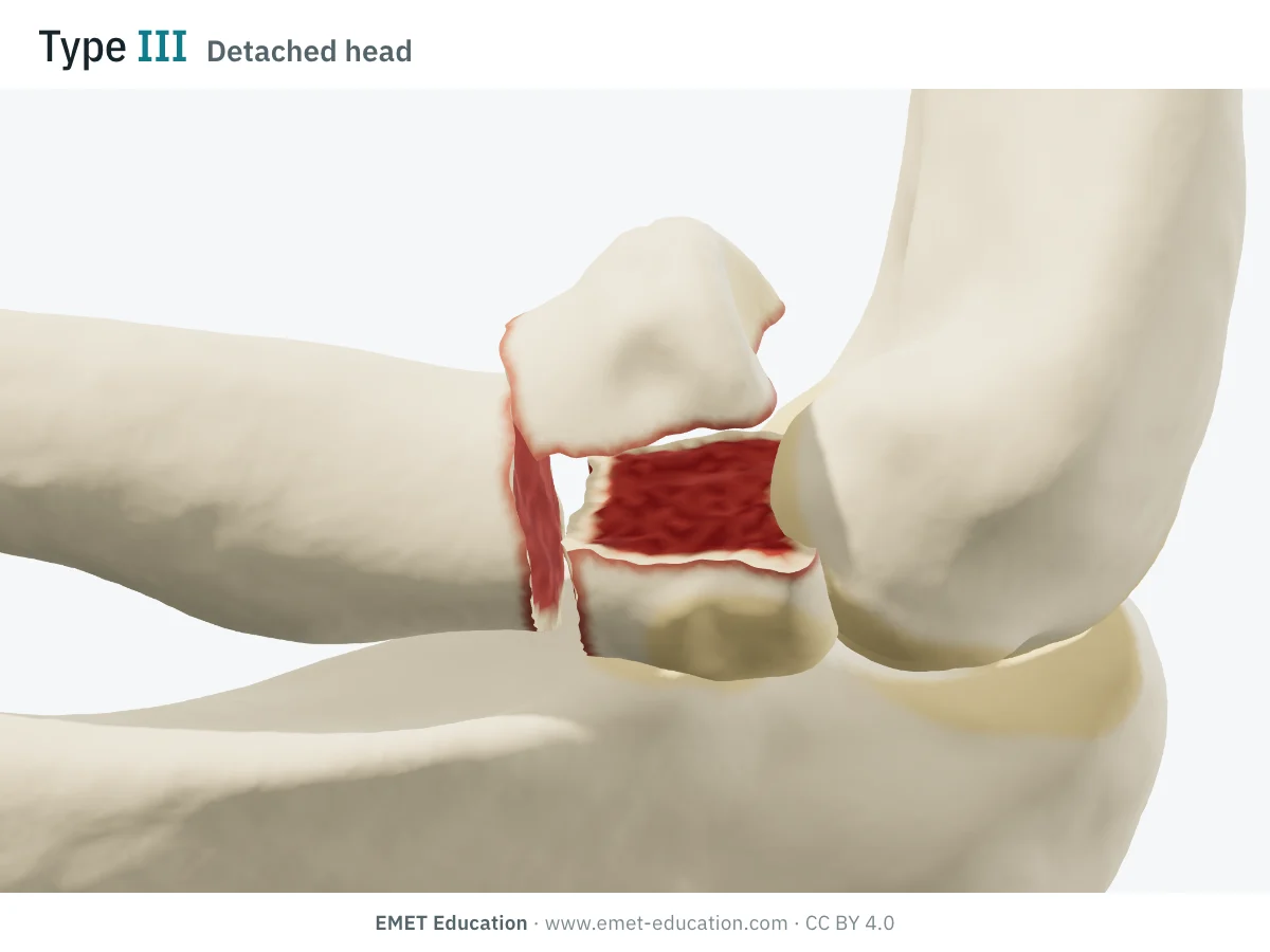
IIIHead detached from the neck
- Definition
- The head, separated from the neck, is also split: a complete articular fracture. Mason does not describe this case separately. EMET Education's reading: type III, since the whole head is involved; in Hotchkiss's scheme, the grade depends on whether it can be reconstructed.
- AO/OTA 2018
- 2R1C1 (complete articular, simple) (AO Surgery Reference)
- In the 3D model
- Fractured neck (3 mm gap, 10° angulation, 1 mm translation) and head split in two. The model's choice: type III.
-
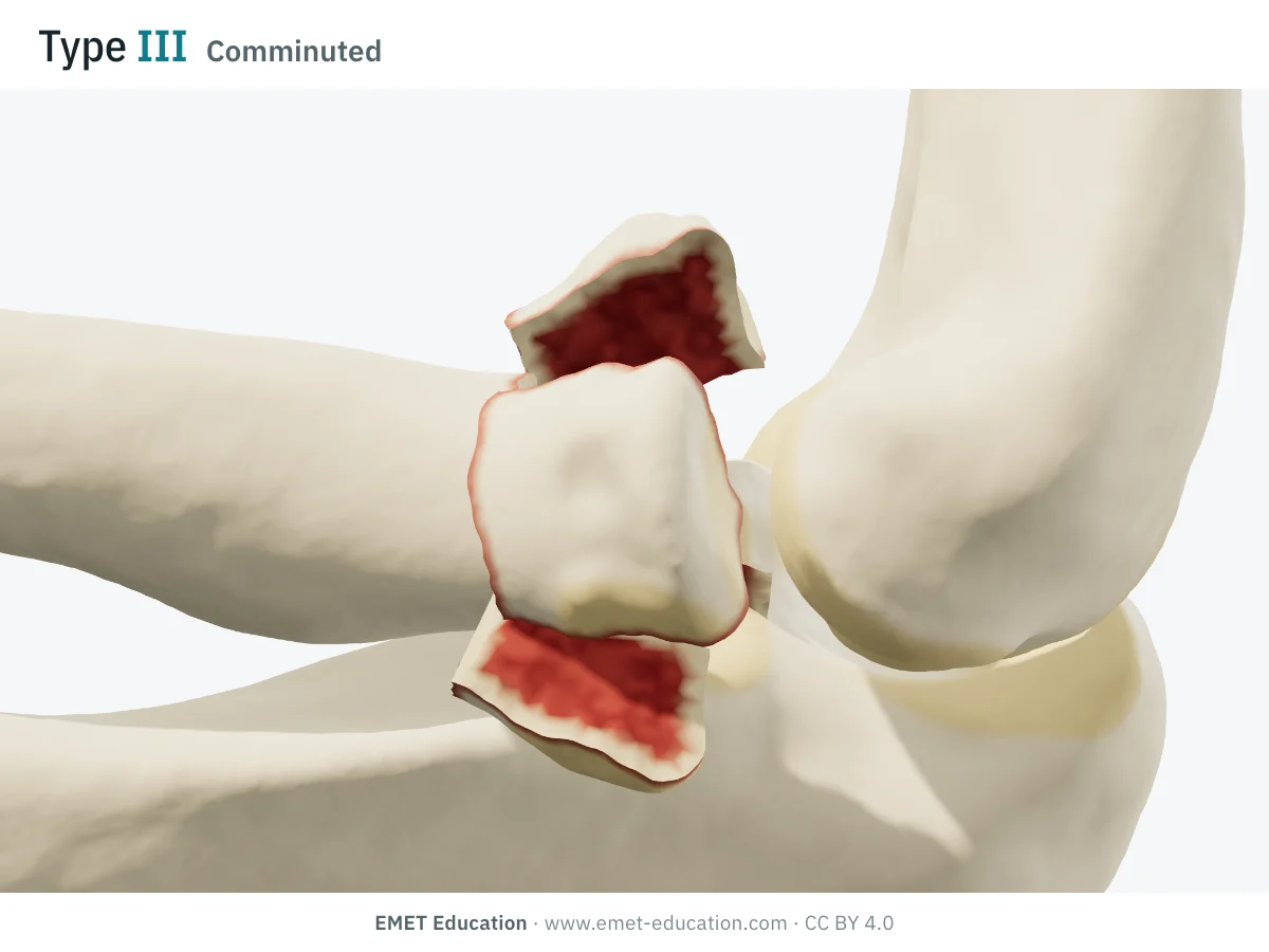
IIIComminuted
- Definition
- Comminuted fracture of the whole head, displaced: Mason's type III, for which the head was then excised. Beyond 3 articular fragments, fixation becomes unreliable (13 of 14 unsatisfactory results: Ring et al.), which Hotchkiss expresses as "not reconstructable".
- AO/OTA 2018
- 2R1C3 (complete articular, multifragmentary) (AO Surgery Reference)
- In the 3D model
- The neck and four fragments of the head (3 to 5 if chosen), displaced 4 mm, tilted 20° and depressed 2 mm.
-
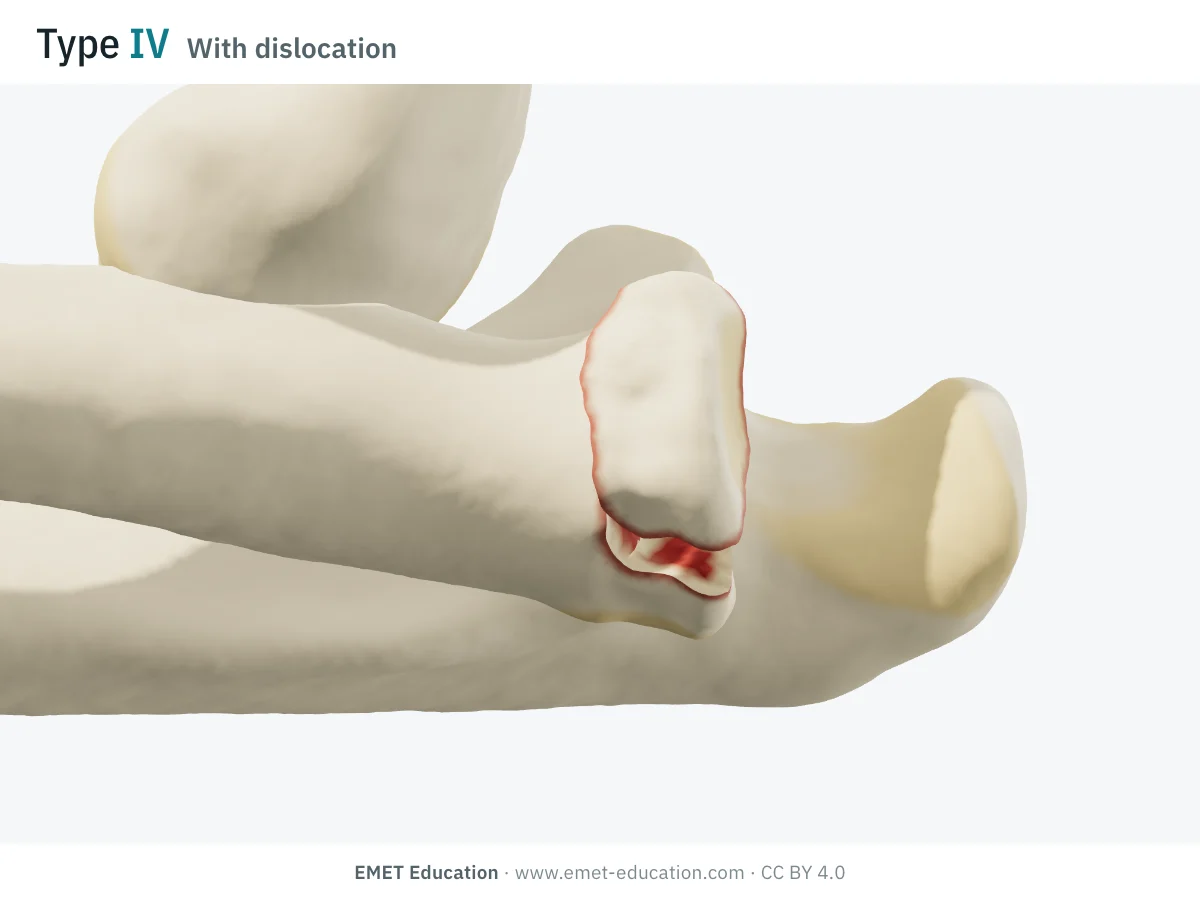
IVWith elbow dislocation
- Definition
- Radial head fracture, of any type, with elbow dislocation. This type is not Mason's: Johnston added it in 1962, whatever the displacement and comminution; Broberg and Morrey took it up in 1987 (Johnston, Broberg and Morrey). It groups injuries of very different severity, from the isolated fracture to the terrible triad.
- AO/OTA 2018
- The AO code of the head fracture, with the universal dislocation modifier [5] (compendium)
- In the 3D model
- Here a displaced 135° partial fragment and the elbow dislocated posteriorly and laterally. The model reads type IV as soon as the elbow is dislocated, whatever the head fracture.
Schematic rendering, for teaching: the model illustrates each type on a single elbow, it does not reproduce a radiograph. Definitions reworded from the reviews (Iannuzzi and Leopold, van der Windt et al., Calderazzi et al.); Mason's figures are not reproduced. The bar shows the type: one segment for I, four for IV.
The original paper
From Cutler (1926) to Mason (1954), then Johnston (1962)
In 1954, Mark L. Mason, of the Isle of Thanet Groups of Hospitals, published in the British Journal of Surgery a review of one hundred radial head fractures (Mason, 1954). Simpler schemes existed: Cutler, in 1926, already separated the undisplaced crack, the single detached fragment and the multiple fragments, with the neck forming a fourth category (Cutler, as reported by Calderazzi et al.). Mason's scheme pairs each type with a course of action.
Mason's three types, as the reviews summarize them, then what his successors added:
- Type I: fissure or marginal fracture, with no displacement;
- Type II: fracture of a marginal sector of the head, with displacement;
- Type III: comminuted fracture of the whole head (Iannuzzi and Leopold, Calderazzi et al.);
- Type IV, added by Johnston (Ulster Medical Journal, 1962): head fracture accompanied by elbow dislocation, whatever the displacement and comminution; taken up by Broberg and Morrey in 1987, in a series of 24 fracture-dislocations (Johnston, Broberg and Morrey, van der Windt et al.);
- Mason's treatment: mobilization for type I, excision of the head for type III, case by case for type II (Iannuzzi and Leopold, Calderazzi et al.). The threshold of 2 mm and 30% of the articular surface is attributed to Broberg and Morrey (1987); it is not in Mason's definition as the reviews report it.
The context has changed since 1954. In 1997, Hotchkiss argued against routine excision of the displaced head in young, active patients: better imaging, exposure and fixation techniques often allow it to be preserved (Hotchkiss). Epidemiologically, radial head fracture affects 2.8 per 10,000 people a year, at a mean age of 48, more often women (Kaas et al.). Distributions vary with recruitment: in a series of 333 adults from a referral center, 67% of fractures were type I, 14% type II and 19% type III, with a dislocation in 14% (van Riet et al., 2005); in a prospective series of 201 head or neck fractures, 51% were type I, 41% type II, 5% type III and 2.5% type IV (Duckworth et al.).
Reliability
How reproducible is the classification?
Moderately, on radiographs. On 90 radiographs read by nine observers, Calderazzi et al. found "moderate" interobserver agreement (κ of 0.49 and 0.50 in the two sessions): κ of 0.40 to 0.45 for types II and III, 0.50 to 0.53 for I and 0.65 to 0.69 for IV, the easiest to recognize. Disagreements almost always oppose two adjacent types (I and II, II and III).
| Study | Measure | Value |
|---|---|---|
| Morgan et al., 1997 | Mason, 20 surgeons, 25 sets of AP and lateral radiographs, two sessions: complete agreement of readers | 16% of cases |
| Matsunaga et al., 2009 | Mason, 4 observers, 3 sessions: inter- then intraobserver kappa (AO/ASIF: 0.25–0.48 · 0.48) | 0.43–0.56 · 0.58 |
| Sheps et al., 2009 | Hotchkiss, 5 observers, 43 patients: interobserver kappa, then with types II and III combined (AO: 0.26 · 0.46) | 0.59 · 0.76 |
| Guitton and Ring, 2011 | Broberg-Morrey, 85 surgeons, 12 fractures: radiographs with 3D CT, then with 2D CT (agreement graded, kappa not given in the abstract) | moderate · fair |
| Ayyaswamy et al., 2019 | Mason, 5 observers, 30 fractures, AP and lateral radiographs: inter- then intraobserver kappa (Charalambous: 0.42 · 0.49) | 0.33 · 0.43 |
| Calderazzi et al., 2025 | Mason-Johnston, 9 observers, 90 radiographs: interobserver Fleiss kappa in the two sessions, four months apart | 0.49 · 0.50 |
3D CT improves agreement only a little: with 3D reconstruction, agreement moves from "fair" to "moderate", and disagreement remains considerable (Guitton and Ring). For Ayyaswamy et al., radiographs alone are not enough to classify these fractures reliably, for practice or for research. Type IV, defined by the dislocation, is the most reproducible; types II and III, which rely on measuring a displacement and counting fragments, are the least.
Variants and related classifications
Johnston, Broberg and Morrey, Hotchkiss, AO/OTA, Mayo
"Mason III" does not mean the same thing for Mason (whole head comminuted), for Broberg and Morrey (likewise) and for Hotchkiss (not reconstructable, whatever the number of fragments). State the version in any publication: one also finds "Mason–Hotchkiss" or "modified Mason" with no further detail.
The main variants and related classifications:
| Classification | What it describes | Note |
|---|---|---|
| Cutler, 1926 | Undisplaced crack; single detached fragment; multiple fragments; the neck apart | Forerunner of Mason, as reported by Calderazzi et al. |
| Mason, 1954 | I: undisplaced fissure or marginal fracture; II: displaced marginal sector; III: whole head comminuted | No numerical threshold in the definition as the reviews report it. |
| Johnston, 1962 (Mason–Johnston) | Adds type IV: head fracture with elbow dislocation | The most reproducible of the four types (Calderazzi et al.), but it groups very different severities. |
| Broberg and Morrey, 1987 | Type II: at least 2 mm of displacement and at least 30% of the articular surface; takes up Johnston's type IV | The version read by the model. Threshold attributed to these authors by the reviews. |
| Hotchkiss, 1997 | I: undisplaced or minimally displaced (under 2 mm), no block; II: displaced (often over 2 mm) or angulated neck, with or without mechanical block, reconstructable; III: comminuted, not reconstructable, excision or replacement | Reasons in terms of management. Interobserver agreement "moderate" (κ 0.59), "substantial" when II and III are combined (0.76). |
| AO/OTA 2018, segment 2R1 (compendium, AO Surgery Reference) | A: extra-articular (including A2, simple neck); B: partial articular (B1 simple, B3 multifragmentary); C: complete articular (C1 simple, C3 multifragmentary) | Morphological code: displacement and dislocation are modifiers. Less reproducible than Mason in two studies (κ 0.26 against 0.59; 0.25–0.48 against 0.43–0.56). The 2R1 codes, one by one, in 3D → |
| Mayo suffixes (van Riet and Morrey, 2008) | The Mason type, followed by letters for associated injuries: c coronoid, o olecranon, l LCL, m MCL, d distal radioulnar joint | Reported reproducibility 98%; 88 associated injuries documented in 333 fractures (26%). The model adds them to its reading. |
| Regan and Morrey, 1989 (coronoid) | I: tip; II: 50% of the process or less; III: more than 50% | Dislocation or associated fracture in 14, 56 and 80% of cases: look for the coronoid in any head fracture with dislocation. |
| Charalambous | Four morphological groups, plus displaced or undisplaced | Classifies more fractures than Mason; interobserver κ 0.42 against 0.33 (Ayyaswamy et al.). |
Use
In research and in clinical practice
In research
The definition of type II sets the inclusion of trials: the RAMBO trial protocol retains partial fractures of at least one third of the articular surface, with at least 2 mm of step-off and less than 2 mm of gap between fragments (Bruinsma et al.). The randomized trial by Mulders et al. (45 adults, isolated type II) shows no functional difference at one year between fixation and nonoperative treatment (DASH at 12 months: 0.0 against 1.7; p = 0.076). In a series of 201 head or neck fractures, age, AO/OTA complexity, radiographic comminution and the choice to operate predicted a poorer result, and 93% of patients had an excellent or good result (Duckworth et al.).
In clinical practice: from type to treatment
Type I. Nonoperative treatment and early mobilization, when forearm rotation is not blocked. A hemarthrosis can mimic a block: an anesthetic injection into the joint, or a repeat examination a few days later, settles it (van der Windt et al.).
Type II. A mechanical block to forearm rotation is a consensus indication for surgery. Without a block, nothing is settled: the review by Kaas et al. (9 level IV series, 224 patients) found 80% success without surgery (114 of 142) and 93% with fixation (76 of 82), without being able to conclude; in a retrospective series, 2 or 3 mm of step-off treated without surgery did not change the results (Furey et al.). Comminution (more than two fragments) is generally an indication for surgery (van der Windt et al.).
Type III. Fix, replace or excise. With more than three articular fragments, fixation almost always fails (Ring et al.); in the meta-analysis by De Mauro et al. (three prospective studies, 169 patients), complications are more frequent after fixation (26 events in 65) than after arthroplasty (8 in 70). Simple excision is not an option in the presence of a coronoid fracture or a ligament injury (van der Windt et al.).
Type IV. Reduce the ulnohumeral joint early and treat the head according to its type; avoid more than four weeks of immobilization (24 elbows, Broberg and Morrey). When the coronoid is fractured, this is the terrible triad: of 11 elbows, 7 redislocated in a splint, and 5 after surgery, including all 4 treated by excision of the head (Ring et al., 2002).
The injuries the type does not tell
The type describes the head, not what surrounds it. In the series by van Riet et al., 16% of fractures had a coronoid fracture and 14% a dislocation. Among 121 type II fractures, complete loss of cortical contact of at least one fragment went with a complex injury in 91% of cases, against 33% without it (odds ratio 21: Rineer et al.). On MRI of 24 type II or III fractures with no known dislocation, the lateral ulnar collateral ligament was not intact in 18, the medial collateral ligament in 13, and the capitellum had an osteochondral defect in 7 (Itamura et al.); the lateral collateral ligament was torn in all 62 elbows operated for dislocation or fracture-dislocation in another series (McKee et al.). Finally, the Essex-Lopresti lesion goes unnoticed: radial shortening of 2 to 4 mm in 22 of the 60 patients with wrist pain, among 237 proximal radius fractures (Duckworth et al., 2011); in cadavers, 3 mm of proximal radial migration under traction, after head excision, indicates disruption of the interosseous membrane (Smith et al.); of 31 injuries, 15 were recognized more than four weeks after the fracture, with a poorer result (Schnetzke et al.). These injuries are recorded with the Mayo suffixes (van Riet and Morrey); the model draws the coronoid, the dislocation, the Osborne-Cotterill lesion and the LCL tear, but neither the interosseous membrane nor Essex-Lopresti.
Grading pitfalls
Five pitfalls when grading a radial head fracture
- Two millimetres and 30%: a convention. These thresholds separate I from II, but they are not proven: 2 or 3 mm of step-off left unoperated did not change results (Furey et al.), and the mean fragment in high-energy fractures covers 42.5% of the surface (Capo et al.). State the threshold and what is measured (step-off or gap). The model applies both: a fragment brought back under 2 mm becomes type I again.
- The radiograph underestimates. On AP and lateral views, the number of fragments, central impaction and loss of cortical contact are not always visible; 3D CT improves agreement by only one notch (Guitton and Ring). State whether the type comes from the radiograph or from CT.
- Between two adjacent types. Disagreements almost always oppose I to II and II to III (Calderazzi et al.). Writing the measurement (millimetres, share of the head, number of fragments) beside the Roman numeral avoids arguments.
- Which version? "Mason III" means the comminuted head in Mason, and the non-reconstructable head in Hotchkiss, where a comminuted but repairable head is a II (van der Windt et al.). The neck is not in Mason: the angulated neck is placed in II by convention. And the model reads the head detached from the neck and split as III, which Mason does not say.
- What the type does not see. Type IV groups very different severities (Iannuzzi and Leopold), and the coronoid, LCL or interosseous membrane are not visible on the radiograph of the head: add the Mayo suffixes, examine the distal radioulnar joint and the wrist.
Self-test
Which Mason type?
The same elbow seen from the front at three-quarters, at the model's seven cases, in random order. Find the Mason type (the neck and the detached head as read on this page); the answer comes with the definition.
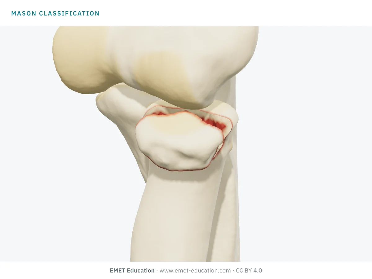
Which Mason type is this?
Download and cite
Free infographics, for teaching and publication
Use them in a course, a presentation, a social media post or a scientific article, under the Creative Commons Attribution 4.0 license: free reuse, including commercial, provided EMET Education is credited with a link to this page. The license covers EMET Education's images, not the classification itself.
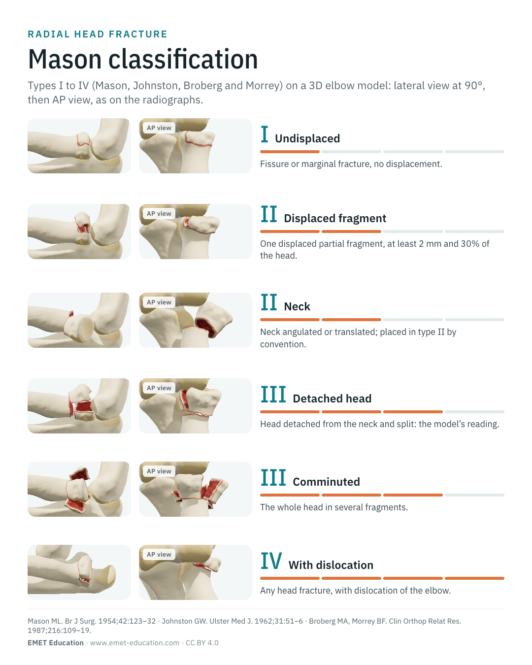
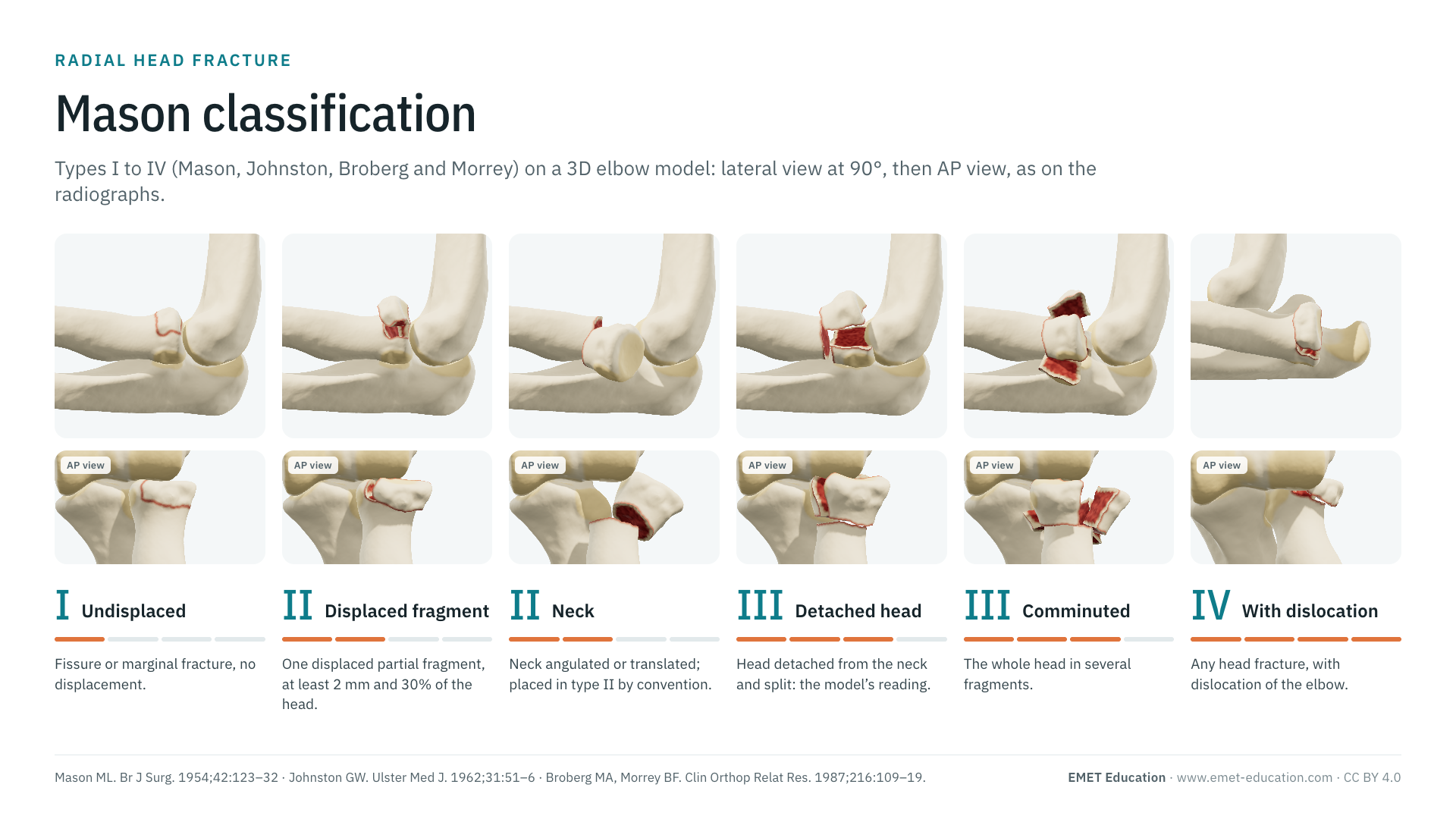
Cite this figure
Ready to paste. For a journal figure, add the original reference (Mason, 1954; Johnston, 1962 for type IV).
EMET Education. Mason classification of radial head fractures: 3D visualization [Internet]. 2026 [cited]. Available from: https://www.emet-education.com/en/classifications/mason-radial-head/. CC BY 4.0.EMET Education. (2026). Mason classification of radial head fractures: 3D visualization [Infographic]. https://www.emet-education.com/en/classifications/mason-radial-head/ (CC BY 4.0)<a href="https://www.emet-education.com/en/classifications/mason-radial-head/"><img src="https://www.emet-education.com/en/classifications/mason-radial-head/img/infographic.webp" alt="Mason classification of radial head fractures in 3D" width="960"></a><br>Figure: <a href="https://www.emet-education.com/en/classifications/mason-radial-head/">EMET Education</a>, CC BY 4.0Questions
Frequently asked questions
What is the difference between Mason II and Mason III?
Type II is a partial fracture: a marginal sector of the head breaks off and is displaced, and the rest of the head remains attached to the neck. Type III involves the whole head, in several displaced fragments. Hotchkiss prefers to reason on whether repair is possible: in his scheme, a comminuted but reconstructable head is a type II, and only the non-reconstructable head is a type III.
What does Mason type IV mean?
Mason described only three types in 1954. Type IV is Johnston's (1962): a radial head fracture accompanied by elbow dislocation, whatever the displacement and the number of fragments. Broberg and Morrey took it up in 1987, hence the names "Mason–Johnston" or "modified Mason". It groups injuries of very different severity; the coronoid and the lateral collateral ligament must be looked for.
How much displacement makes a radial head fracture type II?
In the modification attributed to Broberg and Morrey, a partial fragment is "displaced" (type II) if it has at least 2 mm of displacement and involves at least 30% of the articular surface. This is a convention: in a retrospective series, 2 or 3 mm of step-off treated without surgery did not change the results, and a mechanical block to forearm rotation is the indication for surgery on which there is consensus.
Mason, Hotchkiss, AO/OTA or Mayo: which one to use?
They complement each other. Mason or Hotchkiss to talk about the type and what to do (Hotchkiss: κ 0.59 against 0.26 for AO in the same study); AO/OTA 2R1 for registries, with its modifiers; the Mayo suffixes (c, o, l, m, d) to record associated injuries. Always state the version used.
Where should a radial neck fracture be classified?
Mason described only the head. Reviews place a displaced or angulated neck in type II (Mason–Hotchkiss), and the simple neck is 2R1A2 in AO/OTA 2018. No angulation threshold is established in adults; some authors tolerate up to 4 mm of displacement. The 3D model reads type II from 30° or 2 mm of translation: this is its own choice, not a published threshold.
Can I reuse these images in a course or an article?
Yes. The images and infographics on this page are under the Creative Commons Attribution 4.0 license: free reuse, including commercial, with credit to EMET Education and a link to this page. The "Cite this figure" box gives ready-to-paste citations.
How were the 3D images made? Do they reproduce Mason's drawings?
No, no figure from the papers is reproduced. They are renderings of the EMET Education radial head fracture model, set to each type: the fragments are cut from a radius taken from CT, at the site described by CT series (the anterolateral quadrant, per van Leeuwen et al. and Mellema et al.), then displaced and depressed. It is a schematic model for teaching; it does not reproduce a radiograph and does not draw the interosseous membrane or the annular ligament. EMET Education is affiliated neither with Mason nor with the AO Foundation.
Sources
Author and references
- Mason ML. Some observations on fractures of the head of the radius with a review of one hundred cases. Br J Surg. 1954;42(172):123–32. doi:10.1002/bjs.18004217203
- Johnston GW. A follow-up of one hundred cases of fracture of the head of the radius with a review of the literature. Ulster Med J. 1962;31(1):51–6. PMID 14452145
- Broberg MA, Morrey BF. Results of treatment of fracture-dislocations of the elbow. Clin Orthop Relat Res. 1987;(216):109–19. doi:10.1097/00003086-198703000-00017
- Hotchkiss RN. Displaced fractures of the radial head: internal fixation or excision? J Am Acad Orthop Surg. 1997;5(1):1–10. doi:10.5435/00124635-199701000-00001
- van Riet RP, Morrey BF. Documentation of associated injuries occurring with radial head fracture. Clin Orthop Relat Res. 2008;466(1):130–4. doi:10.1007/s11999-007-0064-8
- Cutler CW. Fractures of the head and neck of the radius. Ann Surg. 1926;83(2):267–78. doi:10.1097/00000658-192602000-00015
- Iannuzzi NP, Leopold SS. In brief: the Mason classification of radial head fractures. Clin Orthop Relat Res. 2012;470(6):1799–802. doi:10.1007/s11999-012-2319-2
- van der Windt AE, Langenberg LC, Colaris JW, Eygendaal D. Which radial head fractures are best treated surgically? EFORT Open Rev. 2024;9(5):413–21. doi:10.1530/EOR-24-0035
- Calderazzi F, Donelli D, Marinelli A, et al. Radial head fractures: is the Mason classification still effective today? A large-sample validation of intra- and inter-observer reliability. J Clin Med. 2025;14(20):7252. doi:10.3390/jcm14207252
- Morgan SJ, Groshen SL, Itamura JM, Shankwiler J, Brien WW, Kuschner SH. Reliability evaluation of classifying radial head fractures by the system of Mason. Bull Hosp Jt Dis. 1997;56(2):95–8. PMID 9220099
- Matsunaga FT, Tamaoki MJ, Cordeiro EF, et al. Are classifications of proximal radius fractures reproducible? BMC Musculoskelet Disord. 2009;10:120. doi:10.1186/1471-2474-10-120
- Sheps DM, Kiefer KR, Boorman RS, et al. The interobserver reliability of classification systems for radial head fractures: the Hotchkiss modification of the Mason classification and the AO classification systems. Can J Surg. 2009;52(4):277–82. PMID 19680511
- Guitton TG, Ring D. Interobserver reliability of radial head fracture classification: two-dimensional compared with three-dimensional CT. J Bone Joint Surg Am. 2011;93(21):2015–21. doi:10.2106/JBJS.J.00711
- Ayyaswamy B, Howell L, Anand A, Charalambous CP. Interobserver and intraobserver variations in radial head fracture classification: assessment of two classification systems. J Orthop. 2019;16(6):463–7. doi:10.1016/j.jor.2019.05.012
- Duckworth AD, Watson BS, Will EM, et al. Radial head and neck fractures: functional results and predictors of outcome. J Trauma. 2011;71(3):643–8. doi:10.1097/TA.0b013e3181f8fa5f
- van Riet RP, Morrey BF, O'Driscoll SW, Van Glabbeek F. Associated injuries complicating radial head fractures: a demographic study. Clin Orthop Relat Res. 2005;(441):351–5. doi:10.1097/01.blo.0000180606.30981.78
- Kaas L, van Riet RP, Vroemen JP, Eygendaal D. The epidemiology of radial head fractures. J Shoulder Elbow Surg. 2010;19(4):520–3. doi:10.1016/j.jse.2009.10.015
- Rineer CA, Guitton TG, Ring D. Radial head fractures: loss of cortical contact is associated with concomitant fracture or dislocation. J Shoulder Elbow Surg. 2010;19(1):21–5. doi:10.1016/j.jse.2009.05.015
- Ring D, Quintero J, Jupiter JB. Open reduction and internal fixation of fractures of the radial head. J Bone Joint Surg Am. 2002;84(10):1811–5. doi:10.2106/00004623-200210000-00011
- Mulders MAM, Schep NWL, de Muinck Keizer RO, et al. Operative vs. nonoperative treatment for Mason type 2 radial head fractures: a randomized controlled trial. J Shoulder Elbow Surg. 2021;30(7):1670–8. doi:10.1016/j.jse.2021.02.025
- Kaas L, Struijs PA, Ring D, van Dijk CN, Eygendaal D. Treatment of Mason type II radial head fractures without associated fractures or elbow dislocation: a systematic review. J Hand Surg Am. 2012;37(7):1416–21. doi:10.1016/j.jhsa.2012.03.042
- Furey MJ, Sheps DM, White NJ, Hildebrand KA. A retrospective cohort study of displaced segmental radial head fractures: is 2 mm of articular displacement an indication for surgery? J Shoulder Elbow Surg. 2013;22(5):636–41. doi:10.1016/j.jse.2013.01.019
- De Mauro D, Chakra SA, Liuzza F, et al. Radial head arthroplasty vs. open reduction and internal fixation in Mason 3 radial head fractures: meta-analysis of prospective trials. JSES Int. 2024;9(1):260–7. doi:10.1016/j.jseint.2024.08.180
- Ring D, Jupiter JB, Zilberfarb J. Posterior dislocation of the elbow with fractures of the radial head and coronoid. J Bone Joint Surg Am. 2002;84(4):547–51. doi:10.2106/00004623-200204000-00006
- McKee MD, Schemitsch EH, Sala MJ, O'Driscoll SW. The pathoanatomy of lateral ligamentous disruption in complex elbow instability. J Shoulder Elbow Surg. 2003;12(4):391–6. doi:10.1016/s1058-2746(03)00027-2
- Itamura J, Roidis N, Mirzayan R, et al. Radial head fractures: MRI evaluation of associated injuries. J Shoulder Elbow Surg. 2005;14(4):421–4. doi:10.1016/j.jse.2004.11.003
- Duckworth AD, Watson BS, Will EM, et al. Radial shortening following a fracture of the proximal radius. Acta Orthop. 2011;82(3):356–9. doi:10.3109/17453674.2011.574563
- Schnetzke M, Porschke F, Hoppe K, et al. Outcome of early and late diagnosed Essex-Lopresti injury. J Bone Joint Surg Am. 2017;99(12):1043–50. doi:10.2106/JBJS.16.01203
- Smith AM, Urbanosky LR, Castle JA, Rushing JT, Ruch DS. Radius pull test: predictor of longitudinal forearm instability. J Bone Joint Surg Am. 2002;84(11):1970–6. PMID 12429757
- van Leeuwen DH, Guitton TG, Lambers K, Ring D. Quantitative measurement of radial head fracture location. J Shoulder Elbow Surg. 2012;21(8):1013–7. doi:10.1016/j.jse.2011.08.056
- Mellema JJ, Eygendaal D, van Dijk CN, Ring D, Doornberg JN. Fracture mapping of displaced partial articular fractures of the radial head. J Shoulder Elbow Surg. 2016;25(9):1509–16. doi:10.1016/j.jse.2016.01.030
- Meinberg EG, Agel J, Roberts CS, Karam MD, Kellam JF. Fracture and Dislocation Classification Compendium—2018. J Orthop Trauma. 2018;32 Suppl 1:S1–S170. doi:10.1097/BOT.0000000000001063
- AO Foundation. AO Surgery Reference: proximal forearm, radius, partial articular (2R1B), complete articular (2R1C) and extra-articular simple (2R1A2) fractures, definitions. surgeryreference.aofoundation.org
- Regan W, Morrey B. Fractures of the coronoid process of the ulna. J Bone Joint Surg Am. 1989;71(9):1348–54. PMID 2793888
- Capo JT, Shamian B, Francisco R, et al. Fracture pattern characteristics and associated injuries of high-energy, large fragment, partial articular radial head fractures: a preliminary imaging analysis. J Orthop Traumatol. 2015;16(2):125–31. doi:10.1007/s10195-014-0331-x
- Bruinsma W, Kodde I, de Muinck Keizer RJ, et al. A randomized controlled trial of nonoperative treatment versus open reduction and internal fixation for stable, displaced, partial articular fractures of the radial head: the RAMBO trial. BMC Musculoskelet Disord. 2014;15:147. doi:10.1186/1471-2474-15-147
3D model
The 3D model and related classifications
The images on this page come from an interactive model, the radial head fracture 3D model. Choose the Mason type (or the AO/OTA 2R1 code), set the fragment size, displacement, tilt and number of fragments from the imaging, add the coronoid, the dislocation, the Osborne-Cotterill lesion or the LCL tear, then play the fixation (headless screws, plate) or the radial head replacement.
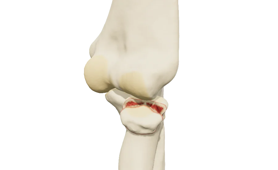 ElbowRadial head fractureMason and AO/OTA, coronoid, dislocation, Osborne-Cotterill lesion, LCL, headless screws, plate and replacement.
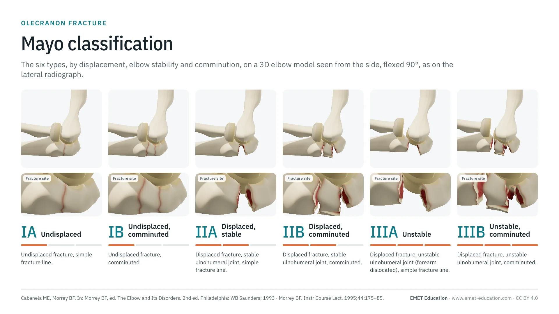 ClassificationsMayo classification of the olecranonSix types of olecranon fractures, in 3D, the neighboring region.
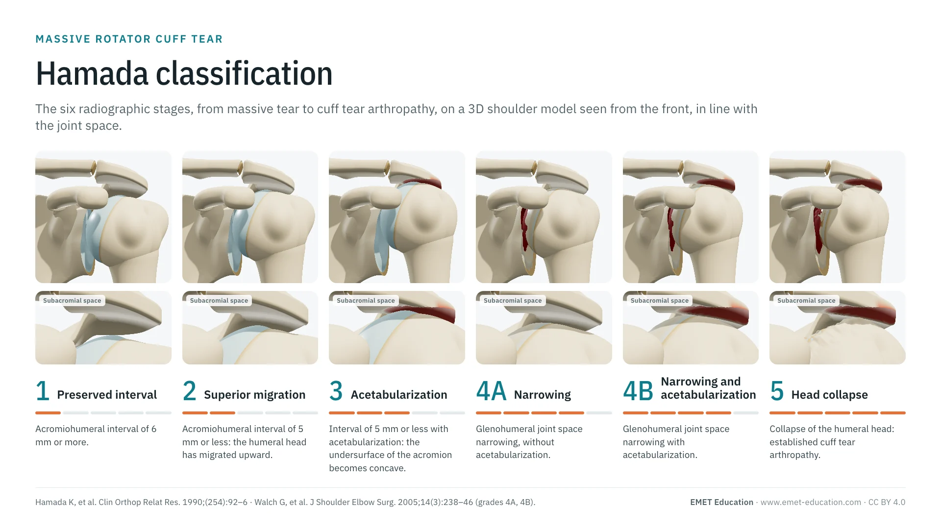 ClassificationsAll 3D classificationsArranged by joint.
Explain the fracture in 3D
The radial head fracture model opens in your browser, at the type you choose, with no sign-up.
Share this page with a colleague or a resident: