- EMET Education
- 3D classifications
- Regan and Morrey classification (coronoid)
Classification · Coronoid fracture
Regan and Morrey Classification of Coronoid Fractures in 3D
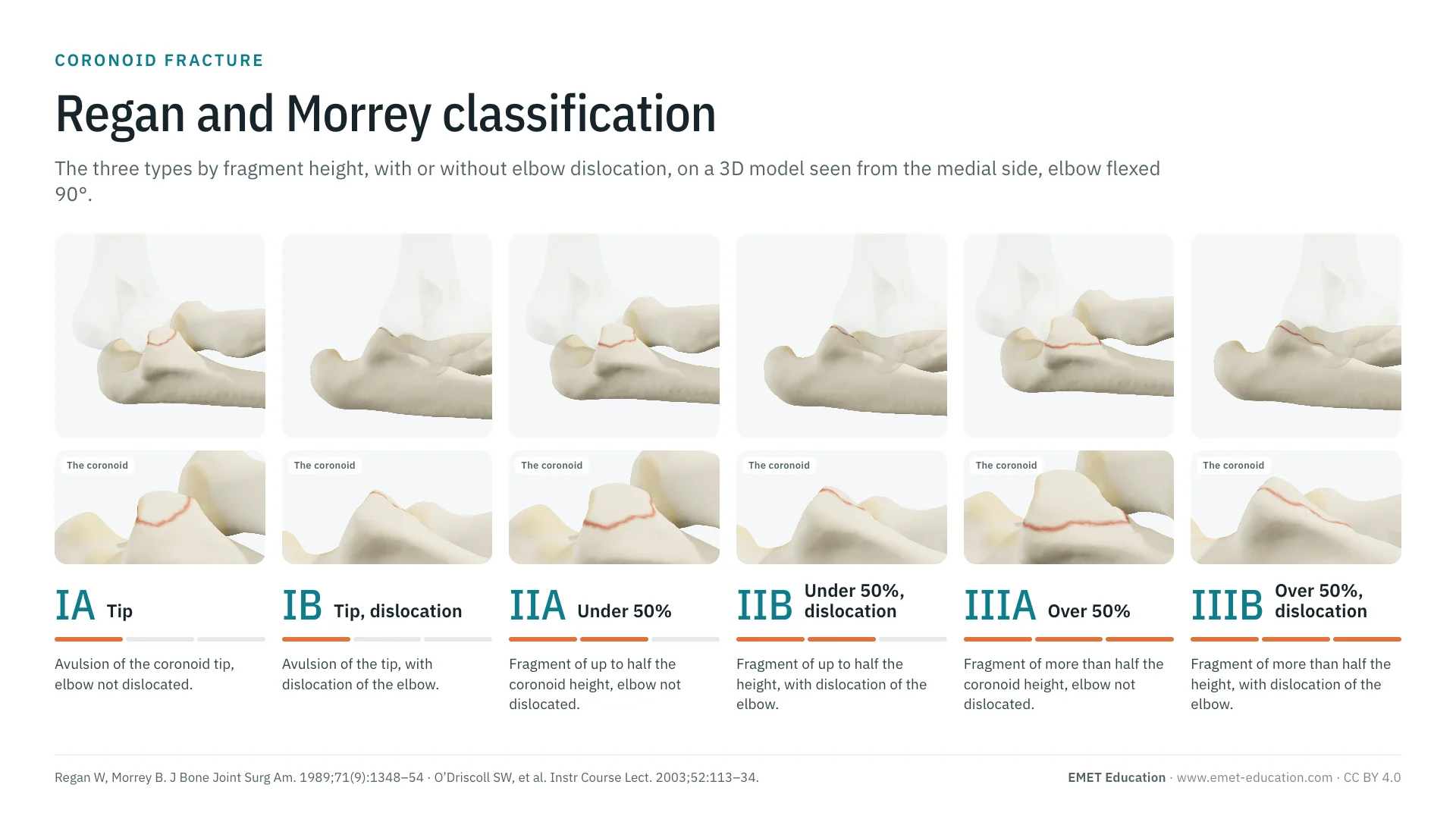
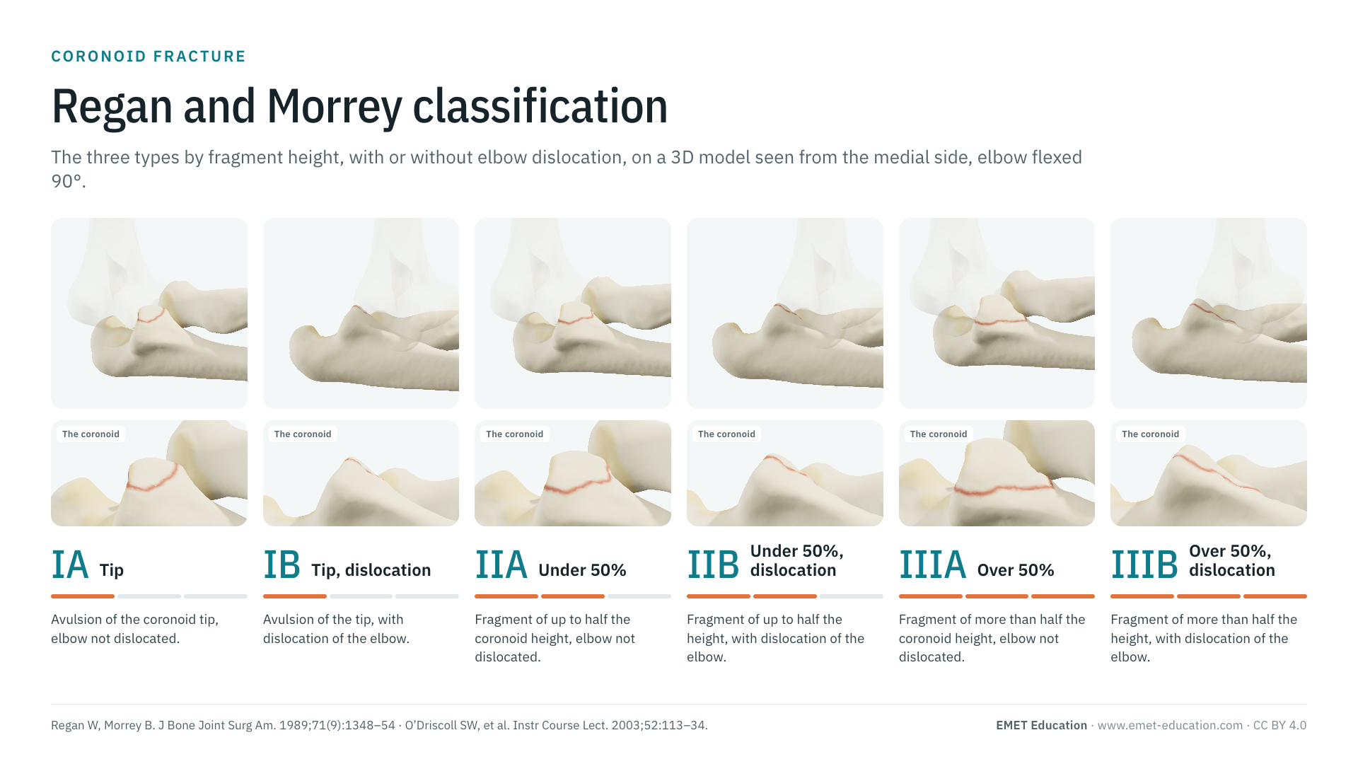
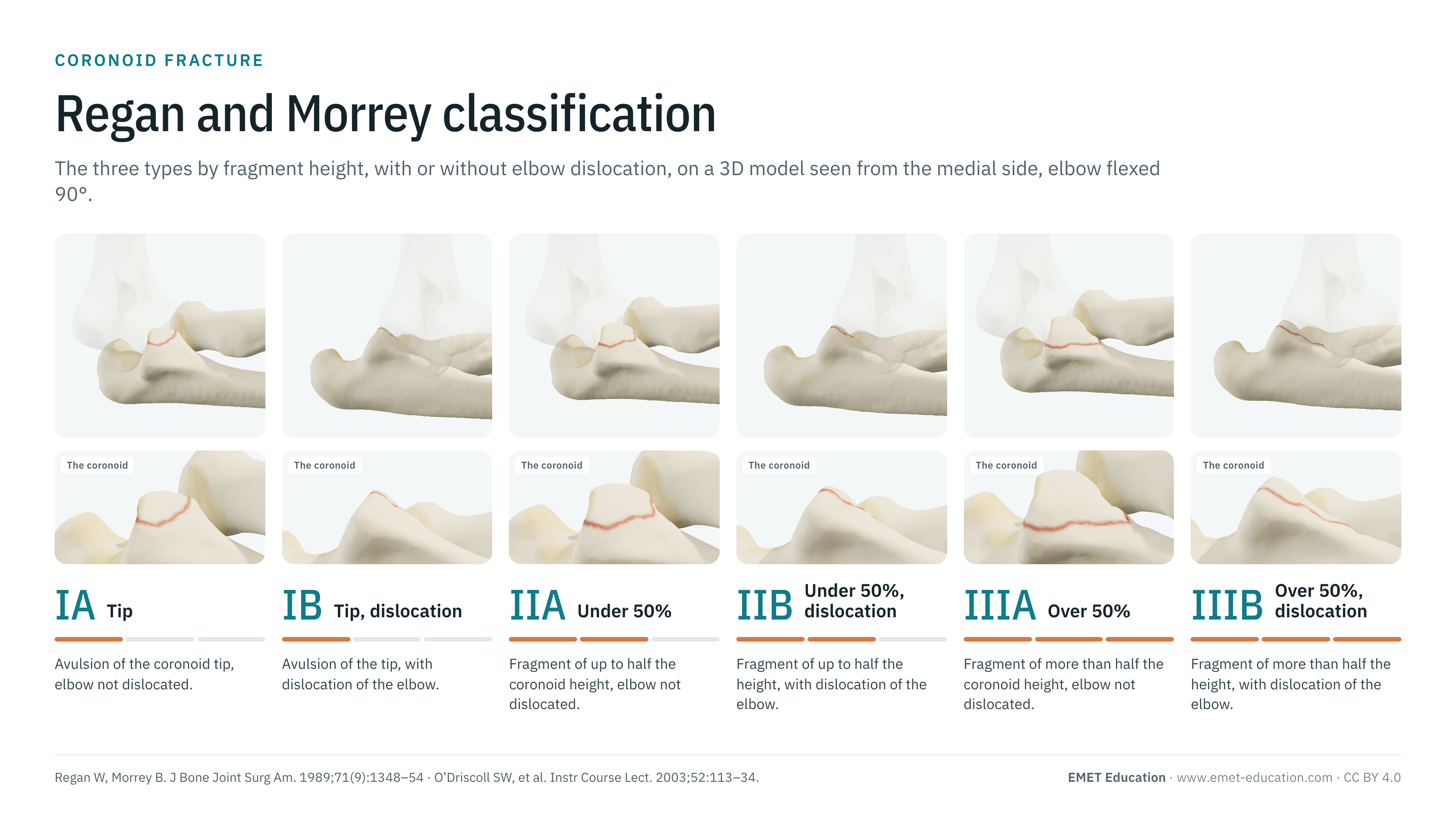
The three Regan and Morrey types, by the height of the coronoid fragment, each with or without dislocation of the elbow, on the radial head fracture 3D model, seen from the medial side, elbow flexed 90°. With the 1989 paper, O'Driscoll's classification, the link with the terrible triad, reliability data, grading pitfalls and free infographics for teaching and publication.
Designed by an orthopedic surgeon
The 3D model opens in your browser, at the type shown, with no sign-up. Infographics are free to reuse under CC BY 4.0.
- 1989Regan and Morrey's paper in the Journal of Bone and Joint Surgery: 35 patients (Regan and Morrey)
- I–III · A/BThree types by fragment height, each with (B) or without (A) dislocation, in the usual form
- 706Citations of the 1989 paper (OpenAlex, November 4, 2026)
- κ 0.40–0.51Interobserver agreement of 29 surgeons, 2D then 3D CT (Lindenhovius)
The types
From type IA to type IIIB, in 3D
Each image is a rendering of the EMET Education radial head fracture model, in which the coronoid is an adjustable associated injury: seen from the medial side, elbow flexed 90°, humerus semi-transparent, with a narrow field to approach the projection of a lateral radiograph. The type is the fragment height (tip, under half, over half); suffix A is a reduced elbow, B a dislocated one, the forearm riding behind the humerus.
-
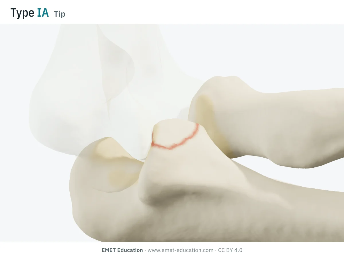
IATip, no dislocation
- Criteria
- Avulsion of the coronoid tip, elbow reduced. In type I, a dislocation or an associated fracture accompanied only 14% of the fractures in the 1989 series, and 92% of patients had a satisfactory result (Regan and Morrey).
- In the 3D model
- A small transverse fragment from the top of the coronoid, a red rim outlining the line; the ulnohumeral joint is reduced.
-
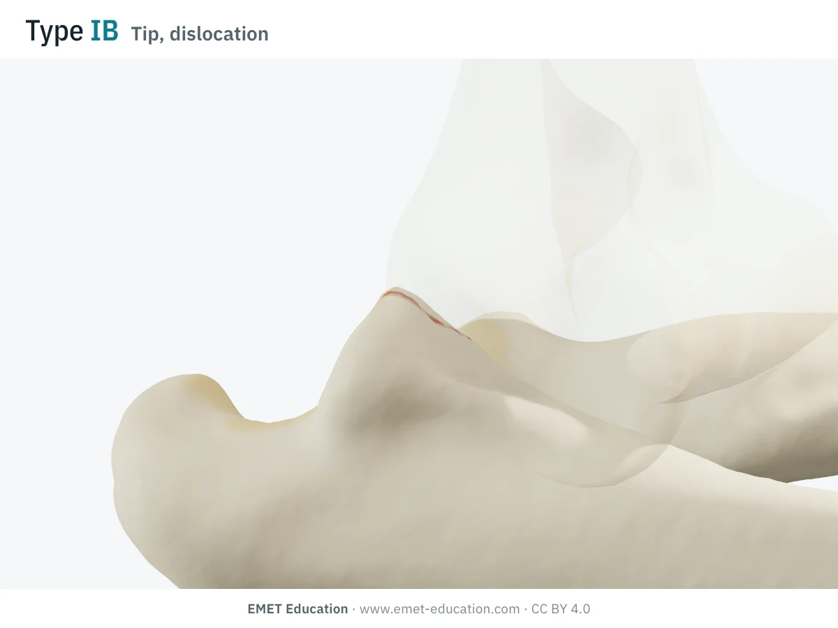
IBTip, with dislocation
- Criteria
- Avulsion of the tip, with dislocation of the elbow. This is the coronoid fracture of the terrible triad when the radial head is also fractured (Doornberg and Ring).
- In the 3D model
- Same tip fragment; the forearm is dislocated behind the humerus, whose trochlea no longer faces the notch.
-
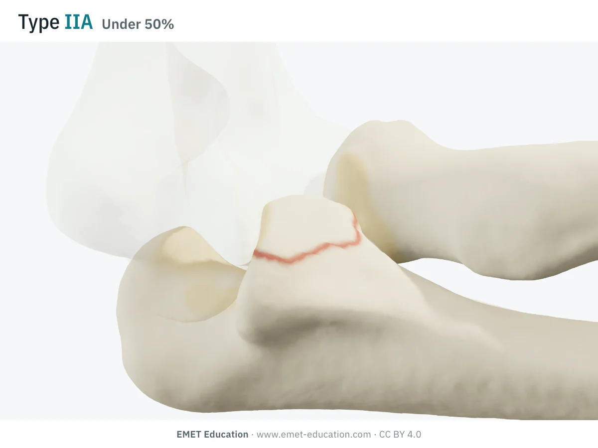
IIA50% or less, no dislocation
- Criteria
- Fragment involving 50% or less of the process, elbow reduced. A dislocation or an associated fracture accompanied 56% of the type II fractures in the 1989 series (Regan and Morrey).
- In the 3D model
- The line cuts the coronoid at under half its height; the fragment, detached from the beak, stays in place.
-
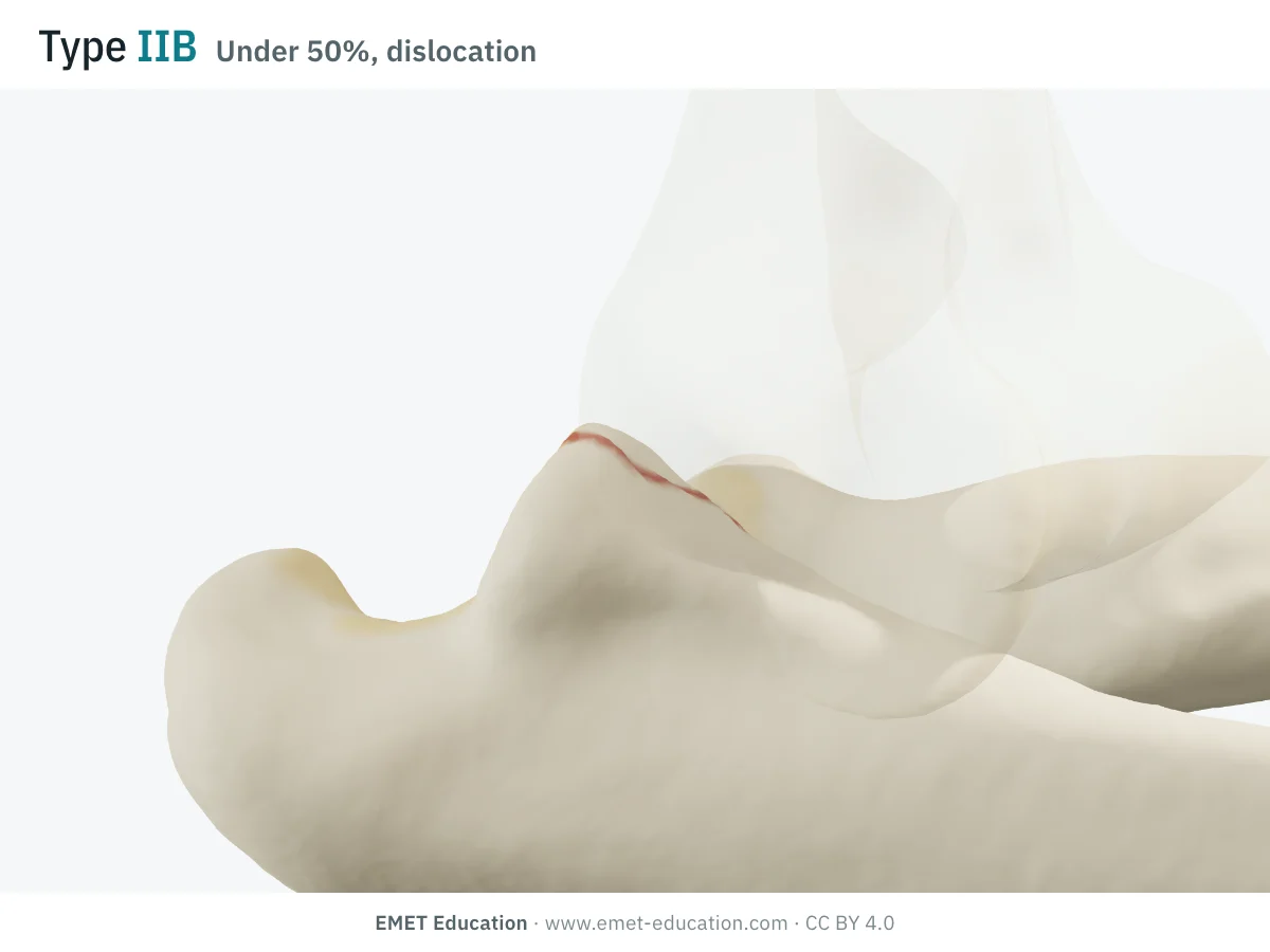
IIB50% or less, with dislocation
- Criteria
- Fragment of 50% or less with dislocation of the elbow. The coronoid fractures of the terrible triad are small transverse fragments, all under 50% in Doornberg and Ring's series (32 elbows); their mean height is 35% (13 CT scans).
- In the 3D model
- The fragment of under half, and the forearm dislocated posteriorly: the coronoid rides behind the trochlea.
-
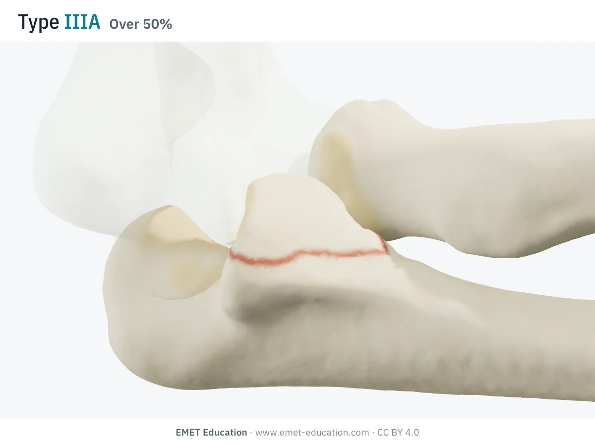
IIIAMore than 50%, no dislocation
- Criteria
- Fragment of more than half the process, elbow reduced. Only type III is large enough to carry the insertion of the anterior bundle of the medial collateral ligament and that of the brachialis (Cage et al.). Result: only 20% satisfactory results in the 1989 series (Regan and Morrey).
- In the 3D model
- The line cuts the coronoid at over half its height: the beak of the ulna, which holds the trochlea anteriorly, is largely detached.
-
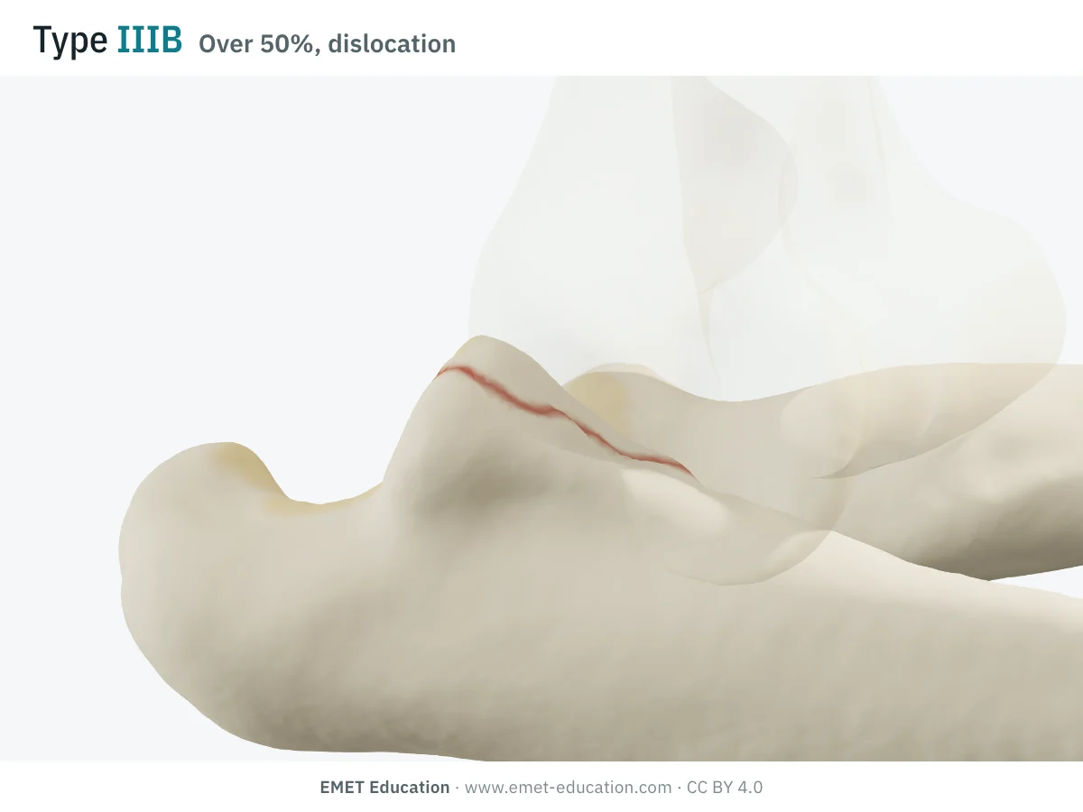
IIIBMore than 50%, with dislocation
- Criteria
- Fragment of more than half with dislocation of the elbow: the coronoid fracture of olecranon fracture-dislocations, where it was large in 22 of 24 cases (Doornberg and Ring). In the 1989 series, a dislocation or an associated fracture accompanied 80% of the type III fractures (Regan and Morrey).
- In the 3D model
- The large fragment left anteriorly, the forearm dislocated posteriorly: the ulna has lost its anterior buttress.
-
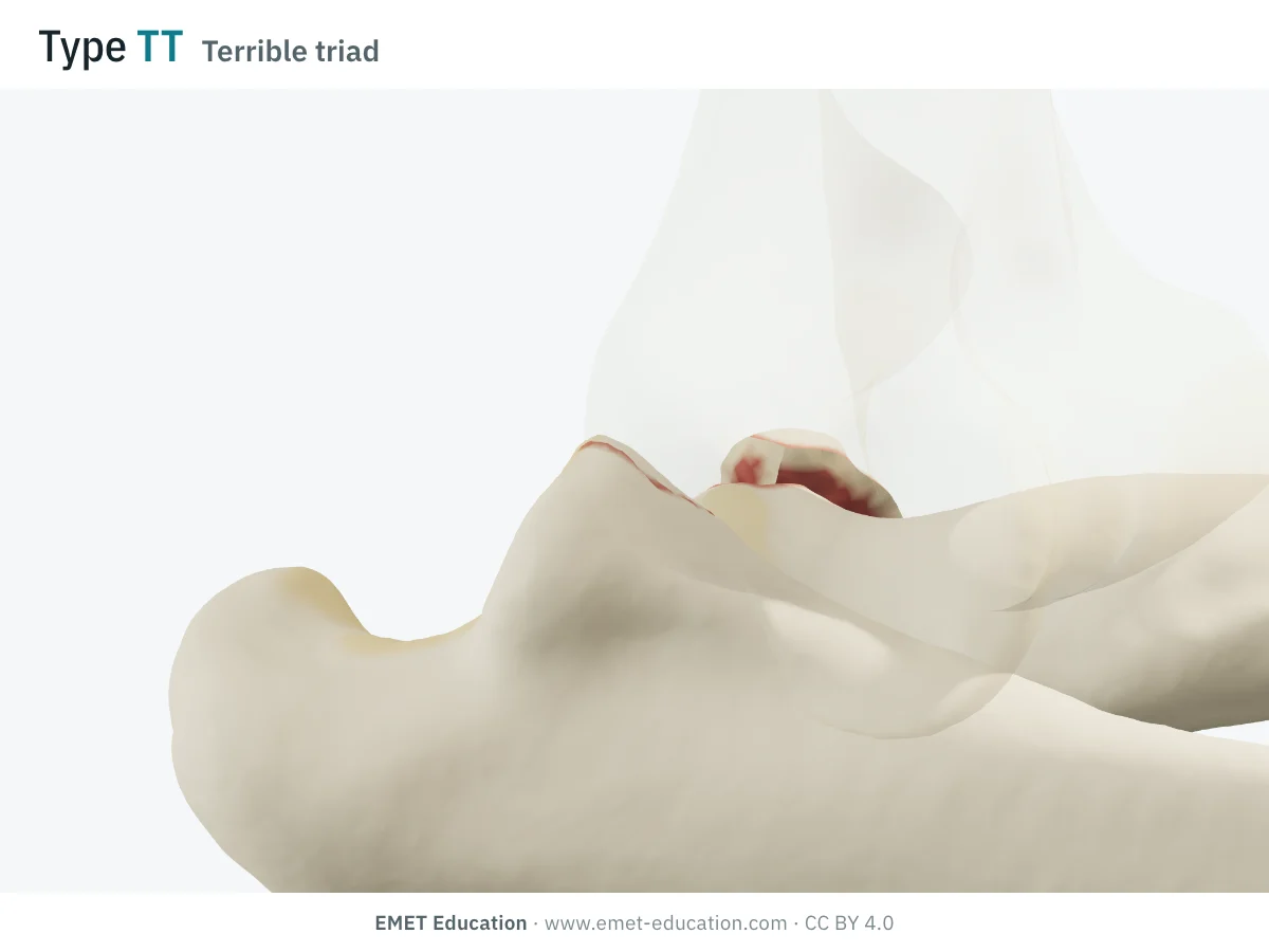
TTTerrible triad
- Criteria
- Elbow dislocation, radial head fracture and coronoid fracture (Fahs et al.). It is not a Regan-Morrey type: the coronoid is graded IB or IIB, the radial head by Mason.
- In the 3D model
- A Mason II radial head, the coronoid tip and the posterolateral dislocation, together; the model calls the combination of the three a terrible triad.
Schematic rendering, for teaching: the model illustrates each type on a single elbow, it does not reproduce a radiograph. The A/B suffix follows the usual textbook form (Burkhart et al.); the abstract of the 1989 paper does not spell it out. Criteria reworded from Regan and Morrey (1989); their figures are not reproduced.
The origin
Regan and Morrey, 1989
W. Regan and B. Morrey, of the Mayo Clinic, described the classification in 1989 in the Journal of Bone and Joint Surgery (71(9):1348–54), from 35 patients with a fracture of the coronoid process of the ulna. The larger the fragment, the more frequent the concomitant dislocation (Cage et al.).
It rests on the height of the fragment, read on the lateral radiograph:
- type I: avulsion of the tip of the process;
- type II: a fragment involving 50% or less of the process;
- type III: a fragment involving more than 50%; the suffix A (no dislocation) or B (with dislocation) is read in the usual form, which the 1989 abstract does not spell out;
- prognosis follows the type: a satisfactory result in 92% of type I patients, 73% of type II and 20% of type III (32 patients reviewed at a mean of 50 months); residual stiffness was most common in type III.
The authors recommended early motion, within three weeks, for types I and II, and thought reduction and fixation followed by early motion preferable for type III. Why size matters: in 20 cadaveric elbows, the anterior bundle of the medial collateral ligament inserted 18.4 mm behind the tip of the coronoid, and the fragment carried it only in type III; the capsule inserted a mean 6.4 mm from the tip (Cage et al., 1995). The 1989 paper has been cited 706 times (OpenAlex, November 4, 2026).
Reliability
How reproducible is the type?
Moderately. On ten fractures, 29 surgeons agreed "moderately" with 3D CT and a little less with 2D CT (Lindenhovius et al.); O'Driscoll's classification was no better. The authors of a study of fragment height in the terrible triad found these fragments hard to place in Regan-Morrey (Doornberg et al.).
| Study | Measure | Value |
|---|---|---|
| Lindenhovius et al., 2009 | Regan-Morrey, interobserver kappa, 29 surgeons, 10 fractures: 2D CT, then 3D CT | 0.40 · 0.51 |
| Lindenhovius et al., 2009 | O'Driscoll, interobserver kappa, same readers: 2D CT, then 3D CT | 0.42 · 0.48 |
| Adams et al., 2012 | Five fracture patterns on CT (52 fractures, 3 readers): inter- and intraobserver reliability | good to very good |
| Doornberg et al., 2006 | Coronoid and fragment height measured on CT (13 terrible triads, 2 readers): inter- and intraobserver reliability | excellent |
CT helps: the Regan-Morrey kappa rose from 0.40 to 0.51 with 3D reconstructions, but agreement on the treatment to choose did not improve (0.27 in 3D against 0.32 in 2D; Lindenhovius et al.). Recognizing a tip fracture was the least reproducible item (kappa 0.19 in 3D, 0.03 in 2D).
Related classifications
O'Driscoll, and what height does not tell
Regan and Morrey grade the size of the fragment on a lateral view. In 2003, O'Driscoll et al. graded the shape: the tip, the anteromedial facet or the base (body) of the coronoid. These patterns predict associated injuries, instability, the approach and the treatment; recent reviews describe O'Driscoll as the reference classification for decisions (Marinelli et al.; Masouros et al.).
The O'Driscoll subtypes, after Marinelli et al.: tip of 2 mm or less (1) or more than 2 mm (2); anteromedial facet, rim alone (1), rim and tip (2), rim and sublime tubercle with or without tip (3); base, isolated (1) or transolecranon fracture-dislocation (2). The two classifications complement more than they replace each other:
| Classification | What it grades | Typical use |
|---|---|---|
| O'Driscoll (2003) | The shape and site of the fragment: tip, anteromedial facet, base; the mechanism (posterolateral rotation, varus posteromedial, axial load) follows from it. | CT; choice of approach and fixation; an anteromedial facet is treated with a buttress plate. |
| Adams (2012) | Five patterns on CT: tip (29%), mid-transverse (24%), basal (23%), anteromedial oblique (17%), anterolateral oblique (7%). | Morphological description, from 52 coronoid fractures among 373 elbow CT scans. |
| Doornberg et Ring (2006) | Not a classification but associations: large fragments with olecranon fracture-dislocations, small transverse fragments with the terrible triad, the anteromedial facet with varus posteromedial instability. | To read with the type: height alone does not tell the ligament injury. |
| Mason | The radial head fracture, graded separately: types I to IV. | The coronoid's companion in the terrible triad; see the Mason classification page. |
Use
In research and in clinical practice
In research
The Regan-Morrey type remains the usual way to describe fragment size in fracture-dislocation series; the Marinelli et al. (2025) review recalls its 50% threshold as an indication for fixation, concluding that elbow stability, more than fragment size, drives the indication. The Adolfsson and Duckworth (2026) annotation notes that the "terrible triad" concept groups very complex fracture-dislocations with more benign injuries, blurring how the literature reads.
In clinical practice
As read in 1989: early motion for types I and II, fixation to be discussed for type III (Regan and Morrey). Today, surgery is discussed when the fragment is at least half the coronoid, when the sublime tubercle is involved or when the ulnohumeral joint is not perfectly reduced (Marinelli et al.).
In the terrible triad, a tip fracture can be left untreated if the elbow is stable after radial head fixation and ligament repair; an anteromedial facet needs a buttress plate, and a large basal fracture screws from posterior to anterior (Masouros et al., 2024). In an early series of 11 terrible triads, seven elbows redislocated in the splint after reduction, and no coronoid had been repaired (Ring et al., 2002).
The terrible triad
Regan-Morrey and the terrible triad
The terrible triad is a posterior dislocation of the elbow with a radial head fracture and a coronoid fracture (Ring et al., 2002; Fahs et al., 2024). The EMET model shows it when the three injuries are present: the coronoid is set to types I to III, the dislocation gives suffix B, and the radial head is graded separately (see Mason).
Its coronoid is most often small: in 32 terrible triads, all had a fragment of under 50%, one of them of the anteromedial facet (Doornberg and Ring, 2006); on 13 CT scans, the coronoid measured a mean 19 mm and the fragment 7 mm, or 35% (Doornberg et al., 2006). The authors concluded that these fractures, of variable height, are not easy to fit into Regan-Morrey, and that classification by morphology and injury pattern might be preferable.
That is the link between the classifications: Regan-Morrey says how much coronoid is missing, O'Driscoll where it is broken, and the terrible triad says why the elbow dislocates. A type IB or IIB with a fractured radial head is a terrible triad; a type III with dislocation points rather to an olecranon fracture-dislocation (Doornberg and Ring).
Grading pitfalls
Five pitfalls when classifying a coronoid fracture
- Height depends on the measuring line. Conventions differ (a line parallel to the ulna through the tip of the olecranon for Morrey; another base for Doornberg) and the percentages are not interchangeable (Wang et al.).
- The boundary between tip and type II is blurred. The paper speaks of an avulsion of the tip; a recent review puts type I at under 25% and type II at 25% to 50% (Marinelli et al.), and Ring judged the limit vague (Wang et al.). State what you measure.
- A small fragment does not mean small damage. In the terrible triad, the coronoid is small and the elbow very unstable: size does not replace examination of the ligaments and the radial head (Doornberg et al.).
- The anteromedial facet escapes Regan-Morrey. It comes from a varus posteromedial rotational injury, often with avulsion of the lateral ulnar collateral ligament, and is poorly seen on the lateral view (Doornberg and Ring; Lanzerath et al.): if in doubt, CT and O'Driscoll's classification.
- Suffix B is sought in the history, not only on the radiograph. An elbow already reduced before the radiograph may have been dislocated: ask the patient and the team that first saw them.
Self-test
Which type?
The same elbow at the six types, in random order, seen from the medial side with the humerus semi-transparent. Find the type of each one; the answer comes with the criteria.
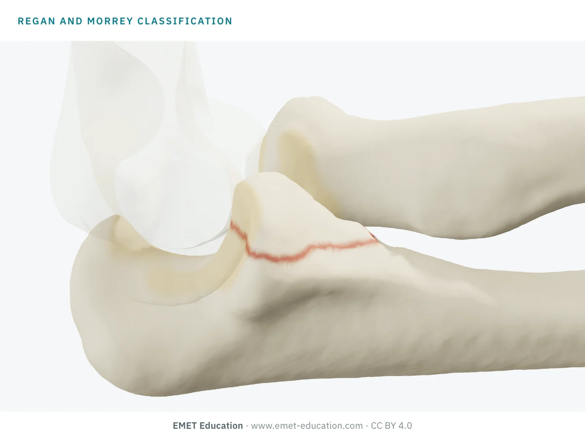
Which Regan-Morrey type is this?
Download and cite
Free infographics, for teaching and publication
Use them in a course, a presentation, a social media post or a scientific article, under the Creative Commons Attribution 4.0 license: free reuse, including commercial, provided EMET Education is credited with a link to this page.
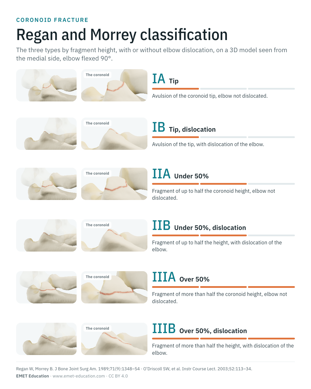
Cite this figure
Ready to paste. For a journal figure, add the original reference (Regan and Morrey, 1989).
EMET Education. Regan and Morrey classification of coronoid fractures in 3D [Internet]. 2026 [cited]. Available from: https://www.emet-education.com/en/classifications/regan-morrey-coronoid/. CC BY 4.0.EMET Education. (2026). Regan and Morrey classification of coronoid fractures in 3D [Infographic]. https://www.emet-education.com/en/classifications/regan-morrey-coronoid/ (CC BY 4.0)<a href="https://www.emet-education.com/en/classifications/regan-morrey-coronoid/"><img src="https://www.emet-education.com/en/classifications/regan-morrey-coronoid/img/infographic.webp" alt="Regan and Morrey classification of coronoid fractures in 3D" width="960"></a><br>Figure: <a href="https://www.emet-education.com/en/classifications/regan-morrey-coronoid/">EMET Education</a>, CC BY 4.0Questions
Frequently asked questions
What is the difference between Regan-Morrey types II and III?
The height of the fragment on the lateral view: 50% or less of the coronoid process in type II, more than 50% in type III. Type III is large enough to carry the insertion of the anterior bundle of the medial collateral ligament and that of the brachialis; it is more often accompanied by a dislocation, and its outcome was worse in the 1989 series.
Regan-Morrey or O'Driscoll?
Regan-Morrey grades the size of the fragment on a lateral radiograph; O'Driscoll grades its shape and site (tip, anteromedial facet, base), preferably on CT, which tells the mechanism, associated injuries and fixation. Recent reviews lean on O'Driscoll to decide; Regan-Morrey remains the usual way to state size, and is also shown in the model.
What do suffixes A and B mean?
In the usual form used by textbooks and by the model, A means an elbow with no dislocation and B a dislocated elbow. The abstract of the 1989 paper does not spell this suffix out: it gives the frequency of a dislocation or an associated fracture by type (14%, 56% and 80%). If a paper uses only I, II and III, that is the base classification.
Is a coronoid tip fracture operated on?
Not necessarily. Isolated with a stable elbow, it is treated with early motion; in a terrible triad, a tip fracture can be left untreated if the elbow is stable after radial head fixation and ligament repair. It is elbow stability, more than fragment size, that drives the indication.
What is the terrible triad?
A dislocation of the elbow with a radial head fracture and a coronoid fracture. The coronoid is most often a small transverse fragment (Regan-Morrey IB or IIB, O'Driscoll tip). The elbow is unstable there and redislocates easily without suitable treatment.
Can I reuse these images in a course or an article?
Yes. The images and infographics on this page are under the Creative Commons Attribution 4.0 license: free reuse, including commercial, with credit to EMET Education and a link to this page. The "Cite this figure" box gives ready-to-paste citations.
How were the 3D images made?
They are renderings of the EMET Education radial head fracture model, in which the coronoid is set to the tip, under 50% or over 50% of its height, with or without posterolateral dislocation. It is a schematic model for teaching and patient explanation; it does not reproduce a radiograph.
Sources
Author and references
- Regan W, Morrey B. Fractures of the coronoid process of the ulna. J Bone Joint Surg Am. 1989;71(9):1348–54. doi:10.2106/00004623-198971090-00011 · PMID 2793888
- O'Driscoll SW, Jupiter JB, Cohen M, Ring D, McKee MD. Difficult elbow fractures: pearls and pitfalls. Instr Course Lect. 2003;52:113–34. PMID 12690844
- Ring D, Jupiter JB, Zilberfarb J. Posterior dislocation of the elbow with fractures of the radial head and coronoid. J Bone Joint Surg Am. 2002;84(4):547–51. doi:10.2106/00004623-200204000-00006
- Lindenhovius A, Karanicolas PJ, Bhandari M, van Dijk N, Ring D; Collaboration for Outcome Assessment in Surgical Trials. Interobserver reliability of coronoid fracture classification: two-dimensional versus three-dimensional computed tomography. J Hand Surg Am. 2009;34:1640–6. doi:10.1016/j.jhsa.2009.07.009
- Doornberg JN, Ring D. Coronoid fracture patterns. J Hand Surg Am. 2006;31(1):45–52. doi:10.1016/j.jhsa.2005.08.014
- Doornberg JN, van Duijn J, Ring D. Coronoid fracture height in terrible-triad injuries. J Hand Surg Am. 2006;31(5):794–7. doi:10.1016/j.jhsa.2006.01.004
- Adams JE, Sanchez-Sotelo J, Kallina CF, Morrey BF, Steinmann SP. Fractures of the coronoid: morphology based upon computer tomography scanning. J Shoulder Elbow Surg. 2012;21(6):782–8. doi:10.1016/j.jse.2012.01.008
- Cage DJ, Abrams RA, Callahan JJ, Botte MJ. Soft tissue attachments of the ulnar coronoid process: an anatomic study with radiographic correlation. Clin Orthop Relat Res. 1995;(320):154–8. PMID 7586820
- Doornberg JN, Ring D. Fracture of the anteromedial facet of the coronoid process. J Bone Joint Surg Am. 2006;88(10):2216–24. doi:10.2106/JBJS.E.01127
- Marinelli A, Riva M, Coliva F, Minerba M, Carbone G, Guerra E. Fractures of the coronoid process: state of the art. J Orthop Traumatol. 2025;27(1):1. doi:10.1186/s10195-025-00891-6
- Masouros P, Christakakis A, Georgiadou A, Kourtzis K, Moustakalis P, Papazotos P. Coronoid fractures and complex elbow instability: current concepts. Orthop Rev (Pavia). 2024;16:118439. doi:10.52965/001c.118439
- Adolfsson L, Duckworth AD. Coronoid fractures: how do we best classify them and what is the 'true terrible triad'? Bone Joint J. 2026;108-B(4):442–6. doi:10.1302/0301-620X.108B4.BJJ-2025-1343.R1
- Wang D, Li J, Xu G, Zhang W, Li L, Tang P, Zhang L. Classification of coronoid process fractures: a pending question. Front Surg. 2022;9:890744. doi:10.3389/fsurg.2022.890744
- Burkhart KJ, Rommens PM, Müller LP. Coronoid fractures. In: Bone and Joint Injuries. Berlin, Heidelberg: Springer; 2014. p. 87–92. doi:10.1007/978-3-642-38388-5_9
- Lanzerath F, Hackl M, Wegmann K, Müller LP, Leschinger T. The treatment of anteromedial coronoid facet fractures: a systematic review. J Shoulder Elbow Surg. 2021;30(4):942–8. doi:10.1016/j.jse.2020.09.008
- Fahs AM, Waldron JE, Afsari A, Best MJ. Management of elbow terrible triad injuries: a comprehensive review and update. J Am Acad Orthop Surg. 2024;32(19):e982–95. doi:10.5435/JAAOS-D-24-00310
3D models
Show the fracture to your patient
The images on this page come from an interactive model. In clinic, set the coronoid, the radial head and the dislocation from your patient's radiograph, then show the proposed fixation.
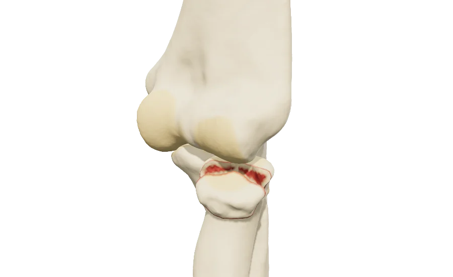
Explain the fracture in 3D
The radial head fracture model, with its coronoid, opens in your browser, at the type you choose, with no sign-up.
Share this page with a colleague or a resident: