EMET Electronic Medical Twin
  1. EMET Education
  2. Musculoskeletal
  3. Knee
  4. ACL tear

3D model · Knee

ACL Tear 3D Model

Reproduce your patient's tear and associated injuries during the visit, then walk through the ACL reconstruction you recommend in 3D. They leave with their model on their phone.

Designed by an orthopedic surgeon

Opens in your browser, no sign-up. For orthopedic surgeons, sports medicine physicians, physiatrists, emergency physicians, radiologists and primary care physicians.

How it works

In the visit, in three steps

Structured information in a few minutes, which the patient can review at home.

  1. 1

    Show

    Open the model and walk through the anatomy: condyles, notch, tibial plateaus, menisci, and the two ACL bundles in the cutaway view. Flex the knee from −5° to 140° to show the ligament at work.

  2. 2

    Reproduce

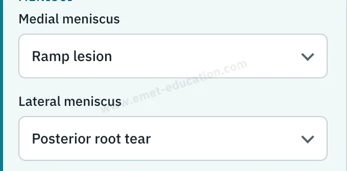
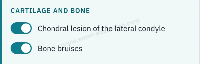
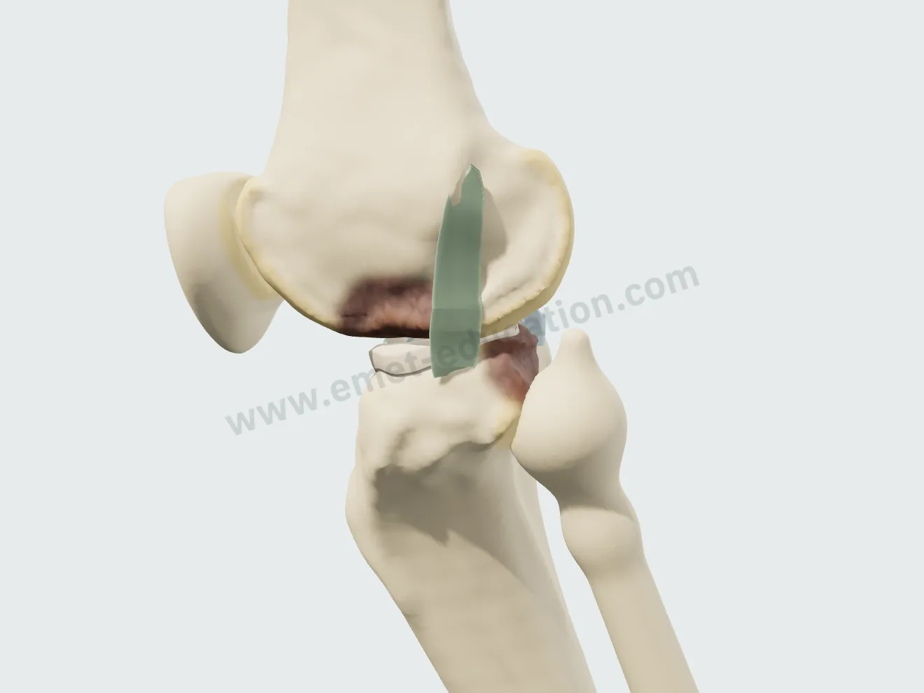
    Set the injuries from the MRI: partial tear, complete tear or tibial spine avulsion, menisci, bone bruises, anterolateral ligament, PCL. Then walk through the proposed reconstruction, step by step.

  3. 3

    Share

    Give the patient the QR code or an image: they find their own case, in their language.

Features

Explain in detail during the visit

Every setting updates the model live: you reproduce the lesions from your patient's imaging in front of them, then walk through the treatment you recommend.

The tear and its associated injuries, in a few clicks

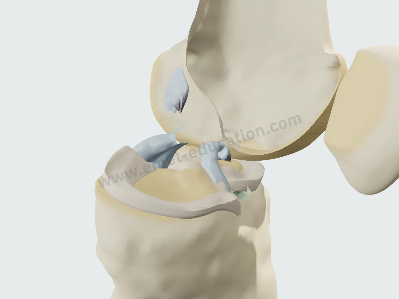
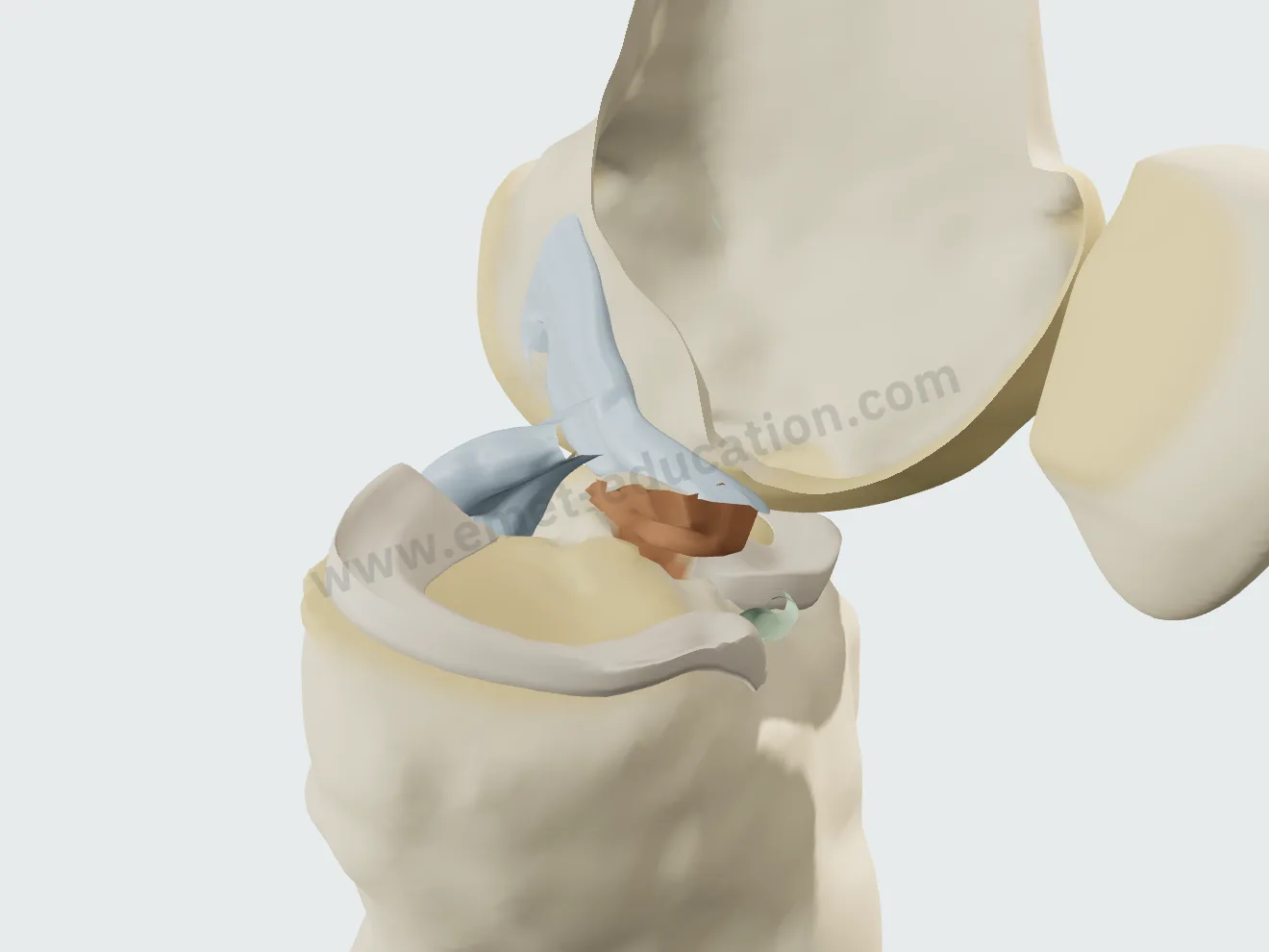
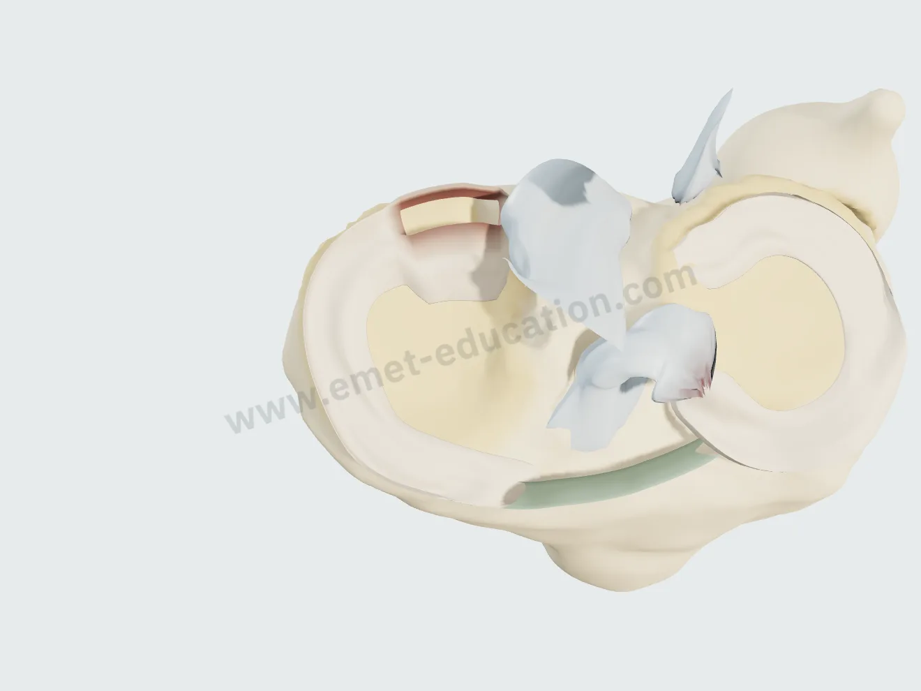
The “Pathology & lesions” panel reproduces the MRI findings. Choosing a tear first plays its mechanism, a deceleration pivot for the ACL, an anterior blow to the flexed knee for the PCL; each injury then forms in front of the patient, in the view that shows it.

Partial, complete, or tibial spine avulsion

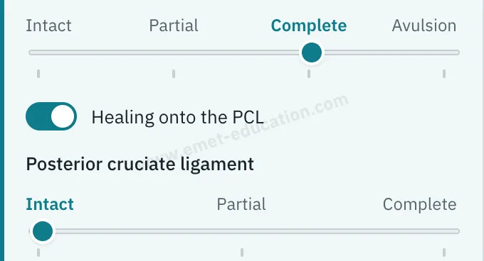
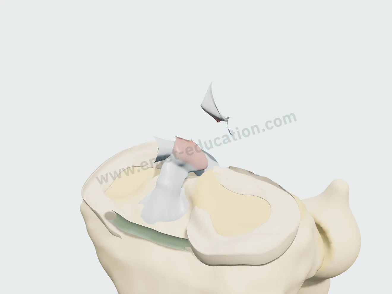
A four-step gauge. Partial: the anteromedial bundle alone, frayed. Complete: proximal-third tear, the tibial stump tipped forward into the notch, the femoral stump retracted. Avulsion: the tibial spine fragment tilts up and takes the intact ligament with it, in a lateral view, as on the radiograph.

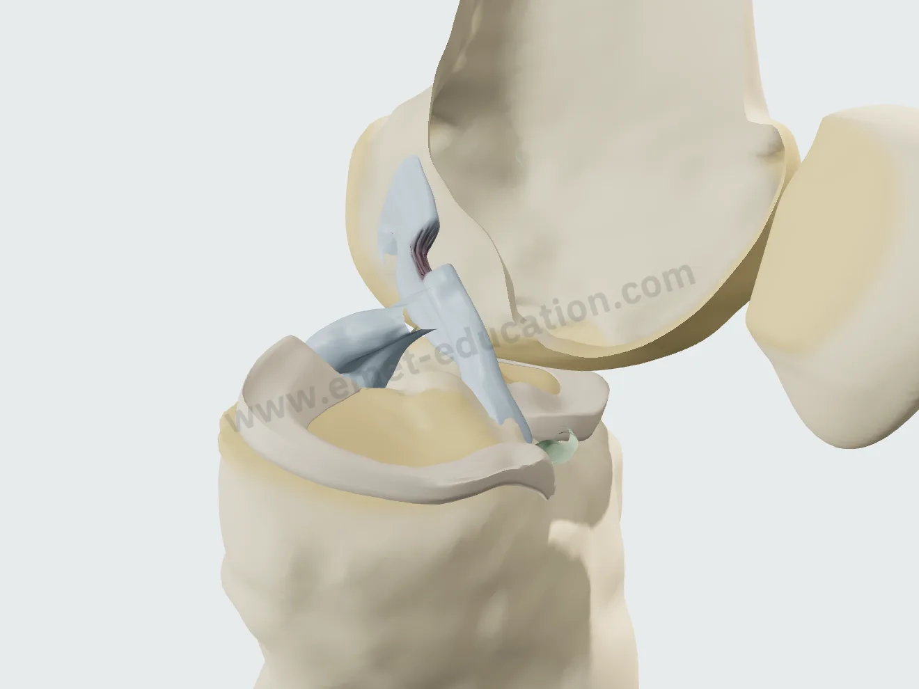
Model panel: “Anterior cruciate ligament” gauge, Intact, Partial, Complete and Avulsion steps.
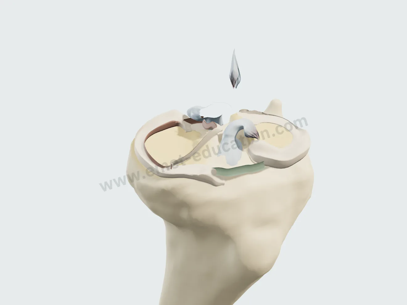
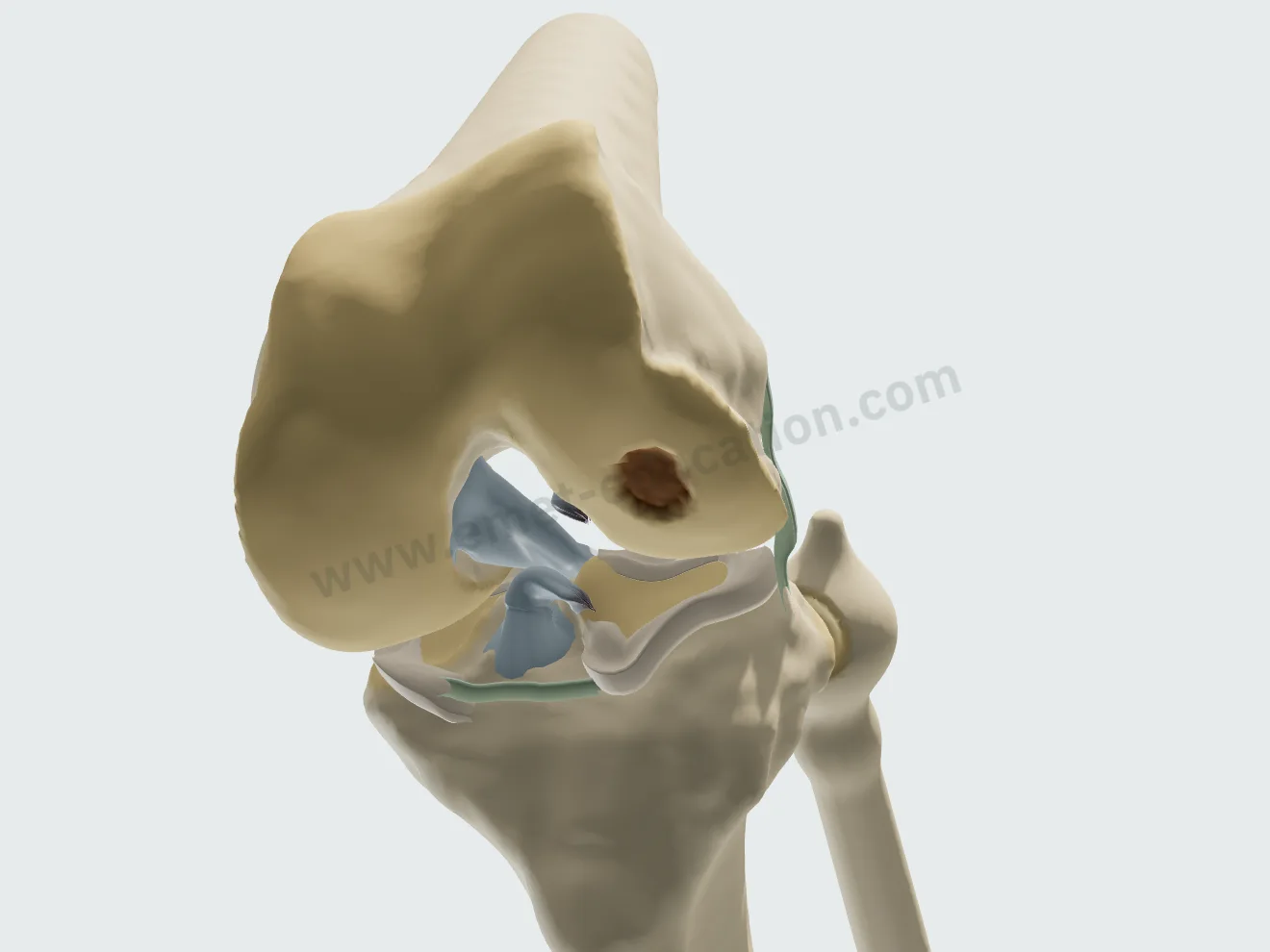
Cutaway view of the notch: the anteromedial ACL bundle torn and frayed, the posterolateral bundle intact.
Partial tear · anteromedial bundle
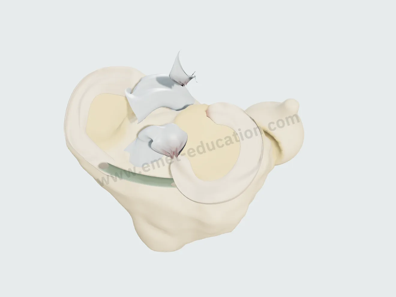
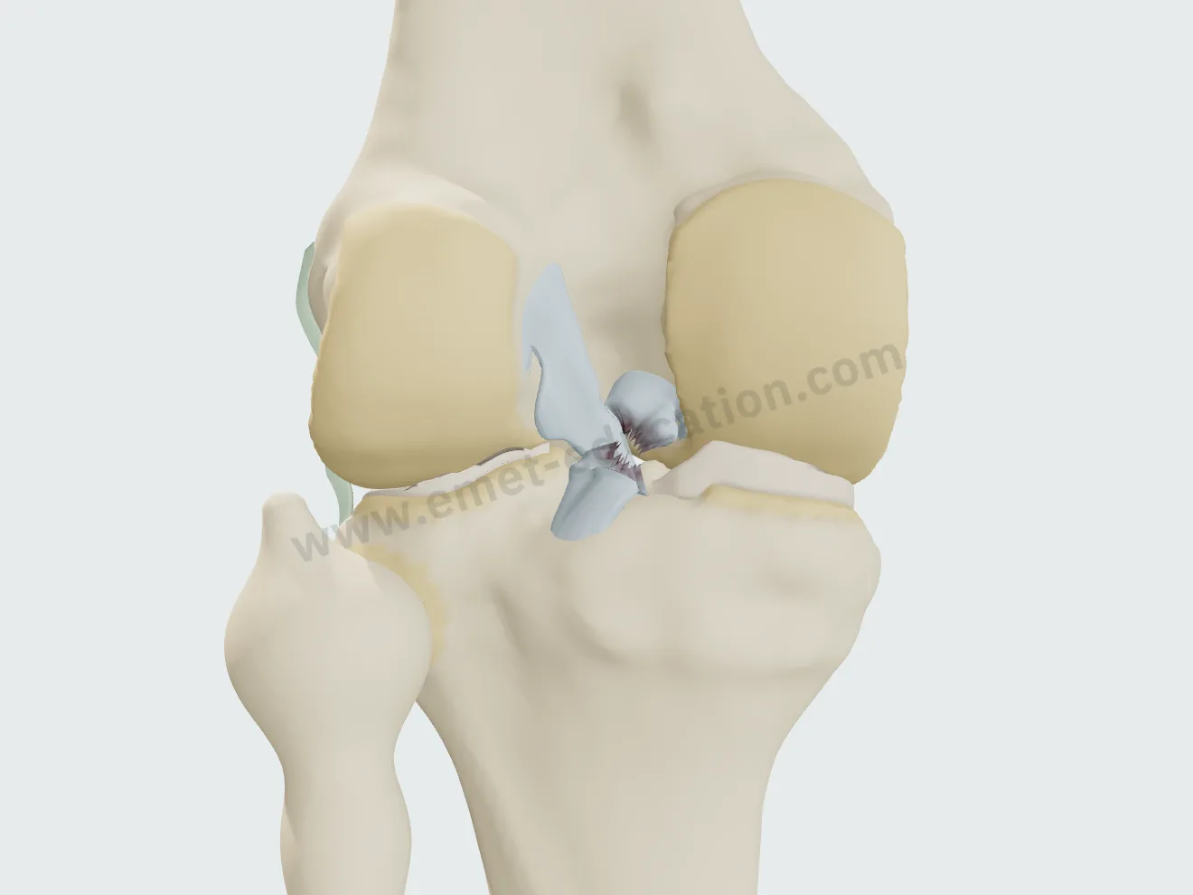
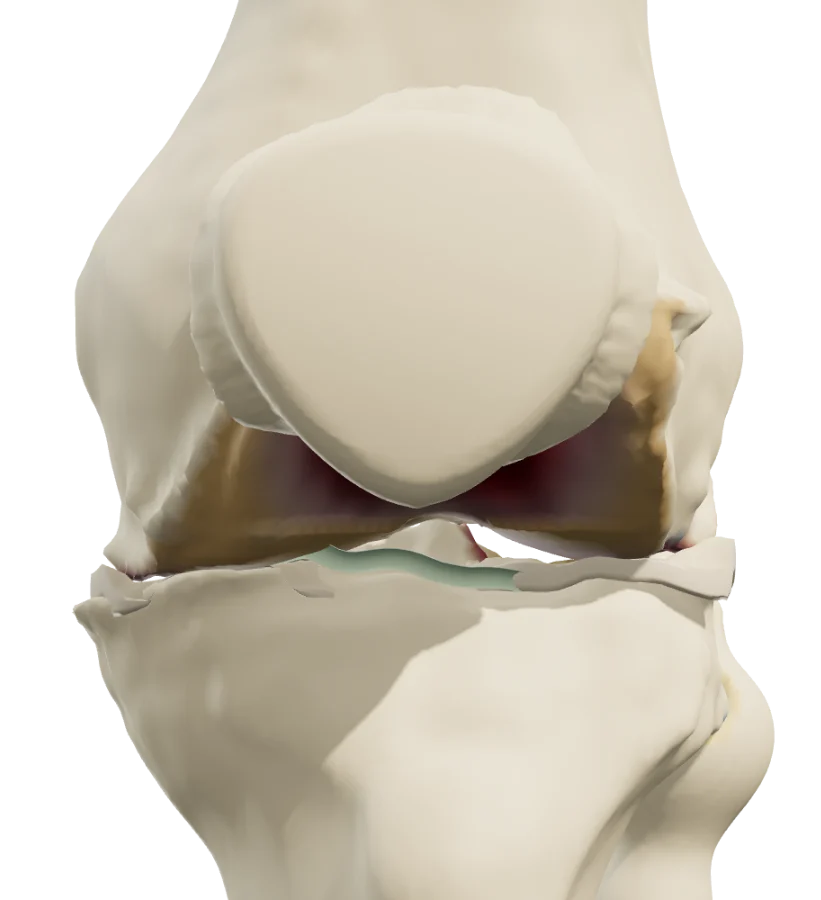
Cutaway view of the notch: complete proximal-third ACL tear, the tibial stump tipped forward, the femoral stump retracted.
Complete tear
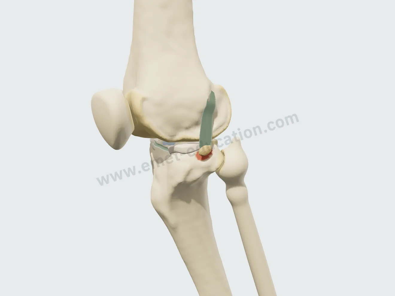
Lateral cutaway view: anterior tibial spine avulsion, the bone fragment tilted up with the intact ACL attached.
Avulsion · tibial spine

ACL reconstruction, step by step

A “Graft” list, an “ACL reconstruction” button: the procedure plays in eight steps, continuously or one step at a time, without skin, with translucent bone during drilling and fixation. The lateral extra-articular tenodesis is a separate procedure, alone or after the graft.

Four-strand hamstring graft

Harvest with a tendon stripper, graft folded over the suspensory button, arthroscopic notch debridement, tibial tunnel, femoral tunnel through the anteromedial portal, graft passage, tibial interference screw.

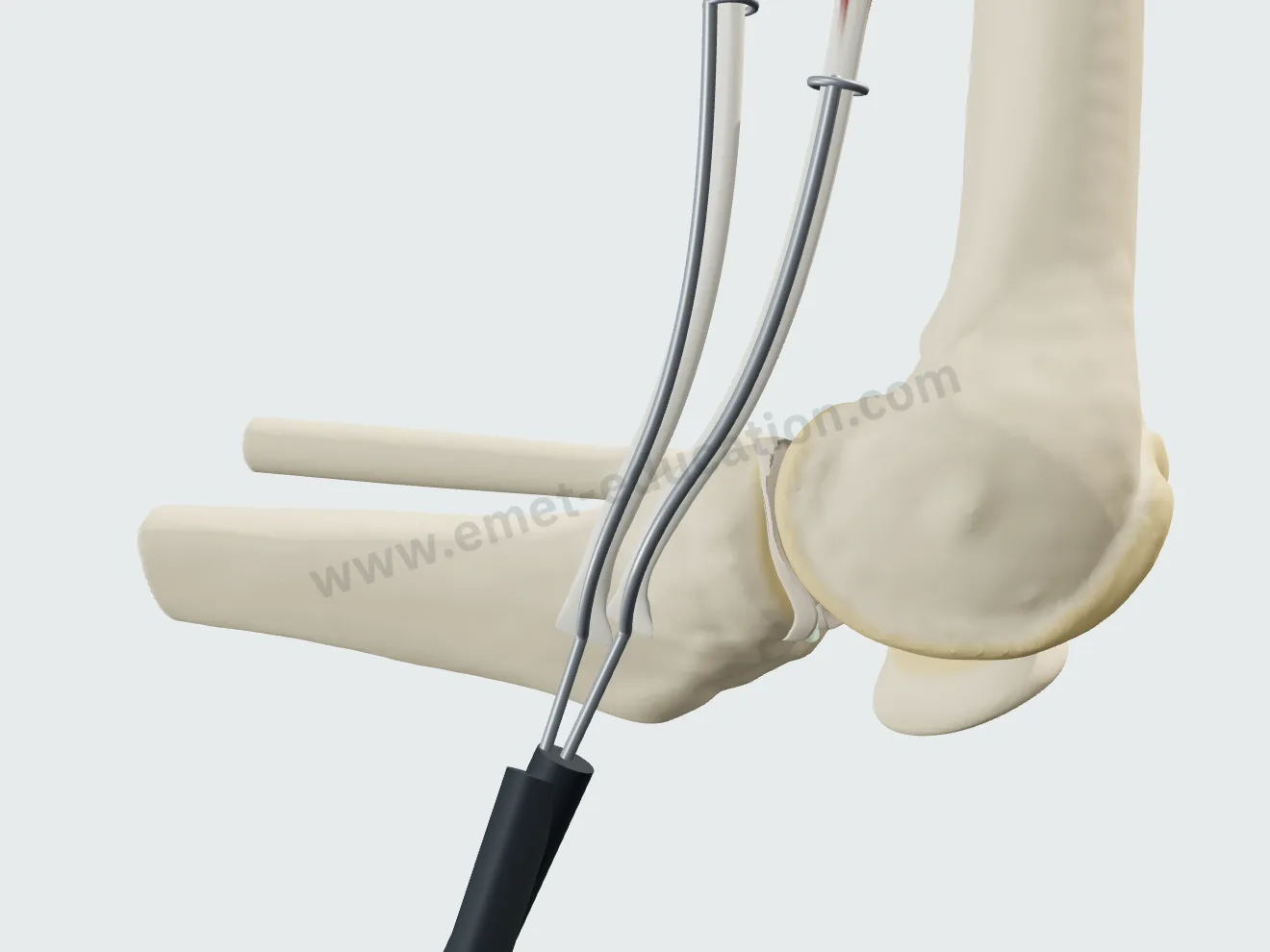
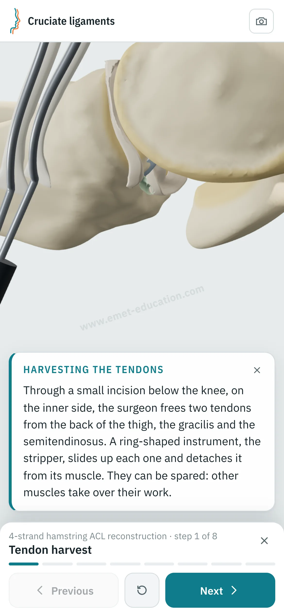
Hamstring graft, step 1: medial view at 90° of flexion, the stripper running up the semitendinosus and gracilis to their musculotendinous junction.
1Harvest
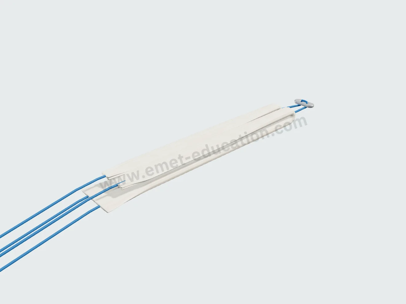
Hamstring graft, step 2: on the back table, both tendons folded over the button loop, four strands of about 8 mm.
2Graft preparation
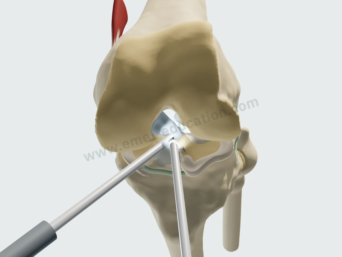
Hamstring graft, step 3: arthroscopic view of the notch, the shaver debriding the ACL stump from its tibial footprint.
3Notch debridement
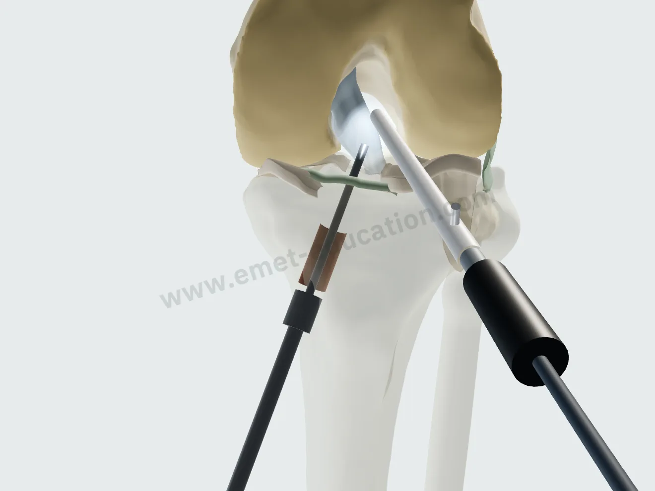
Hamstring graft, step 4: translucent tibia, the 8 mm reamer drilling the tunnel from the anteromedial cortex to the tibial footprint.
4Tibial tunnel
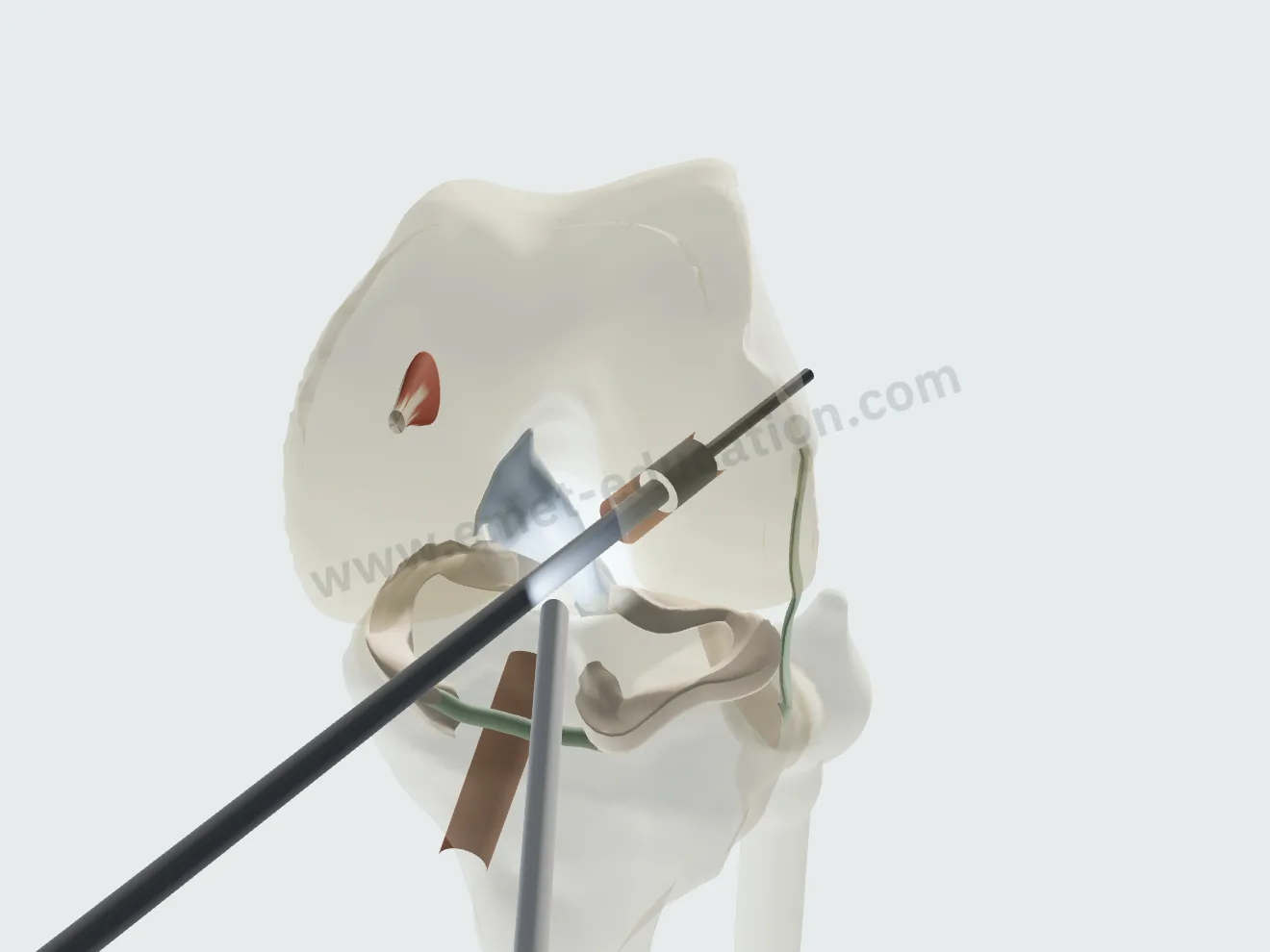
Hamstring graft, step 5: translucent femur, knee at 120°, the femoral socket drilled through the anteromedial portal.
5Femoral tunnel
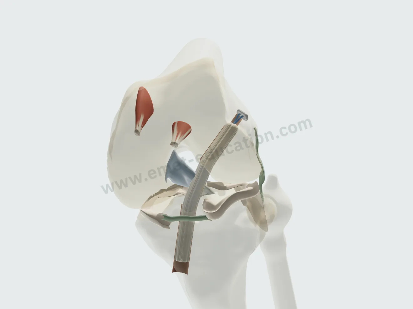
Hamstring graft, step 6: the graft passed from the tibia to the femur, the button flipped onto the lateral cortex.
6Passage, button
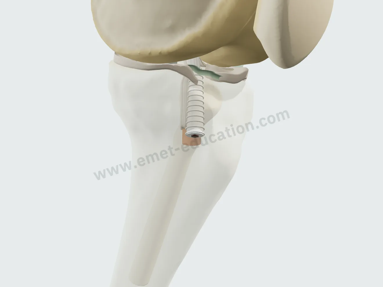
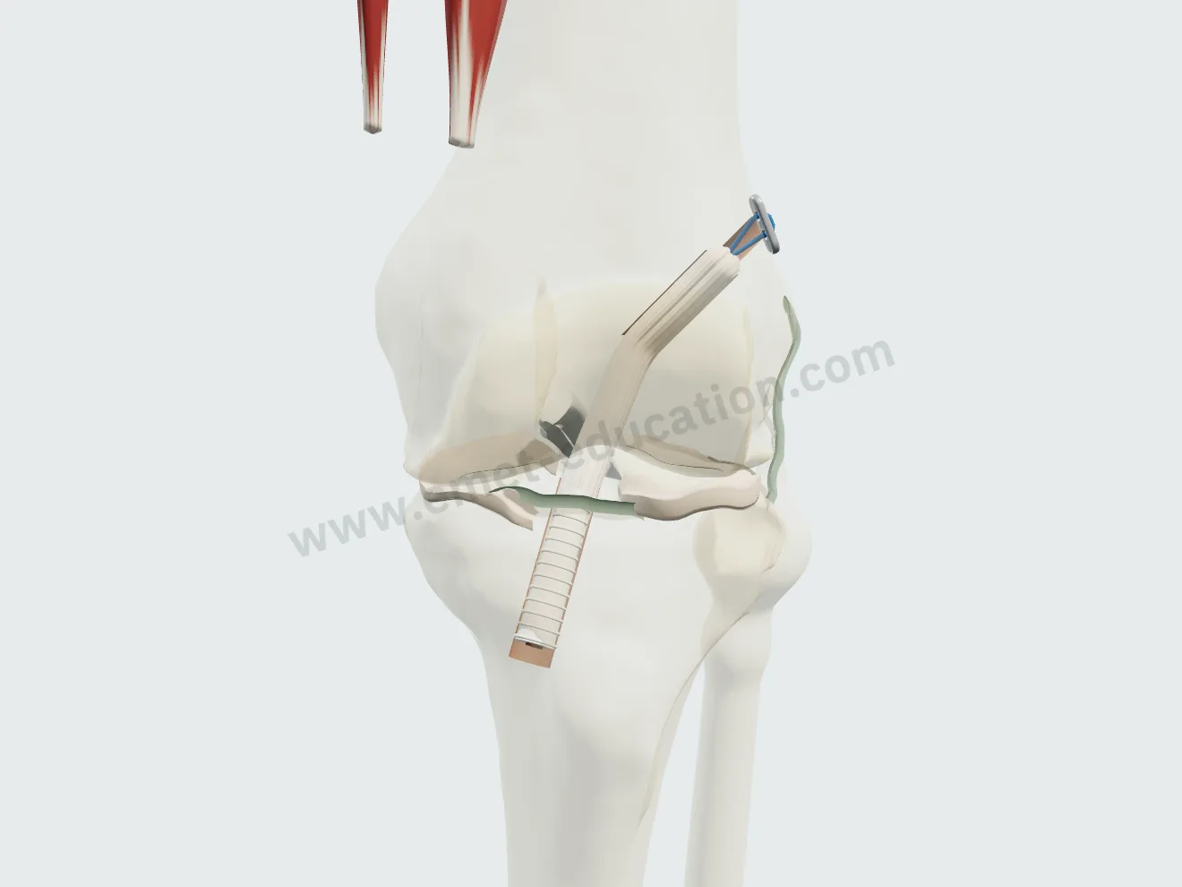
Hamstring graft, step 7: knee at 20°, the bioabsorbable interference screw fixing the graft in the tibial tunnel.
7Tibial screw

Bone–patellar tendon–bone

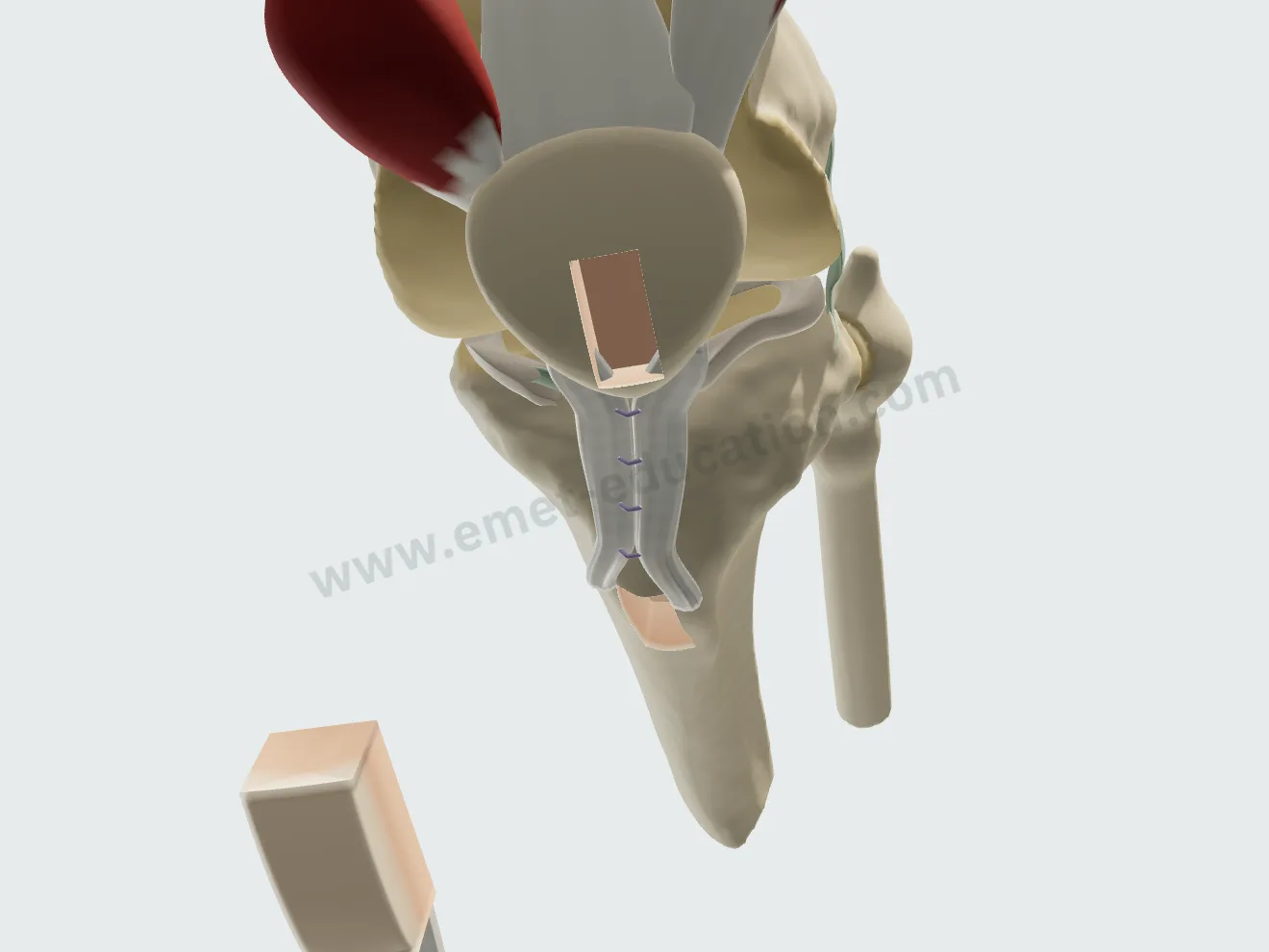
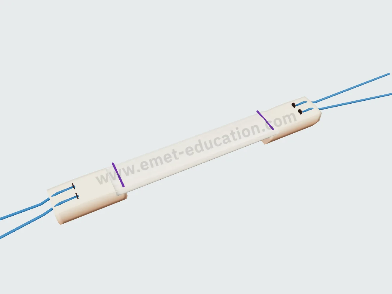
Central third of the patellar tendon with its two bone plugs, 10 mm tunnels, interference screws in the femur and the tibia; the remaining tendon is closed side to side.

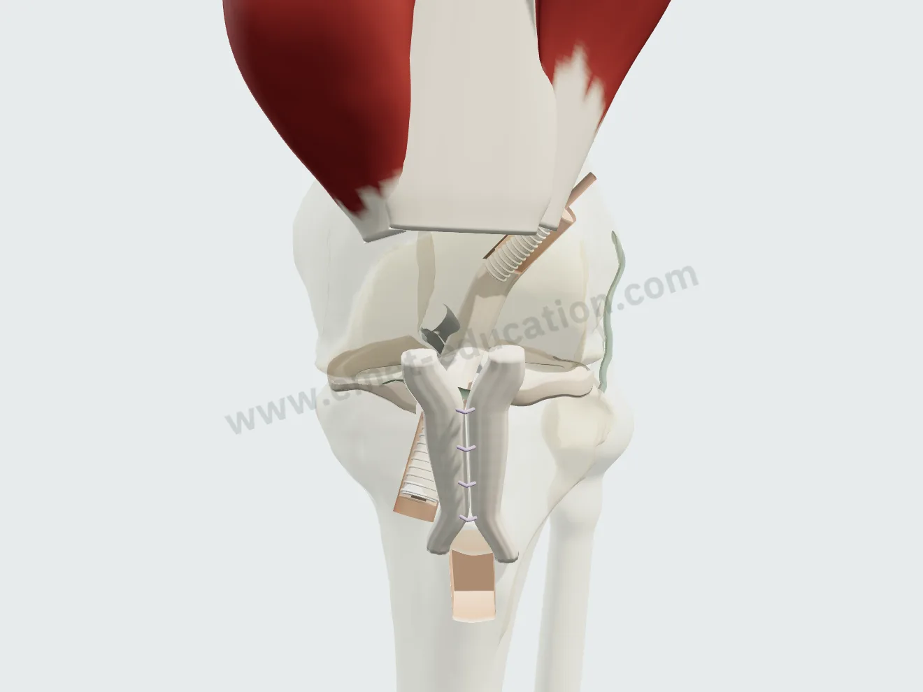
BTB graft, step 1: anterior view at 90° of flexion, the patellar and tibial bone plugs cut and the central third of the patellar tendon harvested.
1Harvest
BTB graft, step 2: on the back table, the bone–tendon–bone graft sized to 10 mm, plugs drilled and sutures passed.
2Graft preparation
BTB graft, step 6: the patellar bone plug at the bottom of the femoral socket, fixed with an interference screw.
3Femoral screw
BTB graft, step 7: the tibial bone plug fully inside the tunnel, fixed with an interference screw.
4Tibial screw

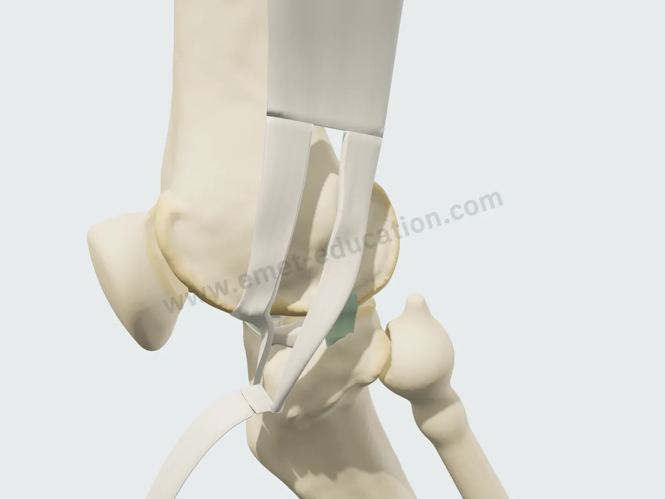
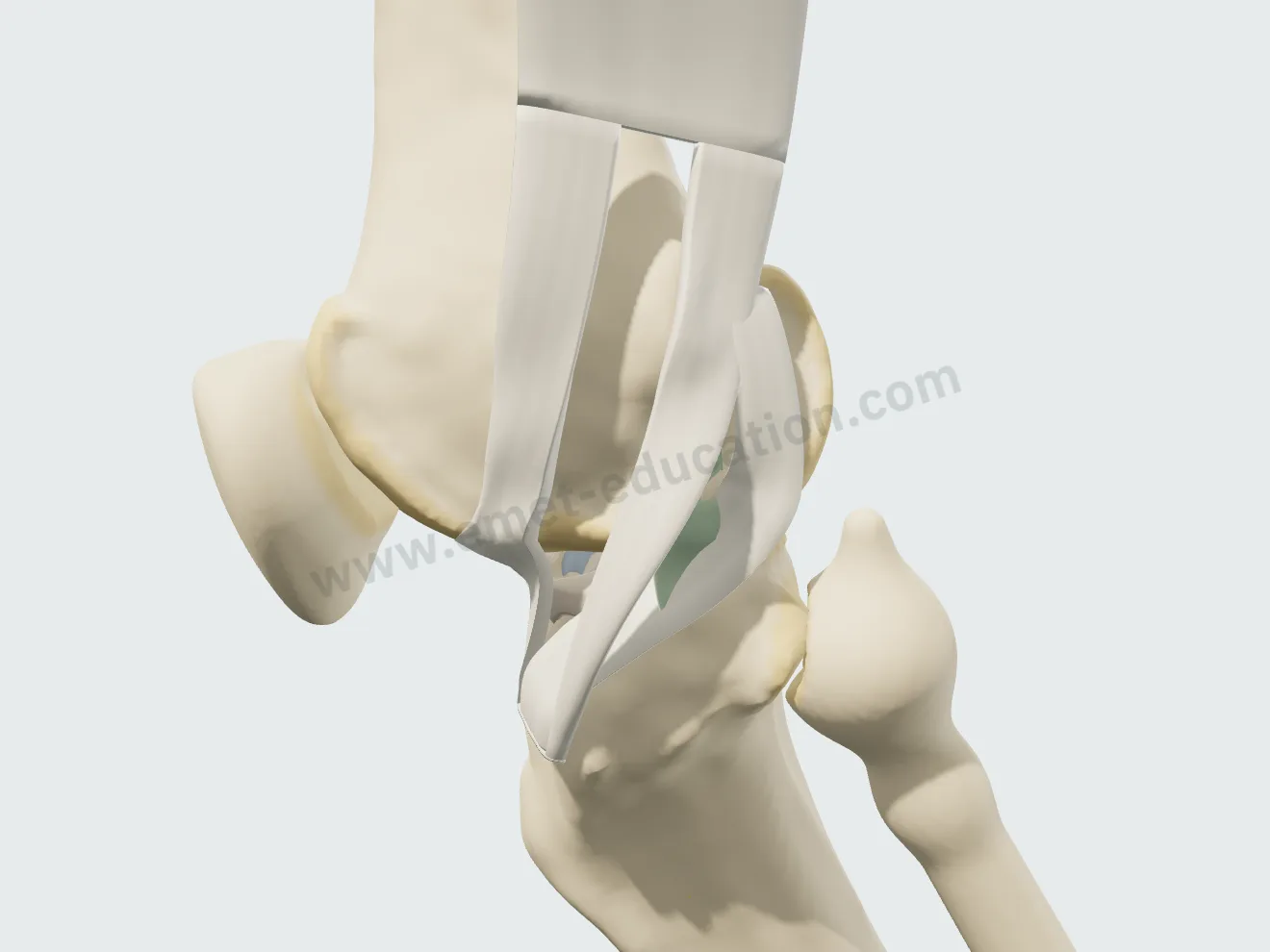
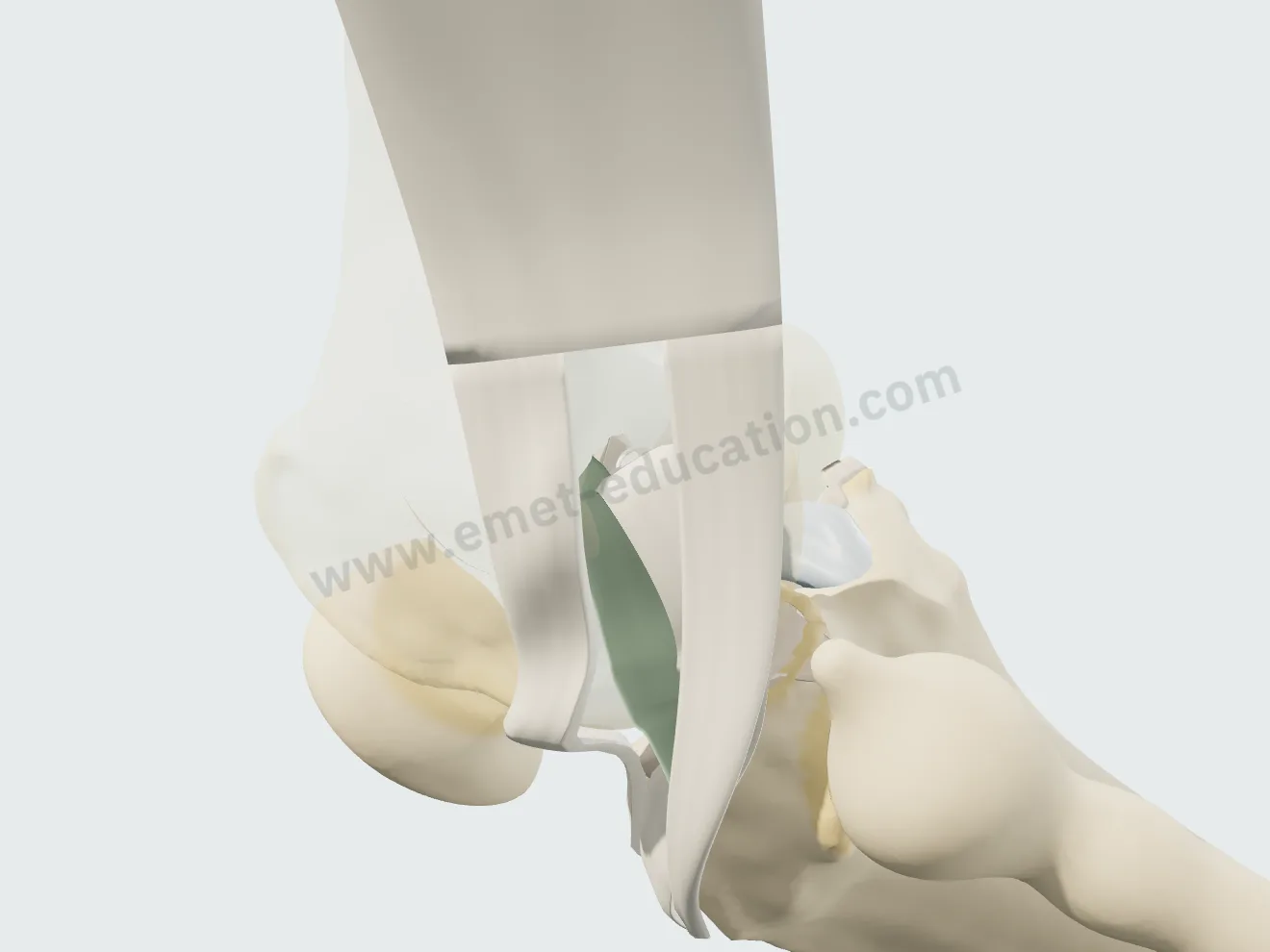
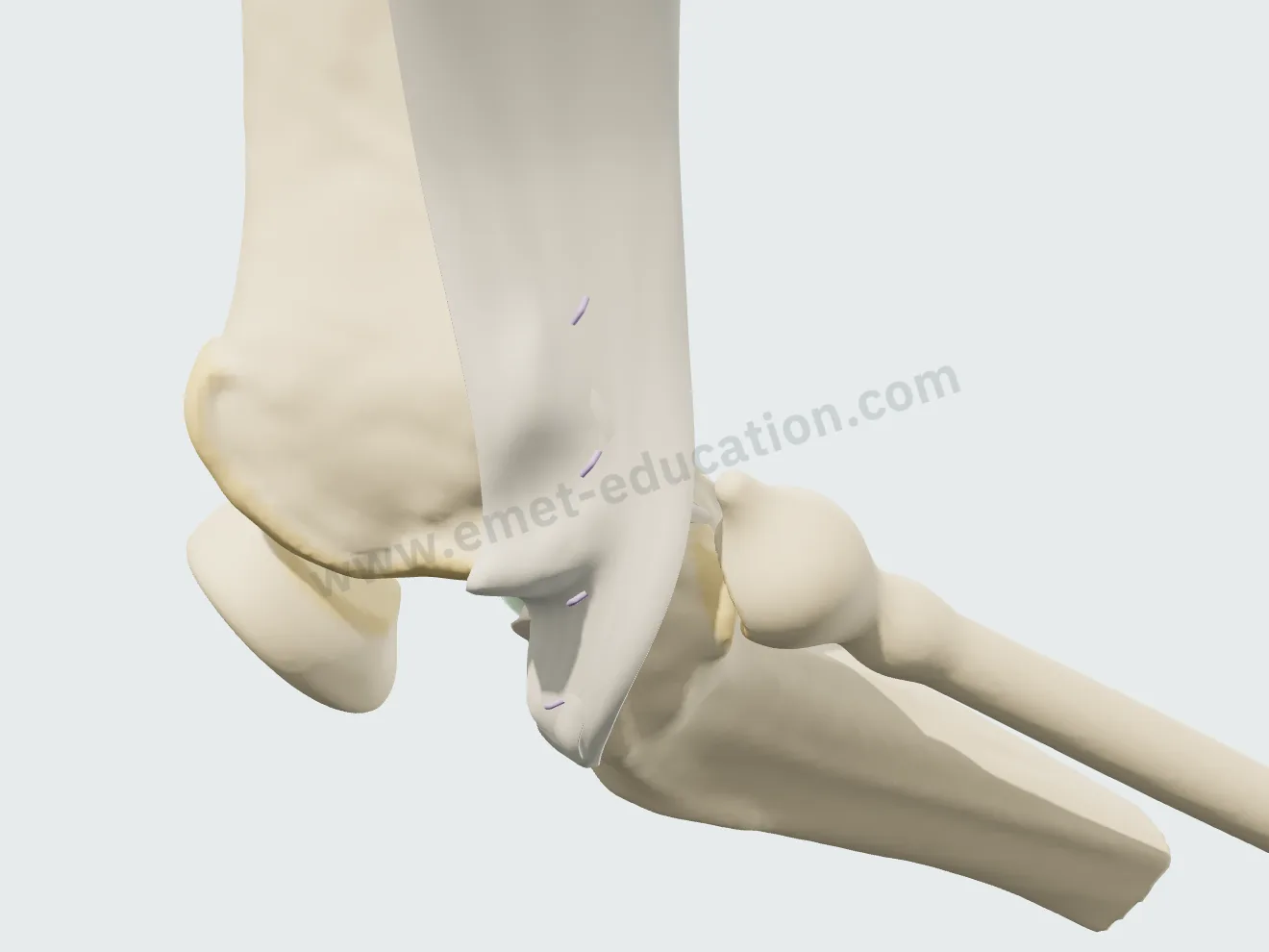
Modified Lemaire tenodesis

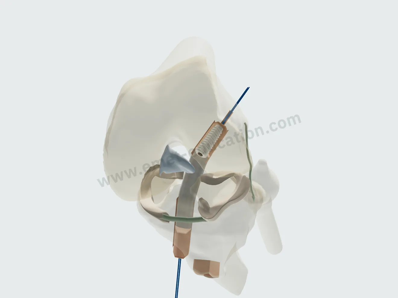
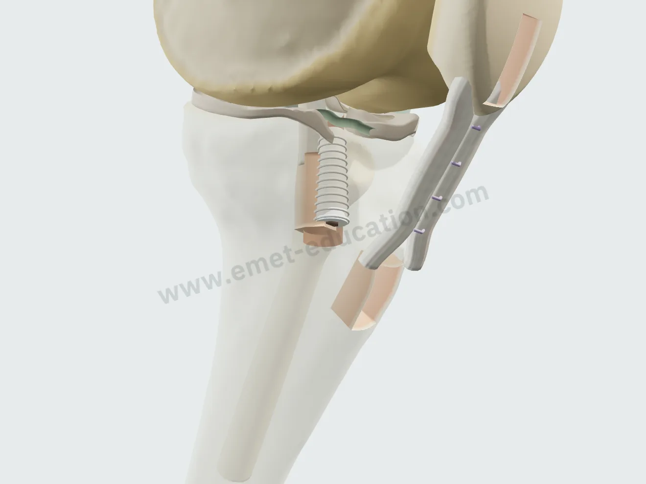
A strip of iliotibial band left attached to Gerdy's tubercle, passed under the lateral collateral ligament, fixed to the femur with an interference screw; the defect is closed.

Lemaire, step 1: lateral view at 30° of flexion, an 80 × 10 mm strip of iliotibial band lifted, still attached to Gerdy's tubercle.
1Harvest
Lemaire, step 2: the strip passed under the lateral collateral ligament, against the capsule.
2Passage
Lemaire, step 3: knee at 60°, translucent femur, the strip fixed posterior and proximal to the lateral epicondyle with an interference screw.
3Femoral fixation
Lemaire, step 4: the iliotibial band defect closed side to side with four sutures.
4Closure

Graft choice

Free or pedicled four-strand hamstring graft, bone–patellar tendon–bone, or a semitendinosus–gracilis graft with anterolateral ligament reconstruction (Sonnery-Cottet). Each construct has its own steps; the final step replays the pivot shift, which the graft now stops.

Model panel: “Graft” list and “ACL reconstruction” button.
Four-strand hamstring graft in place, translucent bone: button on the lateral femoral cortex, interference screw in the tibial tunnel.
Hamstring, 4 strands
Bone–patellar tendon–bone graft in place, translucent bone: an interference screw in each tunnel, the harvest sites visible on the patella and the tubercle.
Bone–patellar tendon–bone
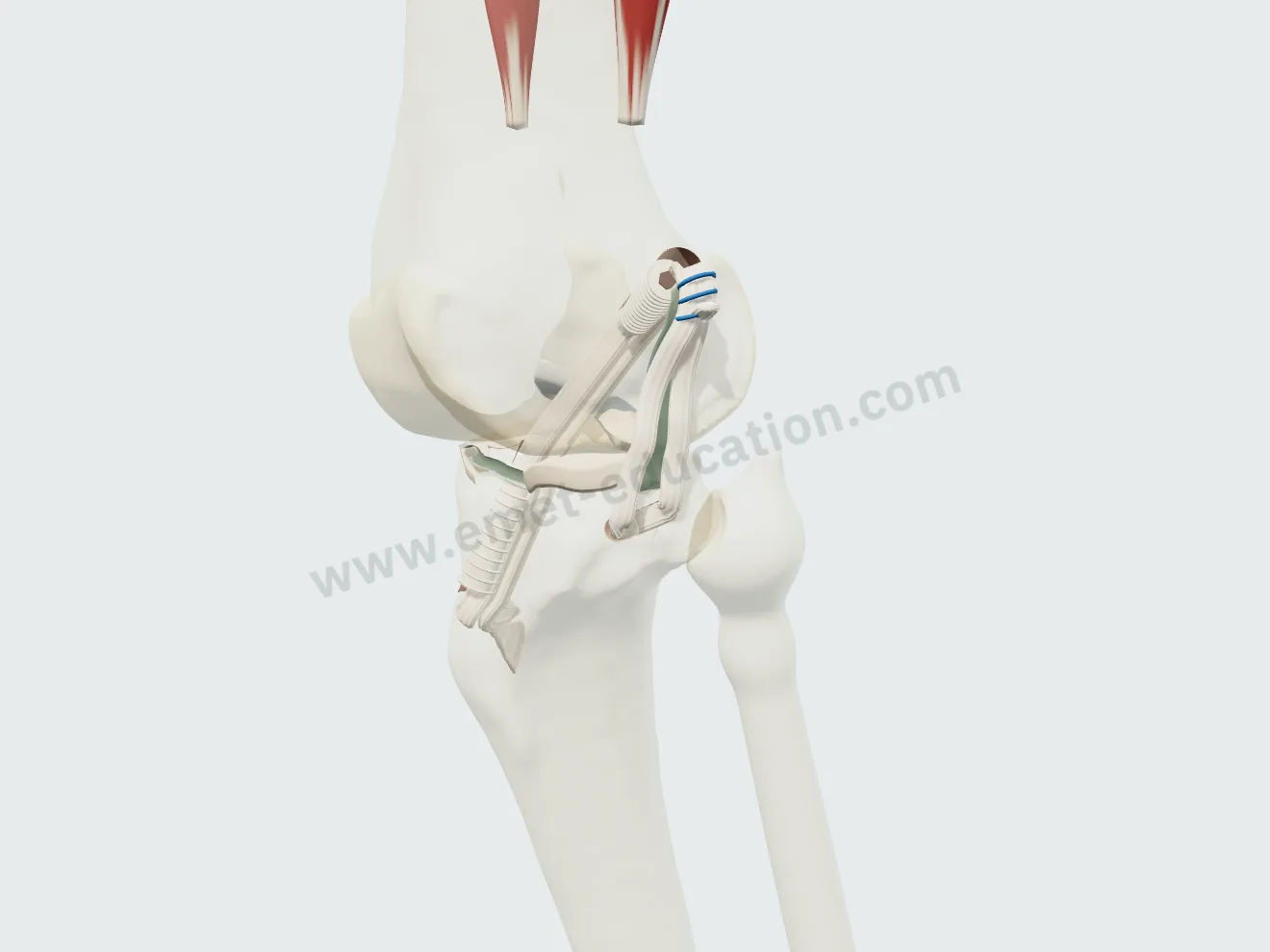
Semitendinosus–gracilis graft in place: the gracilis exits the femoral tunnel and reconstructs the anterolateral ligament as a triangle under the iliotibial band.
Hamstring + ALL

After the visit

The patient leaves with their 3D model

They scan the QR code: the model, their findings and the proposed treatment follow them on their phone, in their language.

The model, exactly as you set it

At the end of the visit, the Consultation panel shows the anatomical findings, the proposed treatment and a QR code. The patient scans it and finds the model on their phone exactly as you set it, in their language.

  • Their lesions, and the proposed treatment to replay step by step.
  • Their anatomical findings and the explanatory card.
  • No identifying data in the link.
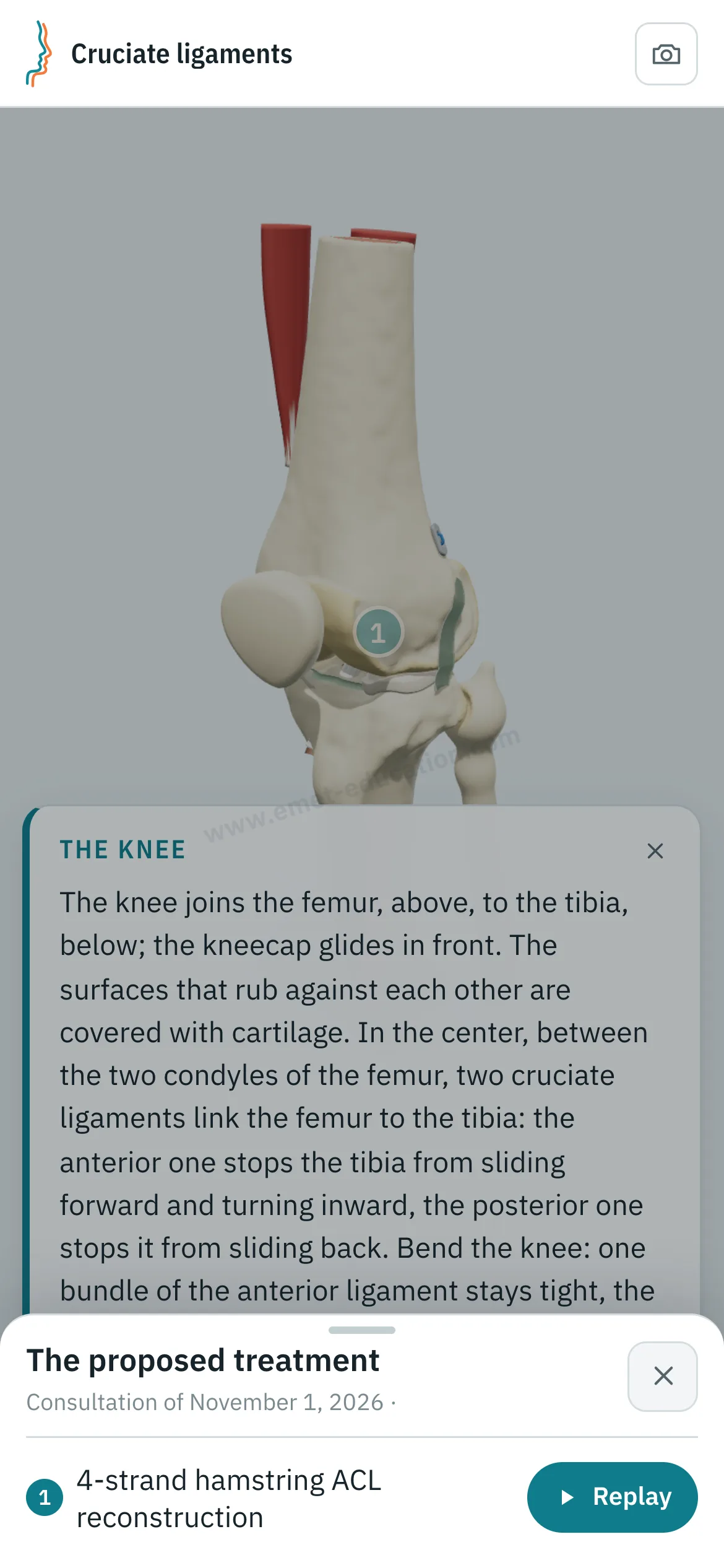
Patient view on a phone: the knee after a four-strand hamstring reconstruction, and the “The proposed treatment” panel open, with the “Replay” button.
What the patient sees after scanning the QR code

Every structure explains itself on tap

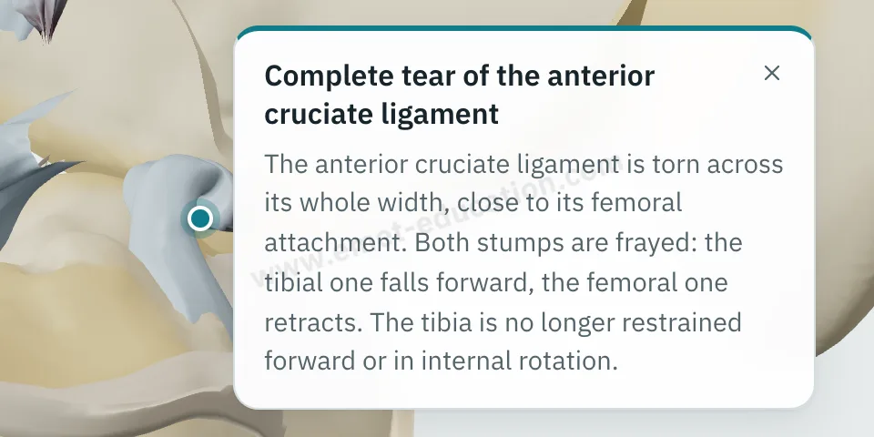
Tap the ligament, a stump, a meniscus, the graft, a tunnel or a screw: its name and an explanation appear.

  • One explanation per structure: bones, ligaments, menisci, injuries, graft, tunnels, screws, button.
  • Written for the patient, in their language.
  • They can reread it on their own, on the tablet or at home.
Explanation bubble “Complete anterior cruciate ligament tear” shown after tapping the stump in the notch.
“What is this?” on tap

They can replay the treatment step by step

From “The proposed treatment,” the patient replays the procedure: each step plays at their own pace, with its explanation, as often as they like.

Patient view on a phone: the four-strand hamstring reconstruction replayed step by step.
“Replay”: the procedure, step by step

Try it with your next case: the model opens in your browser, nothing to install.

Customize the lesions

Patient information

Quality information, documented

Only 12% of U.S. adults have proficient health literacy (National Assessment of Adult Literacy). A patient who sees their own lesions and the proposed procedure understands them, gives truly informed consent, and approaches treatment with confidence.

For the patient

They see their own lesions, reproduced from their imaging, and how the proposed treatment unfolds. They leave with the same material and can review it at home, with their family, as often as they like, before giving consent.

For the physician

Each visit created with an account keeps a record of the information provided: what was shown, when, and whether the patient reviewed it. The model image, with the anatomical findings, the proposed treatment and the QR code, downloads for the patient's chart and prints. Informed consent, as the Joint Commission puts it, is more than getting a signature.

  • Saved and datedThe exact state of the model: injuries shown, treatment proposed.
  • Identical, archivableThe patient receives what you showed; the image goes in the chart and prints.
  • TrackedEach time the patient opens the link is recorded.
  • AnonymousThe link given to the patient carries no identifying data.

“Document the informed consent conversation and the patient’s (or surrogate’s) decision in the medical record in some manner.”

AMA Code of Medical Ethics, Opinion 2.1.1, Informed Consent

The model supports the consent conversation and the written consent form; it does not replace them. Data and privacy: legal information (in French).

Questions

Questions from colleagues

Is the model free?

Partly: the model and the injuries are free. ACL reconstructions, the lateral extra-articular tenodesis and other features are paid options.

Do I need an account?

Not to open the model, set the lesions and walk through the treatment. After about fifteen adjustments, you will be asked to create a free account: it saves your visits, picks them up from one time to the next, and records when the patient opens the link.

What equipment do I need?

Just a web browser, nothing to install: computer, tablet or TV screen in the exam room; the patient opens it on their phone. One click saves an image, without the interface, for the chart.

What happens to patient data?

No patient names are stored. The link given to the patient carries only the state of the model: lesions, treatment, language and visit date. A name written on the image you hand out stays on that image.

Is it a medical device?

No: it is an educational tool. It does not diagnose, does not plan surgery, and does not replace the consent conversation or the written consent form. Among software functions that are not medical devices, the FDA lists those intended for general patient education and interactive anatomy diagrams used for training.

How long does it take in clinic?

Three adjustments and one procedure played: a few minutes, during the visit. The patient reviews it at home, as often as they like.

Can the injury mechanism be shown?

Yes: choosing an ACL tear plays the deceleration pivot, landing, valgus and internal rotation; the ligament turns red as it tightens and gives way, the lateral plateau subluxates and then reduces. Tibial spine avulsion follows the same mechanism, and a PCL tear the anterior blow to the tibia with the knee flexed.

Which grafts are available?

Four-strand hamstring graft, free or pedicled; bone–patellar tendon–bone; semitendinosus–gracilis graft with anterolateral ligament reconstruction. The modified Lemaire tenodesis plays alone or after the graft. A choice outside the usual indications shows a warning, never a block.

Can I embed it on my website?

It is planned for upcoming plans: the model displayed on a practice or hospital website, credited to EMET Education.

Which languages?

English, Spanish, French and Russian. The patient receives the model in the language chosen during the visit, which helps with the roughly 28 million people in the U.S. with limited English proficiency, alongside a qualified interpreter, not instead of one.

Design

Who designed this model

Model designed by Dr. Mikaël Chelli, orthopedic surgeon, based on the reference descriptions. Updated November 1, 2026.

  • Segond P. Recherches cliniques et expérimentales sur les épanchements sanguins du genou par entorse. Progrès Médical, 1879.
  • Jones K.G. Reconstruction of the anterior cruciate ligament: a technique using the central one-third of the patellar ligament. J Bone Joint Surg Am, 1963;45:925-932.
  • Claes S. et al. Anatomy of the anterolateral ligament of the knee. J Anat, 2013. doi:10.1111/joa.12087
  • Sonnery-Cottet B. et al. Outcome of a combined anterior cruciate ligament and anterolateral ligament reconstruction technique with a minimum 2-year follow-up. Am J Sports Med, 2015. doi:10.1177/0363546515571571
  • Getgood A.M.J. et al. Lateral extra-articular tenodesis reduces failure of hamstring tendon autograft anterior cruciate ligament reconstruction: 2-year outcomes from the STABILITY study randomized clinical trial. Am J Sports Med, 2020. PMID 31940222

Try it with your next patient

The model opens in your browser, nothing to install, no sign-up.

Know a colleague who could use it? Email this page ·