- EMET Education
- 3D classifications
- AO/OTA classification (distal radius)
Classification · Distal radius fracture
AO/OTA Classification of Distal Radius Fractures in 3D
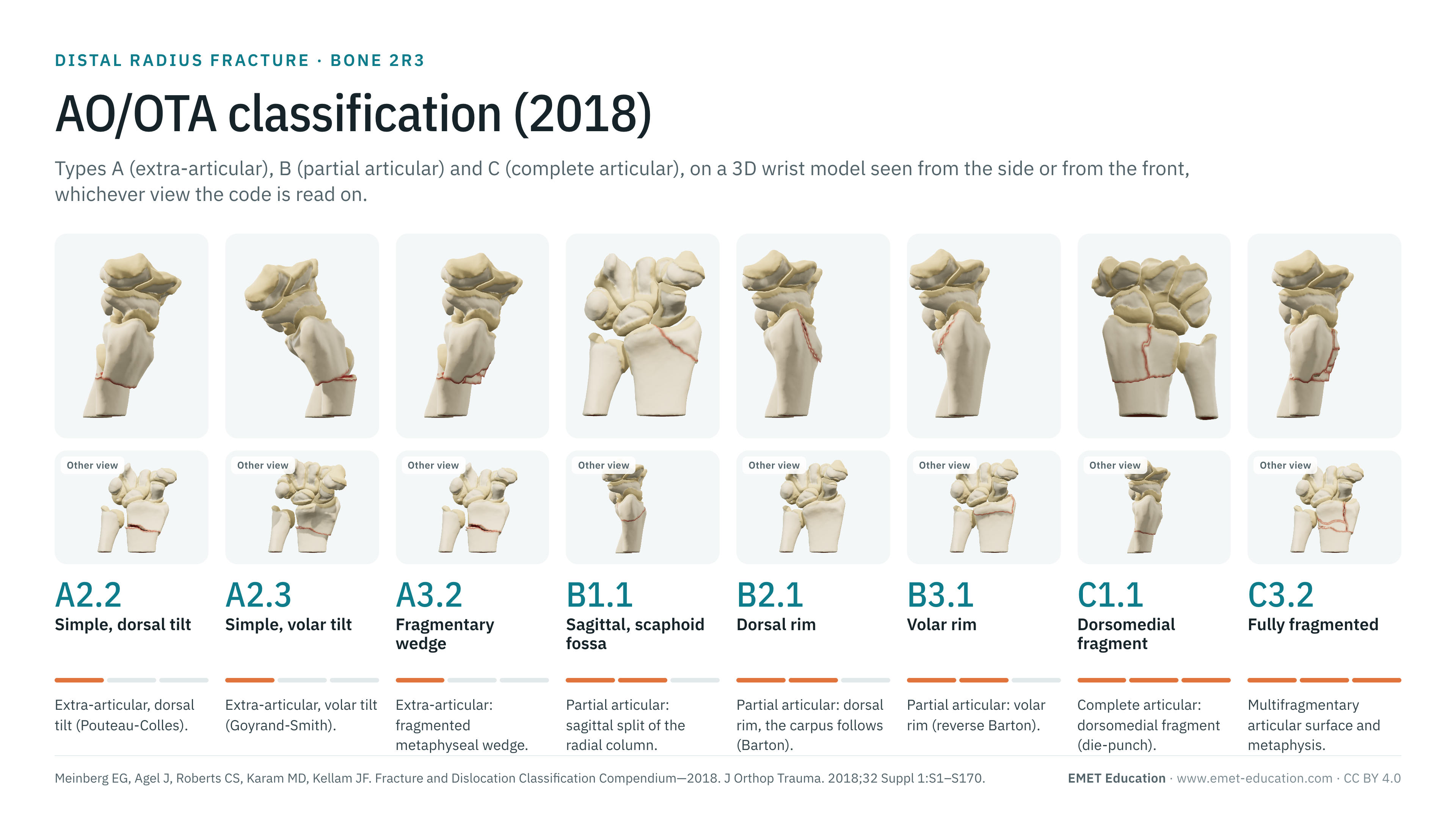
The types, groups and subgroups of the AO/OTA 2018 classification of the distal radius (bone 2R3), on the distal radius fracture 3D model, seen from the side or from the front depending on the radiograph on which the code is read. With how to read the code, the link with eponyms (Pouteau-Colles, Goyrand-Smith, Barton, chauffeur's, die-punch), the ulna 2U3, reliability data, displacement thresholds, grading pitfalls and free infographics for teaching and publication.
Designed by an orthopedic surgeon
The 3D model opens in your browser, at the code shown, with no sign-up. Infographics are free to reuse under CC BY 4.0.
- 2018Revision of the AO/OTA compendium, in the Journal of Orthopaedic Trauma (Meinberg et al.)
- 9Groups of the distal radius, from 2R3A1 to 2R3C3; the ulna now has its own codes (2U3)
- 2,757Citations of the 2018 compendium, all bones (OpenAlex, October 31, 2026)
- κ 0.34Interobserver agreement of the 2018 code among 10 hand surgeons, 50 radiographs (Fernandez: 0.57)
The codes
Types A, B and C, in 3D
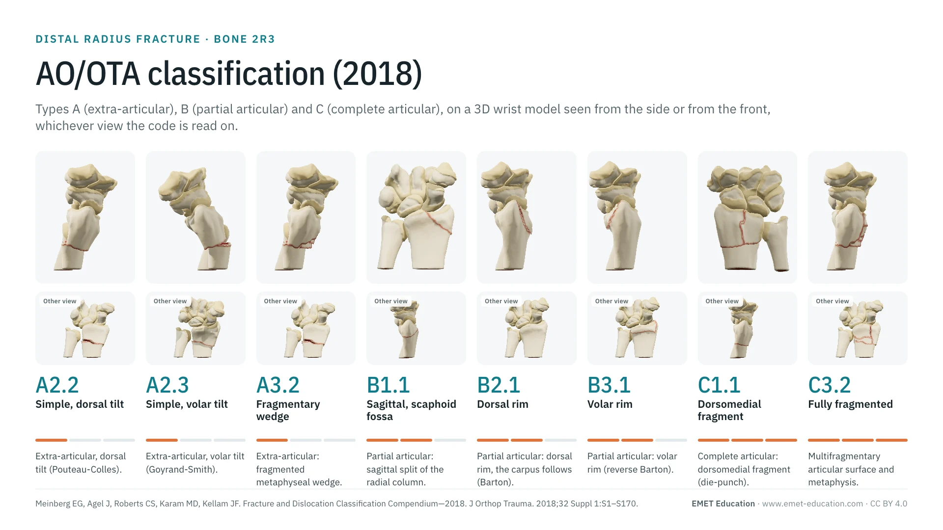
Each image is a rendering of the EMET Education distal radius fracture model at that code, wrist only (the hand is hidden, the carpus stays), with a narrow field to approach the projection of a radiograph. A code is read on the view where its line shows: lateral for tilt, dorsal comminution and frontal lines (type A, dorsal and volar rim, 2R3C1.3, 2R3C2.2, 2R3C3.2), anteroposterior for the styloid and sagittal lines (2R3B1.1, 2R3C1.2, 2R3C2.1, 2R3C3.1), from behind for the dorsomedial fragment (2R3C1.1, 2R3C2), whose line shows on neither of the model's two views; the other view is shown in the infographic. The model draws fifteen of the twenty-four codes described here; the other nine are described without an image. The displacements shown are typical cases: the code itself does not say whether the fracture is displaced (that is the role of the modifiers), except in the A2 subgroups, which separate no tilt, dorsal tilt and volar tilt. Each row gives the usual eponym, when there is one.
-
2R3A1Radial styloid avulsion
- Definition
- Type A, group 1: extra-articular avulsion fracture of the radial styloid. No subgroup. In 2007, group A1 described the distal ulna fracture, which now has its own codes (2U3).
- Eponym
- —
- In the 3D model
- Not drawn: the model's radial styloid fracture enters the joint (2R3B1.1).
-
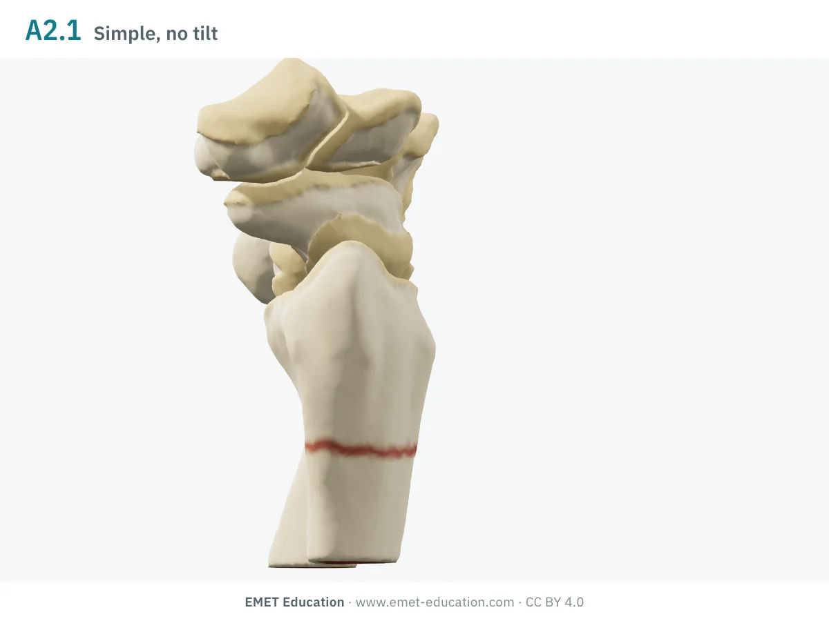
2R3A2.1Simple, no tilt
- Definition
- Type A, group 2: simple extra-articular fracture of the metaphysis, with no displacement or abnormal tilt (impaction or shortening may be present).
- Eponym
- —
- In the 3D model
- Transverse metaphyseal line, fragments aligned: the palmar tilt and radial inclination of the intact radius are kept.
-
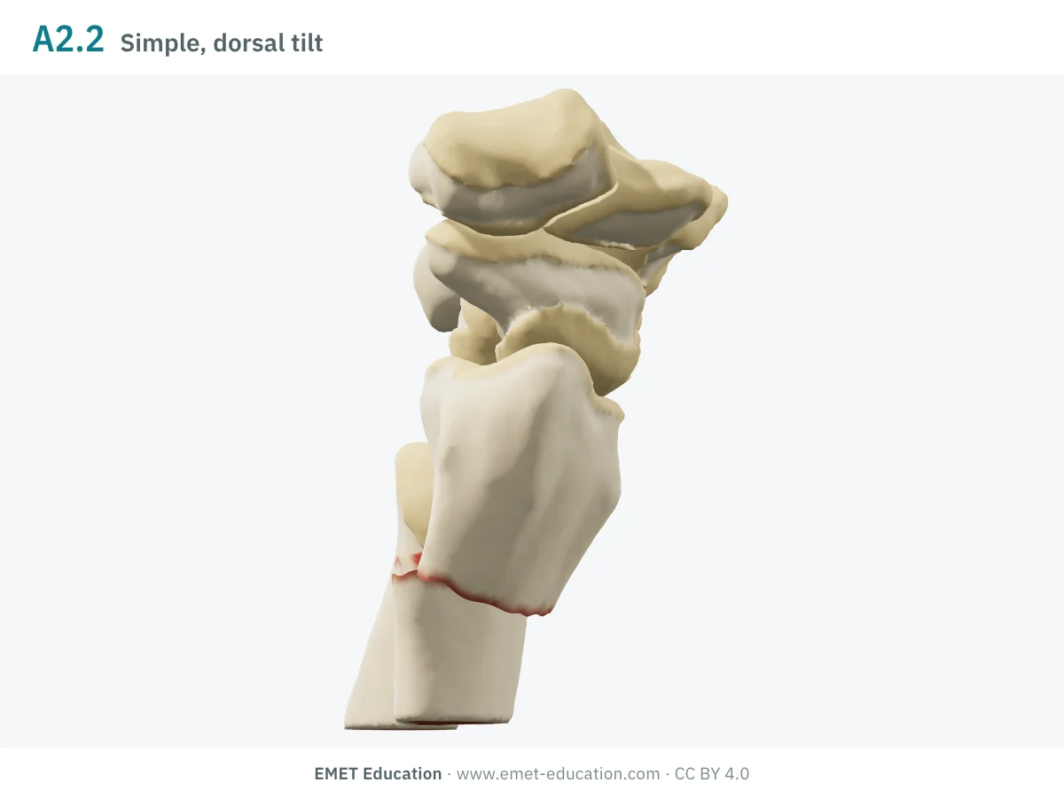
2R3A2.2Simple, dorsal tilt
- Definition
- Type A, group 2: simple extra-articular fracture with impaction and dorsal tilt (or displacement); it is usually stable in its displaced position, which may or may not be acceptable.
- Eponym
- Pouteau-Colles (AO Surgery Reference)
- In the 3D model
- Dorsal tilt of 20° added to that of the intact radius, 9° loss of inclination, 1.8 mm dorsal translation: the distal fragment tips about its palmar hinge.
-
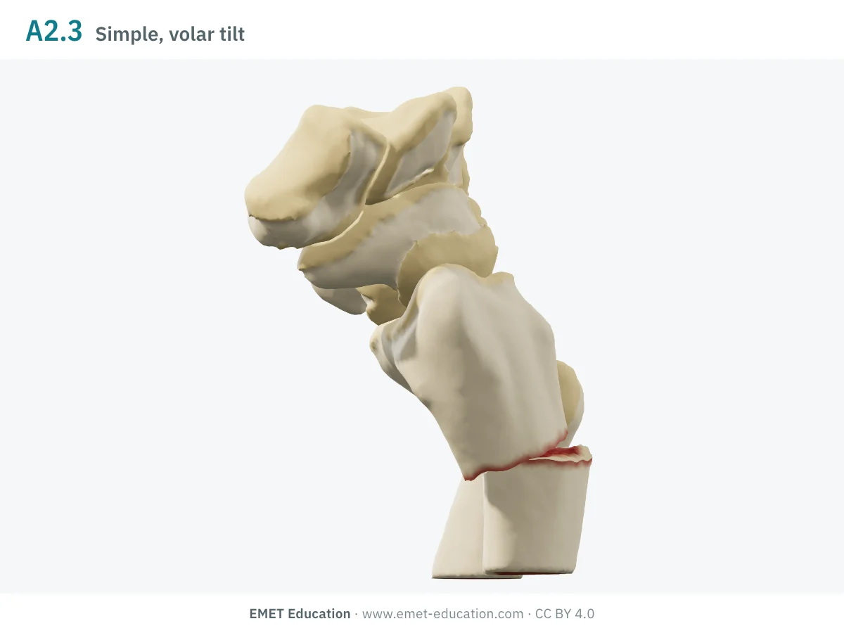
2R3A2.3Simple, volar tilt
- Definition
- Type A, group 2: simple extra-articular fracture with volar tilt (or displacement); markedly less stable than the fracture with dorsal tilt.
- Eponym
- Goyrand-Smith, Smith (AO Surgery Reference)
- In the 3D model
- Volar tilt of 20° added, hinge at the middle of the metaphysis, 1.8 mm volar translation: the carpus follows the fragment.
-
2R3A3.1Intact wedge
- Definition
- Type A, group 3: extra-articular metaphyseal fracture that detaches a wedge in a single fragment.
- Eponym
- —
- In the 3D model
- Not drawn: the model does not cut a single-fragment wedge.
-
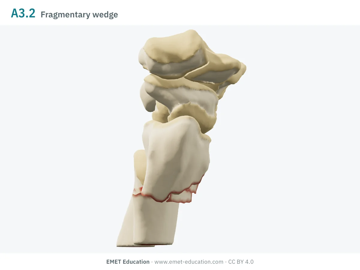
2R3A3.2Fragmentary wedge
- Definition
- Type A, group 3: as 2R3A3.1, but the wedge is itself fragmented.
- Eponym
- —
- In the 3D model
- Dorsal wedge of the metaphysis broken into five splinters, palmar cortex in one piece; dorsal tilt of 20°. EMET Education's reading: the model does not draw a metaphysis fragmented all around (2R3A3.3).
-
2R3A3.3Multifragmentary
- Definition
- Type A, group 3: the metaphysis is fragmented, with no involvement of the articular surface.
- Eponym
- —
- In the 3D model
- Not drawn in the model.
-
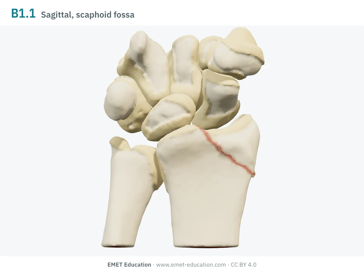
2R3B1.1Sagittal, scaphoid fossa
- Definition
- Type B, group 1 (sagittal): partial articular fracture of the radial column, through the scaphoid fossa; the ulnar part of the surface stays attached to the shaft. A multifragmentary fracture of this fossa is also coded 2R3B1.1.
- Eponym
- Chauffeur's (Hutchinson), EMET Education's reading
- In the 3D model
- Radial styloid detached by a line from the ridge between the fossae to the radial border; 0.8 mm gap, the styloid drops, the carpus stays.
-
2R3B1.3Sagittal, lunate fossa
- Definition
- Type B, group 1: partial articular fracture of the ulnar column, through the lunate fossa; it therefore involves the radiocarpal and the distal radioulnar joints.
- Eponym
- —
- In the 3D model
- Not drawn in the model.
-
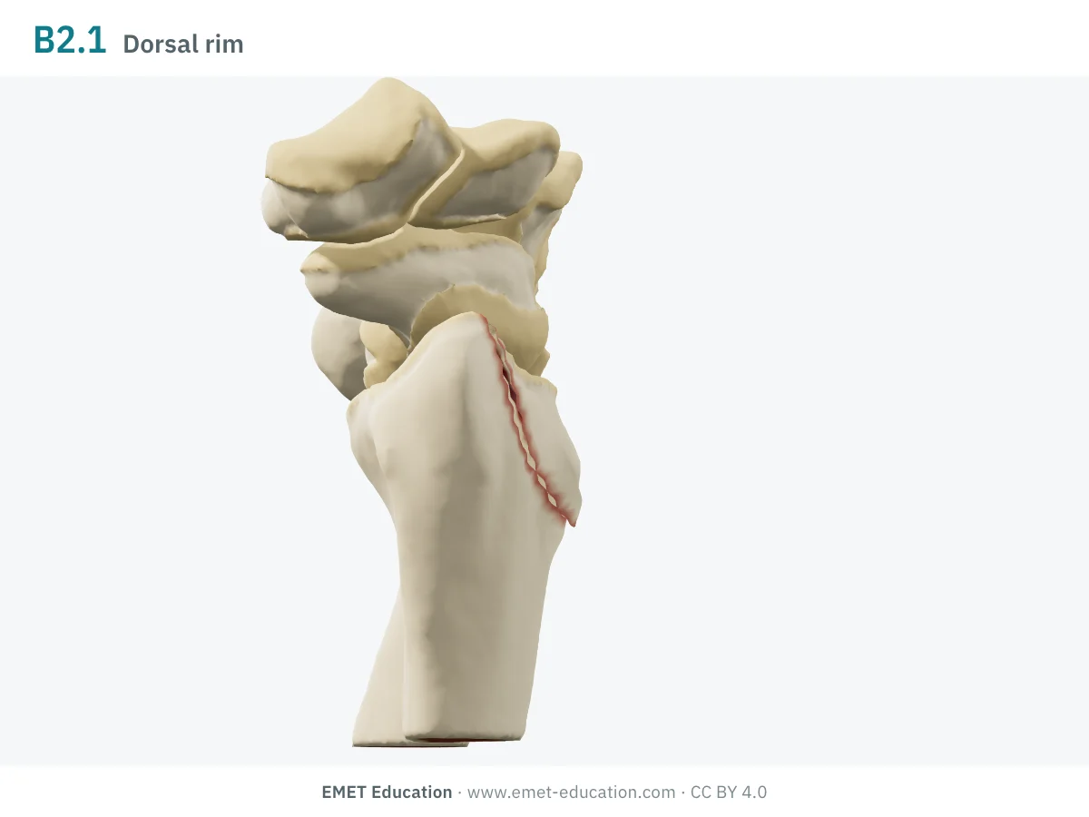
2R3B2.1Dorsal rim
- Definition
- Type B, group 2: simple partial articular fracture of the dorsal rim of the radius: a fragment, often small, close to the rim, which lets the carpus subluxate dorsally.
- Eponym
- Barton
- In the 3D model
- Dorsal third of the surface detached; it slides toward the forearm and tilts, the carpus following it dorsally.
-
2R3B2.2Dorsal rim, fragmentary
- Definition
- Type B, group 2: fragmentary fracture of the dorsal rim, associated with a lateral sagittal fracture (radial styloid); to be reduced and stabilized to restore radiocarpal stability.
- Eponym
- Barton
- In the 3D model
- Not drawn in the model.
-
2R3B2.3Dorsal rim, with dorsal dislocation
- Definition
- Type B, group 2: as the previous subgroups, with greater subluxation of the carpus, often a complete dorsal dislocation; soft-tissue damage is usually significant, with the median nerve at risk.
- Eponym
- Barton
- In the 3D model
- Not drawn in the model.
-
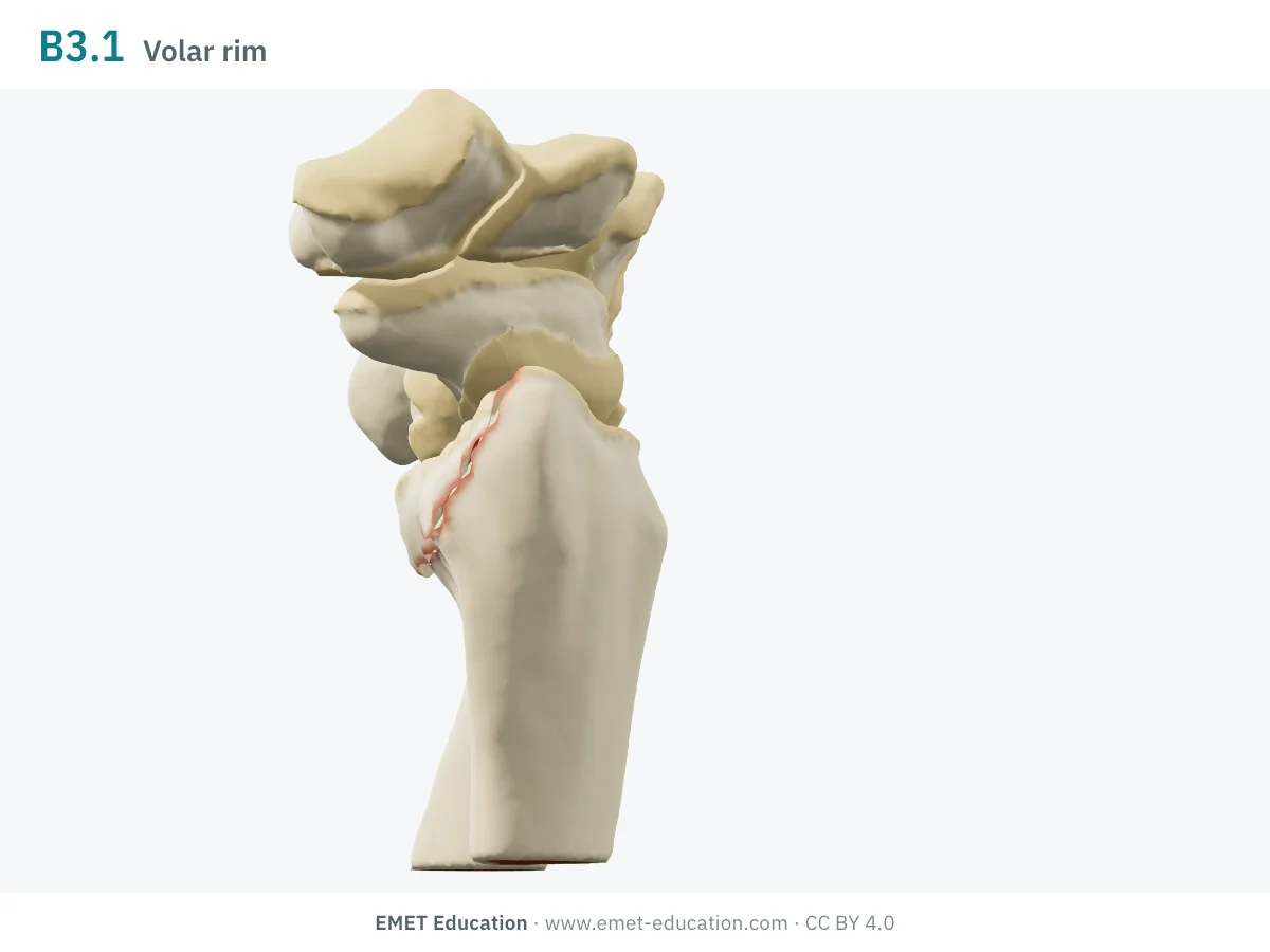
2R3B3.1Volar rim
- Definition
- Type B, group 3: simple partial articular fracture of the volar rim of the radius.
- Eponym
- Reverse Barton, Goyrand-Smith II
- In the 3D model
- Volar third of the surface detached; it slides toward the forearm and tilts, the carpus following it volarly.
-
2R3B3.3Volar rim, fragmentary
- Definition
- Type B, group 3: fragmentary fracture of the volar rim. There is no 2R3B3.2.
- Eponym
- Reverse Barton, Goyrand-Smith II
- In the 3D model
- Not drawn in the model.
-
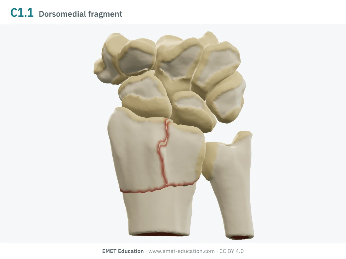
2R3C1.1Dorsomedial fragment
- Definition
- Type C, group 1 (simple articular, simple metaphysis): complete articular fracture whose line isolates a dorsomedial fragment, the dorsal part of the lunate fossa and the sigmoid notch; closed reduction of it is difficult.
- Eponym
- Die-punch (Melone), EMET Education's reading
- In the 3D model
- Dorsomedial fragment cut from the lunate fossa, separated and depressed relative to the rest of the surface: it makes an articular step-off.
-
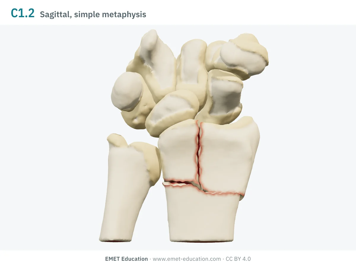
2R3C1.2Sagittal, simple metaphysis
- Definition
- Type C, group 1: complete articular fracture with a single sagittal line, the metaphysis being simple.
- Eponym
- —
- In the 3D model
- Sagittal split between the two columns, above a metaphyseal line; the columns separate 0.8 mm and make a step.
-
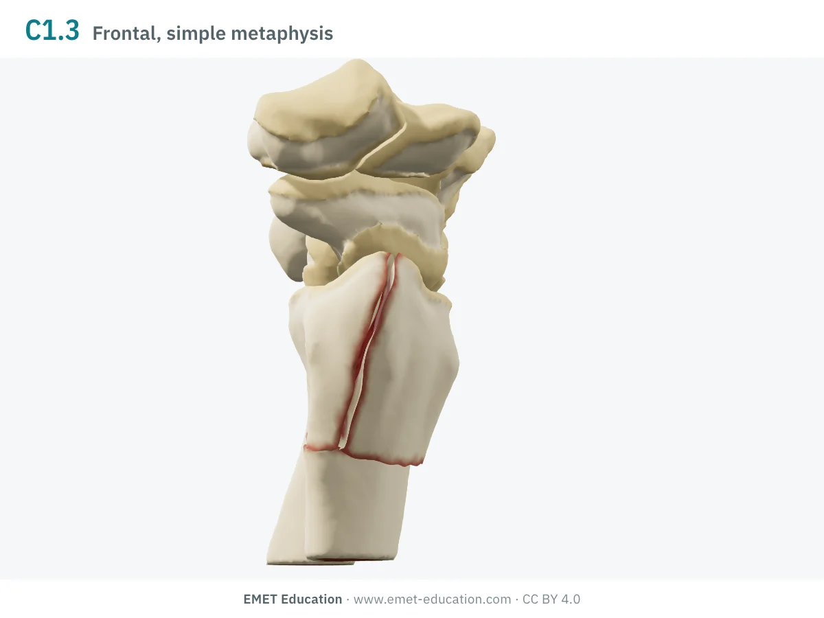
2R3C1.3Frontal, simple metaphysis
- Definition
- Type C, group 1: complete articular fracture with a single frontal (coronal) line, the metaphysis being simple.
- Eponym
- —
- In the 3D model
- Frontal split: the palmar half of the surface separates from the dorsal half, above a metaphyseal line.
-
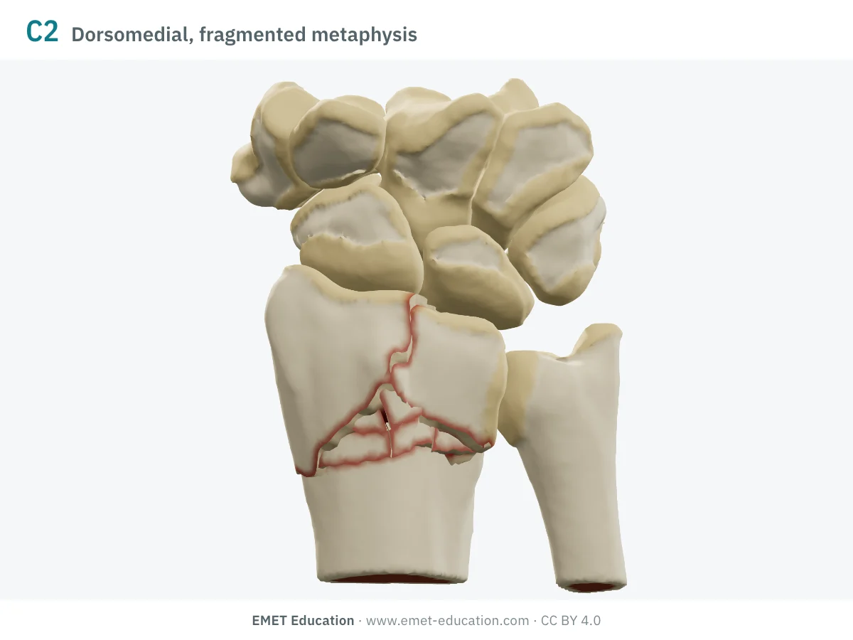
2R3C2Simple articular, multifragmentary metaphysis
- Definition
- Type C, group 2: complete articular fracture with a simple articular line and a multifragmentary metaphysis. The group has three subgroups (sagittal, frontal, diaphyseal extension); a dorsomedial fragment has none: it is coded at group level.
- Eponym
- —
- In the 3D model
- Depressed dorsomedial fragment, with the dorsal metaphyseal wedge broken into splinters; EMET Education's reading, the compendium has no subgroup for it.
-
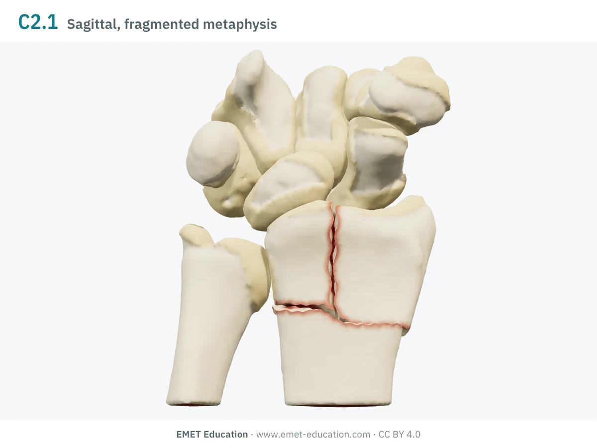
2R3C2.1Sagittal, multifragmentary metaphysis
- Definition
- Type C, group 2: simple sagittal articular line, multifragmentary metaphysis.
- Eponym
- —
- In the 3D model
- Sagittal split of the surface and the dorsal metaphyseal wedge in splinters.
-
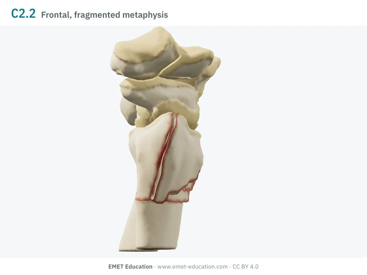
2R3C2.2Frontal, multifragmentary metaphysis
- Definition
- Type C, group 2: simple frontal articular line, multifragmentary metaphysis.
- Eponym
- —
- In the 3D model
- Frontal split of the surface and the dorsal metaphyseal wedge in splinters.
-
2R3C2.3Simple articular, diaphyseal extension
- Definition
- Type C, group 2: simple articular line, multifragmentary metaphysis and a fracture extending into the shaft.
- Eponym
- —
- In the 3D model
- Not drawn: the model does not extend the fracture into the shaft.
-
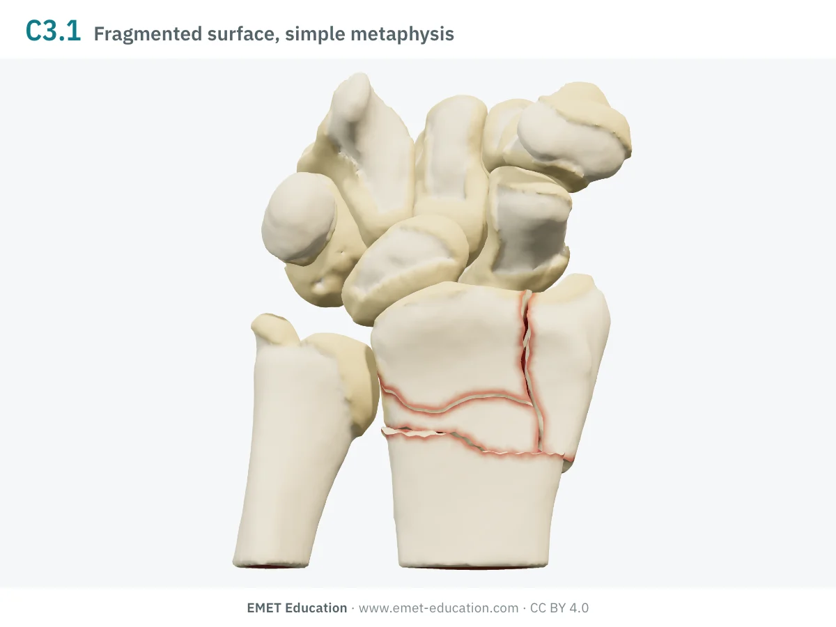
2R3C3.1Fragmented surface, simple metaphysis
- Definition
- Type C, group 3: multifragmentary articular surface, simple metaphyseal line.
- Eponym
- —
- In the 3D model
- Radial column and palmar fragment intact, the dorsal zone under the cartilage burst into six fragments of 39 to 464 mm³, spread dorsally and more or less depressed.
-
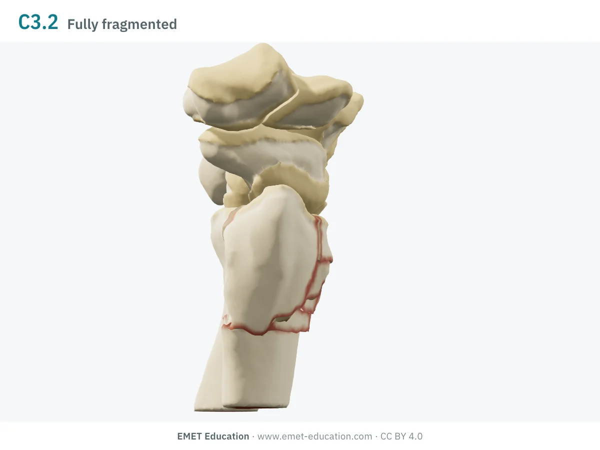
2R3C3.2Fragmented surface and metaphysis
- Definition
- Type C, group 3: multifragmentary articular surface and metaphysis.
- Eponym
- —
- In the 3D model
- As 2R3C3.1, with the dorsal metaphyseal wedge in splinters.
-
2R3C3.3Fragmented surface, diaphyseal extension
- Definition
- Type C, group 3: multifragmentary articular surface, the fracture extending into the shaft.
- Eponym
- —
- In the 3D model
- Not drawn in the model.
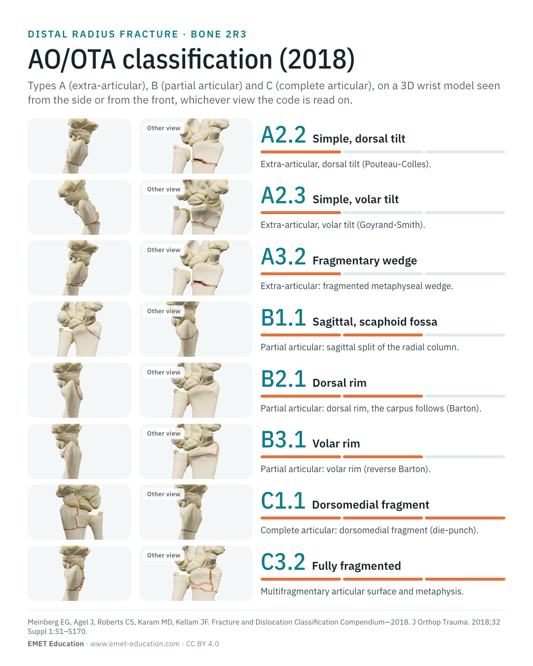
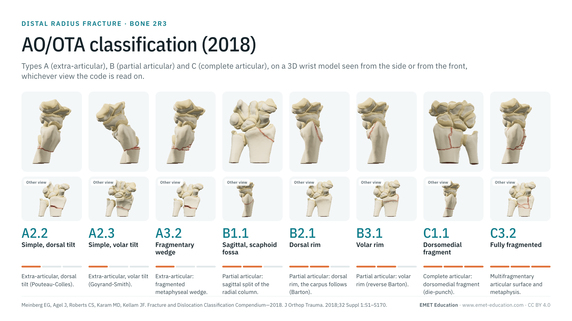
Schematic rendering, for teaching: the model illustrates each code on a single wrist, it does not reproduce a radiograph. Definitions reworded from the 2018 compendium (Meinberg et al.) and the AO Surgery Reference; their figures and pictograms are not reproduced. The bar shows the type: one segment A, two B, three C. Subgroups C1 to C3 carry an optional qualification of distal radioulnar joint stability (t or u), not shown here.
Reading the code
From Müller (1990) to the 2018 compendium
The classification comes from Müller et al.'s classification of long-bone fractures, adopted by the Orthopaedic Trauma Association. In that version, the distal radius and ulna formed a single segment, with nine groups and twenty-seven subgroups (Andersen et al.). The 2007 compendium carries it on; the 2018 compendium separates the radius classification from the ulna one, adds a qualification of distal radioulnar joint stability to complete articular fractures of the radius (Nogueira et al.), and leaves some subgroup numbers vacant without renumbering the others. The page follows the 2018 compendium.
The code reads from left to right:
- 2R3: bone 2, the forearm; the letter R, the radius (U for the ulna: a injury to both bones is coded twice); its segment 3, the distal end. The distal segment is the square whose side is the widest part of the epiphysis;
- the type: A, extra-articular fracture; B, partial articular (part of the joint surface stays in continuity with the shaft); C, complete articular (no part of the surface stays attached to it);
- the group and subgroup: A1 radial styloid avulsion, A2 simple line, A3 wedge or multifragmentary; B1 sagittal, B2 dorsal rim, B3 volar rim; C1 simple articular and simple metaphysis, C2 simple articular and multifragmentary metaphysis, C3 multifragmentary articular. The missing numbers (2R3B1.2, 2R3B3.2) do not exist in 2018;
- the qualification, a lower-case letter in round brackets, optional, on C subgroups: t, distal radioulnar joint stable; u, unstable. Fractures of the distal ulna are coded separately: 2U3A1.1 (tip of the styloid) and 2U3A1.2 (base of the styloid), 2U3A2 (simple), 2U3A3 (multifragmentary);
- universal modifiers, as numbers in square brackets, shared by all bones (displacement, impaction, bone quality…): see the proximal humerus page, which lists them. Thus 2R3A2.2[2]: simple fracture with dorsal tilt, displaced.
In the series by Nogueira et al. (322 adult distal forearm fractures, a single hospital, coded in 2018), type C was the most common in the radius (48.4%), ahead of A (32.3%) and B (18.0%); the distal ulna was fractured in 41.9% of cases, most often at the tip of the styloid (2U3A1.1, 30.7%). A single-centre series is not an incidence: the proportions depend on recruitment.
Eponyms
Pouteau-Colles, Goyrand-Smith, Barton, chauffeur's, die-punch
Before classifications, wrist fractures bore the names of those who described them; the AO classification sorts them into types A, B and C (Thurston). The correspondence is not always one to one: the table gives the code where each eponym most often fits.
| Eponym | AO/OTA 2018 code | Note |
|---|---|---|
| Pouteau-Colles (Colles) | 2R3A2.2 | Simple extra-articular fracture with impaction and dorsal tilt, usually stable in its displaced position (AO Surgery Reference). Described by Colles in 1814 (Colles) and also by Pouteau (Thurston). With dorsal comminution: A3. |
| Goyrand-Smith (Smith) | 2R3A2.3 | Simple extra-articular fracture with volar tilt, markedly less stable than the previous one (AO Surgery Reference). Described by Goyrand before Smith (1847) (Thurston). |
| Barton | 2R3B2 (2R3B2.1 to 2R3B2.3) | Fracture of the dorsal rim of the radius, with dorsal subluxation of the carpus: the compendium names it so ("Barton's") at group B2. |
| Reverse Barton, Goyrand-Smith II | 2R3B3 (2R3B3.1, 2R3B3.3) | Fracture of the volar rim: the compendium gives both names at group B3. Usage wavers between "Smith II" and "reverse Barton" (Thurston). |
| Chauffeur's (Hutchinson) | 2R3B1.1 | Oblique fracture of the radial styloid entering the joint, described by Hutchinson in 1866, named in 1904 (Andreotti et al.). EMET Education's correspondence: the radial column is the scaphoid fossa. |
| Die-punch (Melone) | 2R3C1.1 (or C2, C3) | For Melone, most articular fractures of the distal radius result from a "die-punch" mechanism (Melone). EMET Education's correspondence: the dorsomedial fragment of 2R3C1.1. |
Reliability
How reproducible is the code?
The type is far more reproducible than the group, and the group than the subgroup. As early as 1996, Kreder et al. found high agreement on the AO type and on articular displacement, and growing difficulty agreeing on the group and then the subgroup; Andersen et al. did not improve agreement by cutting the 27 categories to 9, but did by keeping only the three types. In the only study of the 2018 version that we found, agreement is lower than for Fernandez.
| Study | Measure | Value |
|---|---|---|
| Kreder et al., 1996 | AO classification of the time, 36 assessors of varying experience, 30 radiographs; kappas are not in the abstract | type: high; group, subgroup: difficult |
| Andersen et al., 1996 | Frykman, Melone, Mayo, AO: inter- then intraobserver agreement of the AO, 4 readers, 55 radiograph sets (Mayo: moderate); the three types alone: "substantial" | fair · fair |
| Illarramendi et al., 1998 | Inter- then intraobserver kappa of the AO, 6 readers, 200 fractures (Frykman: 0.43 · 0.61) | 0.37 · 0.57 |
| Belloti et al., 2008 | Mean intraobserver kappa of the AO, 5 readers, 98 displaced fractures (Universal: 0.61; Fernandez: 0.59; Frykman: 0.55); interobserver agreement "unsatisfactory" for all | intra 0.49 |
| Wæver et al., 2018 | Interobserver kappa of the AO/OTA, 4 readers, 573 radiographs: types (A, B, C), then groups (Frykman: 0.41; Older: 0.10) | 0.45 · 0.24 |
| Carula et al., 2022 | 2018 version, inter- then intraobserver kappa, 10 hand surgeons, 50 radiographs (Fernandez: 0.57 · 0.55) | 0.34 · 0.31 |
The authors conclude that the 2018 code has low reproducibility (Carula et al.) and prefer Fernandez for routine use; Wæver et al. find the AO/OTA the most reliable of the three systems they compare, with low group-level kappas. In practice: the type to communicate, the group or subgroup for research, with written thresholds, and a CT if one needs to tell B from C.
Neighbouring classifications
Frykman, Melone, Fernandez, Universal, Older
Several classifications of the distal radius coexist: none has prevailed. The AO/OTA is the most used in research and registries because its code fits on one line; the others describe an aspect the AO/OTA does not tell, or steer treatment.
The neighbouring classifications, and how they relate to the AO/OTA:
| Classification | Principle | Relation to the AO/OTA |
|---|---|---|
| Frykman (1967) | Eight types, depending on whether the fracture involves the radiocarpal joint, the distal radioulnar joint, both or neither, and whether an ulnar styloid fracture is associated (even types) (Frykman). | Describes neither displacement nor comminution. Reliability of the same order as the AO/OTA (κ 0.41 against 0.45 for the types, Wæver). |
| Melone (1993) | Articular fractures, by the "die-punch" mechanism: type I stable, type II unstable (IIA reducible, IIB irreducible), III with a spike fragment, IV with wide separation of articular fragments, V "explosion" (Melone). | Overlaps types B and C; a single AO/OTA code covers several Melone types. |
| Fernandez (2001) | Five groups by mechanism of injury, with associated distal radioulnar lesions, management recommendations and an alphanumeric code for computer documentation (Fernandez). | Better agreement than the AO/OTA 2018 in Carula et al. (κ 0.57 against 0.34), and in Belloti et al. for interobserver agreement (0.44). |
| Universal (Cooney, 1993) | A treatment-based classification: it directs management, with possible subclassifications (Cooney). | Best mean intraobserver agreement of four classifications compared (0.61, against 0.49 for the AO, Belloti). |
| Older (1965) | Classification of Colles' fractures, to choose treatment (Older et al.). | The least reproducible of the three systems of Wæver et al. (κ 0.10). |
Use
In research and in clinical practice
In research
The AO/OTA code is the language of registries, databases and multicentre trauma studies: it fits on one line, sorts and groups. To read literature published before 2018, one needs to know which version was used: group A1 does not designate the same fracture there and the qualification of the distal radioulnar joint does not exist (Nogueira et al.).
In clinical practice
The type, A, B or C, sums up the essentials: extra-articular fracture, partial articular fracture (a fragment stays attached to the shaft) or complete. The code alone does not tell whether the fracture is stable. The AO Surgery Reference notes that a fracture with dorsal tilt (2R3A2.2) is usually stable in its displaced position, that one with volar tilt (2R3A2.3) is much less so, and that a fragmentary dorsal rim (2R3B2.2) is reduced and stabilized to restore carpal stability (AO Surgery Reference).
It is the radiographic parameters, more than the code, that steer treatment. The AAOS guideline (moderate strength) proposes surgical fixation rather than casting when, after reduction, shortening exceeds 3 mm, dorsal tilt 10° or articular step-off 2 mm (Lichtman et al.). In an earlier review, Nana et al. accepted shortening up to 5 mm, radial inclination above 15°, tilt from 15° dorsal to 20° volar and a step-off under 2 mm: thresholds vary between sources, and the model uses the strictest (dorsal tilt > 10°, inclination < 15°, shortening > 3 mm, step ≥ 2 mm). For the risk of secondary displacement, Lafontaine et al. summed dorsal tilt over 20°, dorsal comminution, articular involvement, an associated ulnar fracture and age over 60; Mackenney et al. found age, metaphyseal comminution and ulnar variance the most consistent predictors, and not dorsal tilt for displaced fractures.
Grading pitfalls
Five pitfalls when coding a distal radius fracture
- Type first. Each level of the code adds disagreement between readers. For decisions and discussion, the type is often enough; the full code serves research, with written rules.
- The radius and the ulna are coded separately. An ulnar styloid fracture is coded separately, with its own code (2U3A1.1 or 2U3A1.2). Do not confuse 2R3A1 (radial styloid avulsion) with the 2007 A1 (ulna); state the version in any publication.
- B or C? The question is whether any part of the articular surface stays attached to the shaft. On plain radiographs, agreement on the type is only weak (Wæver et al.): when in doubt, CT helps. A sagittal split of the scaphoid fossa stays a B1.1, even if fragmentary; C begins when the surface is detached from the shaft.
- A2 or A3? A simple line with dorsal tilt is an A2.2; with a wedge or a fragmented metaphysis, it is an A3, whatever the deformation. The names of the A3 subgroups differ between sources (intact wedge, fragmentary wedge, multifragmentary in the 2018 compendium; impaction, angulated wedge, multifragmentary in the AO Surgery Reference): cite the compendium (Meinberg et al.).
- The t or u qualification. It applies only to C subgroups, and it is optional: a code without a letter does not mean the distal radioulnar joint is stable. It supposes assessing the joint, the 2018 version integrating the physical examination (Nogueira et al.); without that assessment, do not write a letter.
Self-test
Which code?
The same wrist seen from behind, at ten of the model's codes, in random order. Find the code of each one; the answer comes with the definition.
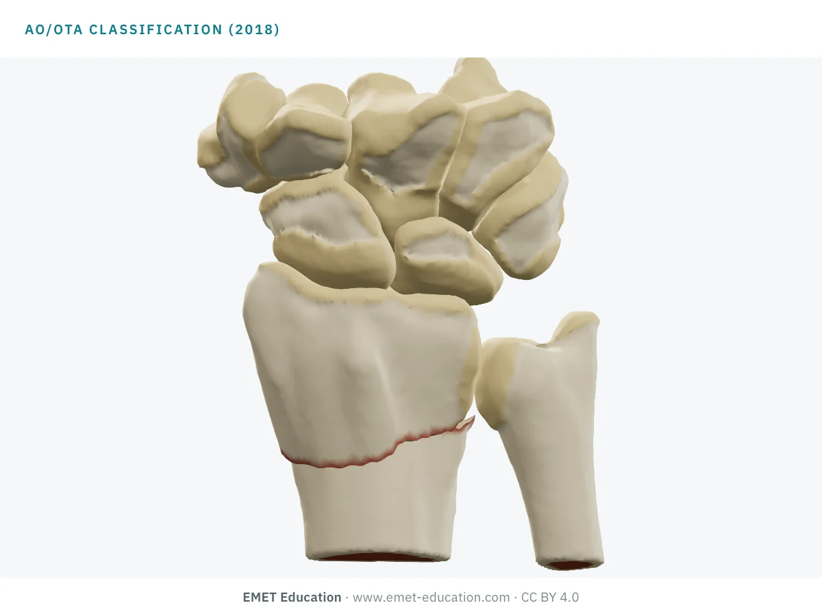
Which AO/OTA code is this?
Download and cite
Free infographics, for teaching and publication
Use them in a course, a presentation, a social media post or a scientific article, under the Creative Commons Attribution 4.0 license: free reuse, including commercial, provided EMET Education is credited with a link to this page. The license covers EMET Education's images, not the classification itself.
Cite this figure
Ready to paste. For a journal figure, add the original reference (Meinberg et al., 2018).
EMET Education. AO/OTA classification of distal radius fractures: 3D visualization [Internet]. 2026 [cited]. Available from: https://www.emet-education.com/en/classifications/ao-distal-radius/. CC BY 4.0.EMET Education. (2026). AO/OTA classification of distal radius fractures: 3D visualization [Infographic]. https://www.emet-education.com/en/classifications/ao-distal-radius/ (CC BY 4.0)<a href="https://www.emet-education.com/en/classifications/ao-distal-radius/"><img src="https://www.emet-education.com/en/classifications/ao-distal-radius/img/infographic.webp" alt="AO/OTA classification of distal radius fractures in 3D" width="960"></a><br>Figure: <a href="https://www.emet-education.com/en/classifications/ao-distal-radius/">EMET Education</a>, CC BY 4.0Questions
Frequently asked questions
What does "2R3A2.2" mean in the code?
The 2 is the forearm, the letter R the radius (U: the ulna), the 3 the distal segment. A is the type (extra-articular fracture), 2 the group (simple line) and .2 the subgroup (dorsal tilt, the Pouteau-Colles). Modifiers, in square brackets, and the distal radioulnar joint qualification (t or u, on C subgroups) come after.
What is the difference between the 2007 and 2018 AO classifications of the distal radius?
In 2018, the radius classification (2R3) is separated from the ulna one (2U3); an optional qualification describes the stability of the distal radioulnar joint (t or u) in complete articular fractures; and the numbers of some subgroups disappeared without the others being renumbered (no 2R3B1.2 or 2R3B3.2). The descriptive criteria of articular fractures are maintained.
Which AO codes for Pouteau-Colles, Goyrand-Smith and Barton?
Pouteau-Colles: 2R3A2.2 (simple extra-articular fracture, dorsal tilt). Goyrand-Smith: 2R3A2.3 (volar tilt). Barton: 2R3B2 (dorsal rim); reverse Barton, or Goyrand-Smith II: 2R3B3 (volar rim). Chauffeur's is usually read 2R3B1.1 and the die-punch 2R3C1.1, EMET Education's correspondences. Dorsal comminution moves a Colles to 2R3A3.
AO/OTA, Frykman or Fernandez: which one to use?
None has high reliability. The AO/OTA is the most used in research; in the study by Carula et al. on the 2018 version, Fernandez agreed better (κ 0.57 against 0.34 between readers). Frykman describes neither displacement nor comminution. Whatever the choice, state the version and measure the radiographic parameters (tilt, inclination, shortening, step-off).
Does the AO/OTA code say whether the fracture is stable or unstable?
Not by itself. The subgroup gives a lead (Colles is usually stable, Smith much less so), but instability is judged on age, dorsal comminution, tilt, shortening, articular involvement and the associated ulnar fracture (Lafontaine, Mackenney). The AAOS guideline proposes surgery for shortening over 3 mm, dorsal tilt over 10° or a step-off over 2 mm after reduction.
Can I reuse these images in a course or an article?
Yes. The images and infographics on this page are under the Creative Commons Attribution 4.0 license: free reuse, including commercial, with credit to EMET Education and a link to this page. The "Cite this figure" box gives ready-to-paste citations.
Do the images reproduce the AO drawings? How were they made?
No: no figure or pictogram from the compendium is reproduced, and the definitions are reworded. They are renderings of the EMET Education distal radius fracture model, set to each code: the radius and ulna of a real hand, taken from CT, are cut along the lines of the code and displaced so that they never cross each other. It is a schematic model for teaching; it does not reproduce a radiograph. EMET Education is not affiliated with the AO Foundation or the Orthopaedic Trauma Association.
Sources
Author and references
- Meinberg EG, Agel J, Roberts CS, Karam MD, Kellam JF. Fracture and Dislocation Classification Compendium—2018. J Orthop Trauma. 2018;32 Suppl 1:S1–S170. doi:10.1097/BOT.0000000000001063
- AO Foundation. AO Surgery Reference, orthopedic trauma, adult trauma, distal forearm (fracture types and AO/OTA codes). surgeryreference.aofoundation.org. Consulté le 31 octobre 2026.
- Marsh JL, Slongo TF, Agel J, et al. Fracture and dislocation classification compendium – 2007: Orthopaedic Trauma Association classification, database and outcomes committee. J Orthop Trauma. 2007;21(10 Suppl):S1–133. doi:10.1097/00005131-200711101-00001
- Müller ME, Nazarian S, Koch P, Schatzker J. The Comprehensive Classification of Fractures of Long Bones. Berlin: Springer; 1990.
- Nogueira AF, Moratelli L, Martins MDS, et al. Evaluation of distal forearm fractures using the AO 2018 classification. Acta Ortop Bras. 2019;27(4):220–2. doi:10.1590/1413-785220192704218467
- Carula BC, Giostri GS, Bebber F, Batista MFDS, da Silva FB, Novak EM. Evaluation of intra- and interobserver reproducibility of the new AO/OTA classification for distal radius fractures compared with the Fernandez classification. Rev Bras Ortop (Sao Paulo). 2022;57(6):917–23. doi:10.1055/s-0041-1740471
- Kreder HJ, Hanel DP, McKee M, Jupiter J, McGillivary G, Swiontkowski MF. Consistency of AO fracture classification for the distal radius. J Bone Joint Surg Br. 1996;78(5):726–31.
- Andersen DJ, Blair WF, Steyers CM Jr, Adams BD, el-Khouri GY, Brandser EA. Classification of distal radius fractures: an analysis of interobserver reliability and intraobserver reproducibility. J Hand Surg Am. 1996;21(4):574–82. doi:10.1016/s0363-5023(96)80006-2
- Illarramendi A, González Della Valle A, Segal E, De Carli P, Maignon G, Gallucci G. Evaluation of simplified Frykman and AO classifications of fractures of the distal radius. Assessment of interobserver and intraobserver agreement. Int Orthop. 1998;22(2):111–5. doi:10.1007/s002640050220
- Belloti JC, Tamaoki MJ, Franciozi CE, et al. Are distal radius fracture classifications reproducible? Intra and interobserver agreement. Sao Paulo Med J. 2008;126(3):180–5. doi:10.1590/s1516-31802008000300008
- Wæver D, Madsen ML, Rölfing JHD, et al. Distal radius fractures are difficult to classify. Injury. 2018;49 Suppl 1:S29–32. doi:10.1016/S0020-1383(18)30299-7
- Frykman G. Fracture of the distal radius including sequelae—shoulder-hand-finger syndrome, disturbance in the distal radio-ulnar joint and impairment of nerve function. A clinical and experimental study. Acta Orthop Scand. 1967;Suppl 108:3+. doi:10.3109/ort.1967.38.suppl-108.01
- Melone CP Jr. Distal radius fractures: patterns of articular fragmentation. Orthop Clin North Am. 1993;24(2):239–53.
- Fernandez DL. Distal radius fracture: the rationale of a classification. Chir Main. 2001;20(6):411–25. doi:10.1016/s1297-3203(01)00067-1
- Cooney WP. Fractures of the distal radius. A modern treatment-based classification. Orthop Clin North Am. 1993;24(2):211–6.
- Older TM, Stabler EV, Cassebaum WH. Colles fracture: evaluation and selection of therapy. J Trauma. 1965;5:469–76.
- Lafontaine M, Delince P, Hardy D. [Instability of fractures of the lower end of the radius: apropos of a series of 167 cases]. Acta Orthop Belg. 1989;55(2):203–16.
- Mackenney PJ, McQueen MM, Elton R. Prediction of instability in distal radial fractures. J Bone Joint Surg Am. 2006;88(9):1944–51. doi:10.2106/JBJS.D.02520
- Lichtman DM, Bindra RR, Boyer MI, et al. Treatment of distal radius fractures. J Am Acad Orthop Surg. 2010;18(3):180–9. doi:10.5435/00124635-201003000-00007
- Nana AD, Joshi A, Lichtman DM. Plating of the distal radius. J Am Acad Orthop Surg. 2005;13(3):159–71. doi:10.5435/00124635-200505000-00003
- Thurston AJ. 'Ao' or eponyms: the classification of wrist fractures. ANZ J Surg. 2005;75(5):347–55. doi:10.1111/j.1445-2197.2005.03414.x
- Andreotti M, Tonon F, Caruso G, et al. The "chauffeur fracture": historical origins of an often-forgotten eponym. Hand (N Y). 2020;15(2):252–4. doi:10.1177/1558944718792650
- Colles A. On the fracture of the carpal extremity of the radius. Edinb Med Surg J. 1814 (reprinted: Clin Orthop Relat Res. 1972;(83):3–5).
3D models
Show the fracture to your patient
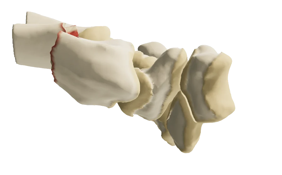
The images on this page come from an interactive model, the distal radius fracture 3D model. In clinic, choose the type and the line, set displacement and shortening from your patient's imaging, add the ulnar styloid fracture or distal radioulnar instability, and read the AO/OTA code and the radiographic parameters live.
 Hand and wristDistal radius fractureTypes A, B and C, displacement, shortening, ulnar styloid, distal radioulnar instability, live AO/OTA code; cast, Kapandji pinning, screw fixation, volar plate.
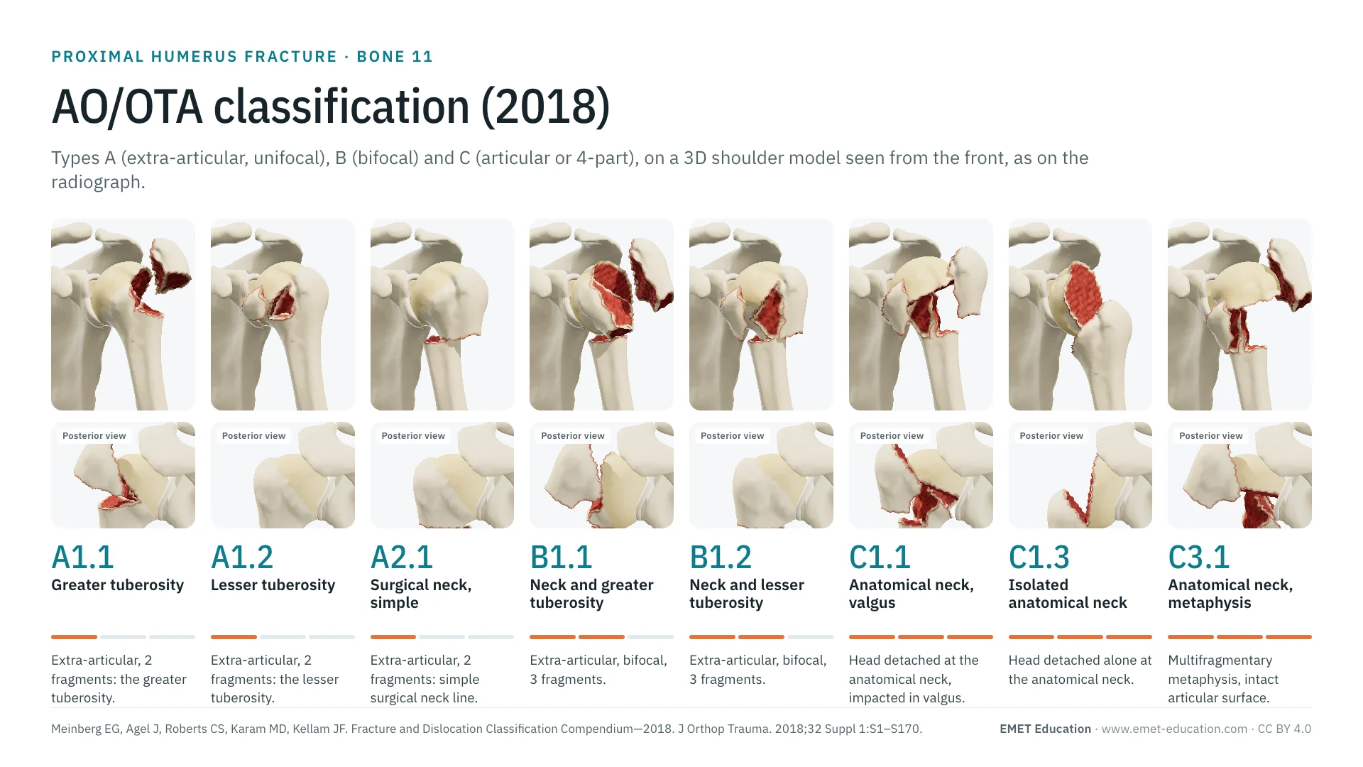 ClassificationsAO/OTA classification of the proximal humerusThe same classification, for the shoulder: types A, B and C, in 3D.
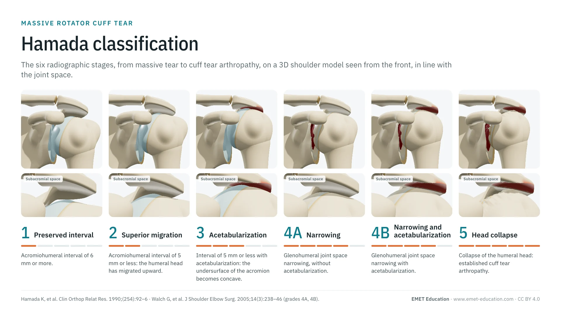 ClassificationsAll 3D classificationsArranged by joint.
Explain the fracture in 3D
The distal radius fracture model opens in your browser, at the code you choose, with no sign-up.
Share this page with a colleague or a resident: