- EMET Education
- 3D classifications
- AO/OTA classification (clavicle)
Classification · Clavicle fracture
AO/OTA Classification of Clavicle Fractures in 3D
The codes of the AO/OTA 2018 classification of the clavicle (bone 15), for the shaft (15.2) and the lateral end (15.3), on a 3D shoulder model seen from the front, beam tilted upward, as on the radiograph. With how to read the code, what the shaft does not subdivide, the link with Robinson and Neer, reliability data, grading pitfalls and free infographics for teaching and publication.
Designed by an orthopedic surgeon
The 3D model opens in your browser, at the code shown, with no sign-up. Infographics are free to reuse under CC BY 4.0.
- 2018Revision of the AO/OTA compendium, in the Journal of Orthopaedic Trauma (Meinberg et al.)
- 15.2 · 15.3Segments: 15.2, the shaft, in three types A, B, C with no subgroups; 15.3, the lateral end, in A and B (AO Surgery Reference)
- 2,771Citations of the 2018 compendium, all bones (OpenAlex, November 4, 2026)
- κ 0.34–0.42Interobserver agreement of the OTA classification for the lateral clavicle, in two studies (none for the shaft)
The codes
Shaft and lateral end, in 3D
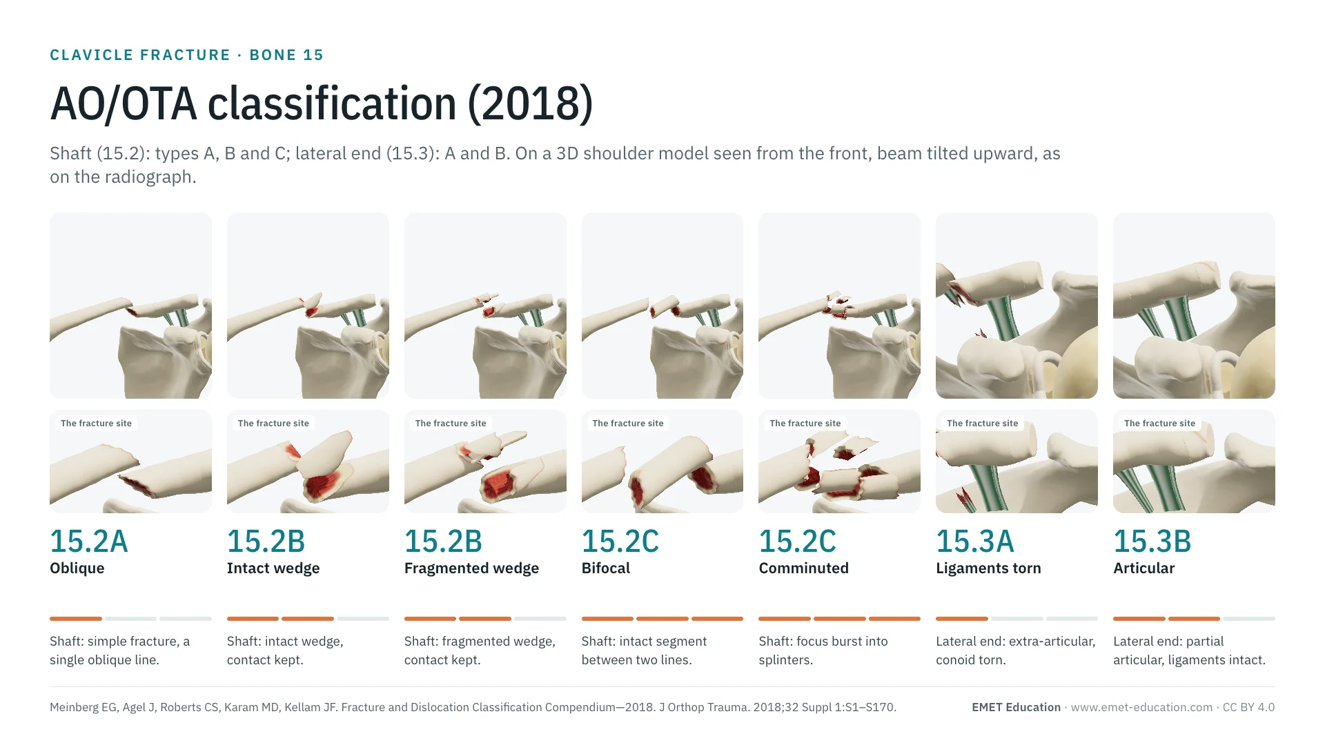
Each image is a rendering of the EMET Education clavicle fracture model at that code, seen from the front, beam tilted 12° upward (Zanca view), with a narrow field to approach the projection of a radiograph; under the code, a close-up of the fracture. For the shaft, the classification does not subdivide the types: the page shows under each letter the patterns the model draws (three simple lines under A, two wedges under B, two multifragmentary foci under C), which carry the same code. Fractures are shown displaced by 100% of the bone's width, the model's first choice: the code itself does not say whether the fracture is displaced. For the lateral end, the model draws three cases: Neer I and Neer IIB under 15.3A, Neer III under 15.3B.
Shaft: 15.2
-
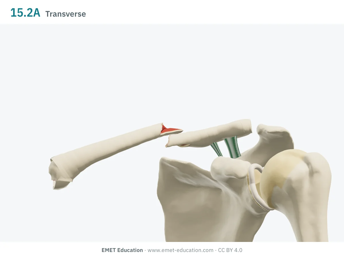
15.2ASimple, transverse
- Definition
- Shaft type A: simple fracture, a single line crossing the bone. Its direction (transverse, oblique, spiral) does not change the code.
- In the 3D model
- Planar line about 10° from the right section, at the junction of the middle and lateral thirds; the two ends are offset by one bone width and no longer touch.
-
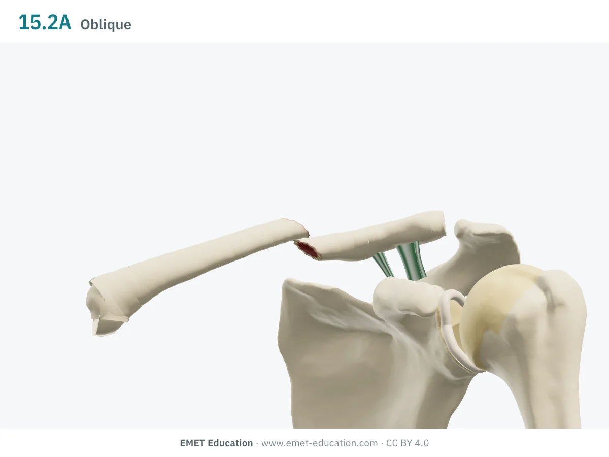
15.2ASimple, oblique
- Definition
- Shaft type A: simple fracture with an oblique line; same code as the transverse or spiral fracture.
- In the 3D model
- Planar line at about 50°, rising medially on the superior surface; the medial fragment overlaps the lateral one as shortening increases.
-
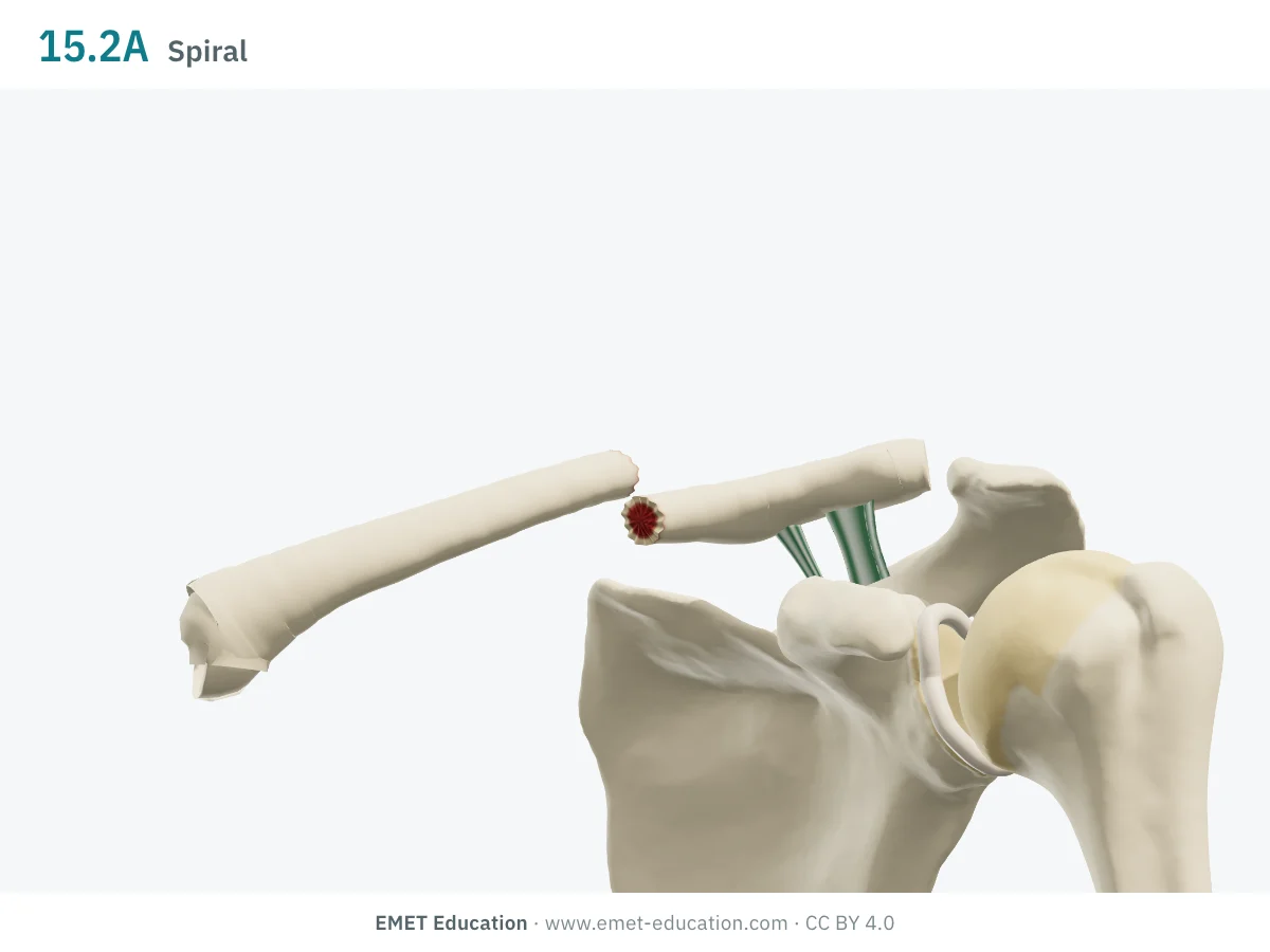
15.2ASimple, spiral
- Definition
- Shaft type A: simple fracture with a spiral line that winds around the bone; still 15.2A.
- In the 3D model
- A one-turn helix with a pitch of about 26 mm and a longitudinal split anteriorly and inferiorly: the fracture is long.
-
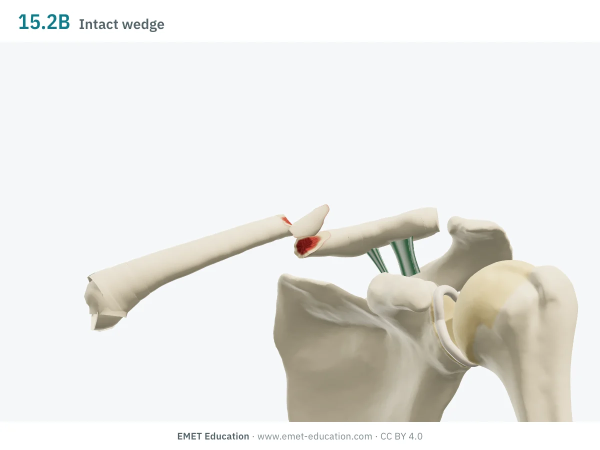
15.2BIntact wedge
- Definition
- Shaft type B: wedge fracture, that is, an intermediate fragment in one piece; the two main fragments keep a contact.
- In the 3D model
- Almost transverse main line and an anterosuperior wedge with a 20 mm base, in one fragment; the wedge moves apart and tips up.
-
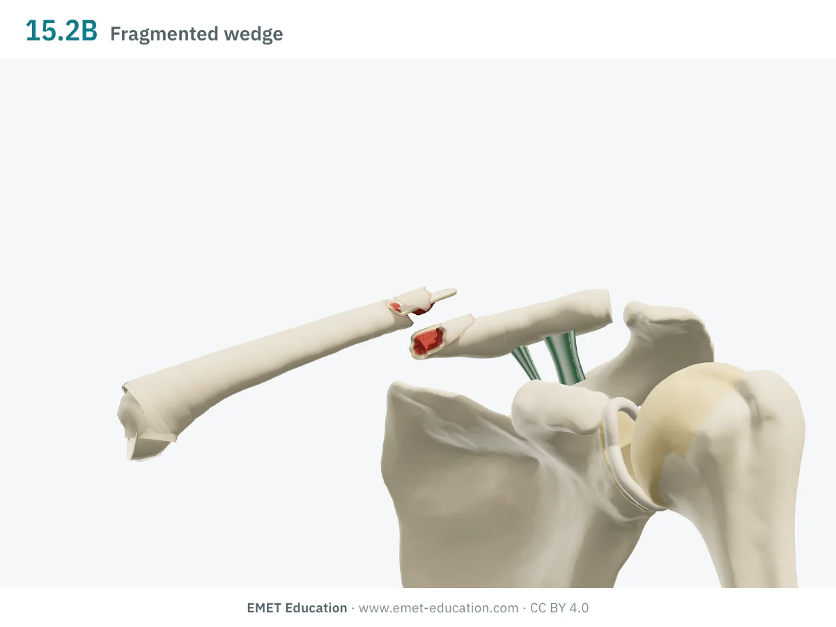
15.2BFragmented wedge
- Definition
- Shaft type B: as the intact wedge, but the wedge is itself fragmented; same code 15.2B, because the classification does not subdivide wedges.
- In the 3D model
- The same wedge, with a 24 mm base, in three splinters; the two main fragments are offset by one bone width.
-
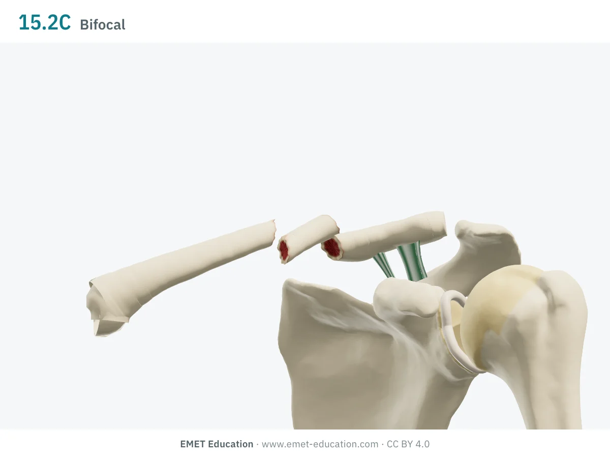
15.2CBifocal, intact segment
- Definition
- Shaft type C: multifragmentary fracture. An intact segment between two lines (bifocal or segmental fracture) is coded 15.2C, as is a fragmented spiral line or a fragmented segment.
- In the 3D model
- Two oblique lines isolate an intact segment of about 28 mm; the segment tilts by 16°.
-
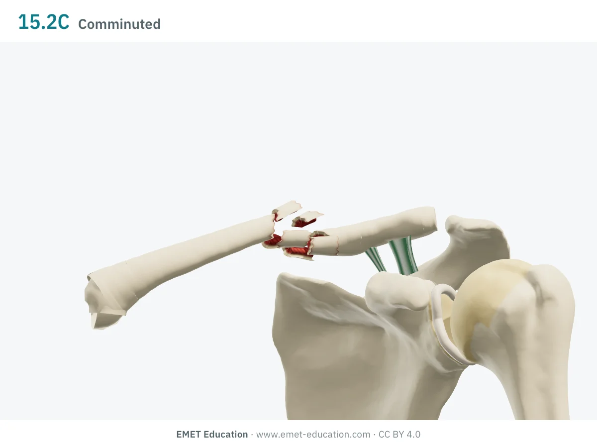
15.2CComminuted
- Definition
- Shaft type C: multifragmentary fracture, the focus burst into several splinters; it is coded 15.2C with no further detail.
- In the 3D model
- A 22 mm focus burst into six splinters (three sectors over two levels); the splinters move apart by 0.35 to 0.7 bone width.
Lateral end: 15.3, and medial segment: 15.1
-
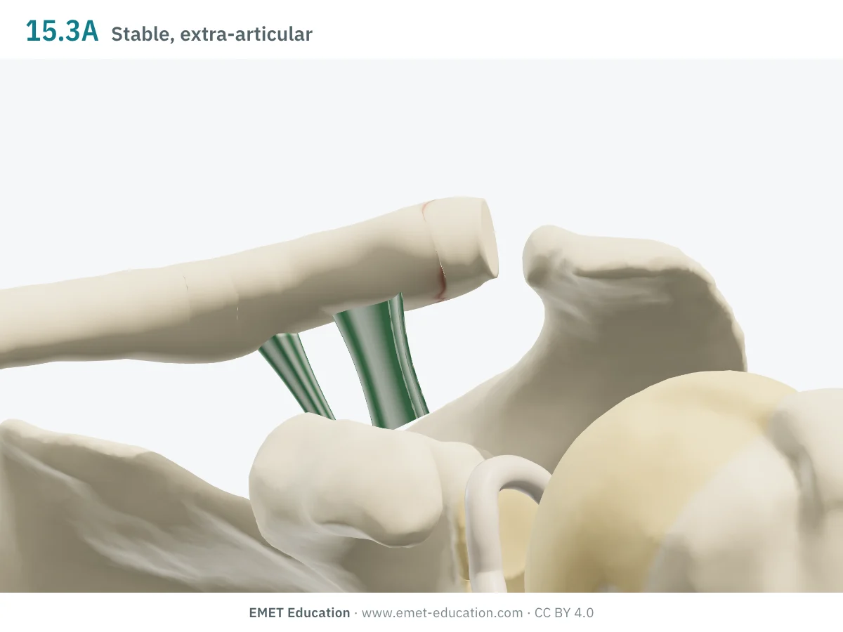
15.3AExtra-articular, stable
- Definition
- Lateral segment type A: extra-articular fracture, lateral to a line perpendicular to the medial cortex of the coracoid base (AO Surgery Reference). Here, the coracoclavicular ligaments are intact.
- In the 3D model
- Line about 4 mm lateral to the trapezoid; both ligaments stay on the medial fragment, which does not move. It is a Neer type I.
-
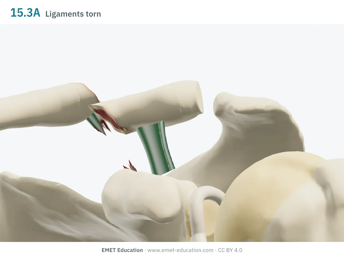
15.3AExtra-articular, ligaments torn
- Definition
- Lateral segment type A, with a line that tears the coracoclavicular ligaments: the AO Surgery Reference codes a displaced, extra-articular fracture with torn ligaments as 15.3A(c) (AO Surgery Reference). The model does not carry the (c) qualification.
- In the 3D model
- Line between the conoid and trapezoid footprints: the conoid is torn, the trapezoid stays on the lateral fragment; the medial fragment rises about 9 mm at 100%. It is a Neer type IIB.
-
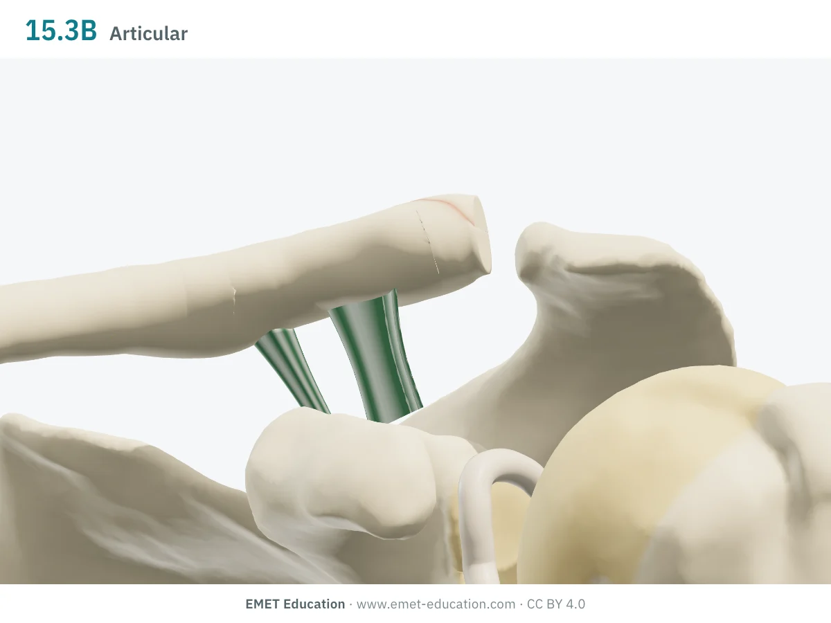
15.3BPartial articular
- Definition
- Lateral segment type B: partial articular fracture, the line entering the acromioclavicular joint (AO Surgery Reference). Here, the ligaments are intact.
- In the 3D model
- Upper half of the lateral end detached, from the articular facet to about 8 mm medially; the ligaments are intact and the fracture is not displaced. It is a Neer type III.
-
15.1Medial segment
- Definition
- The medial segment of the clavicle, 15.1, is the first of the three segments; the AO Surgery Reference details no type for it (AO Surgery Reference). It accounts for a small minority of fractures: 2% in a series of 535 fractures (Postacchini et al.).
- In the 3D model
- Not drawn: the model covers the shaft and the lateral end, not the medial third.
Schematic rendering, for teaching: the model illustrates each code on a single shoulder, it does not reproduce a radiograph. Definitions reworded from the 2018 compendium (Meinberg et al.) and the AO Surgery Reference; their figures and pictograms are not reproduced. The bar shows the type letter: one segment A, two B, three C.
Reading the code
The 2018 AO/OTA compendium
The classification is that of the AO Foundation and the Orthopaedic Trauma Association, whose compendium was revised in 2018 for all bones (Meinberg et al.). The clavicle is bone 15. The compendium allows educational use of its classification, not commercial use without the publisher's agreement; the page reproduces neither its figures nor its sentences.
The code reads from left to right:
- 15: the clavicle, bone 15;
- .1, .2, .3: the segment, medial, shaft, lateral. The shaft runs from the attachment of the coracoclavicular ligament, laterally, to the costoclavicular ligament, medially; the lateral segment lies lateral to a line perpendicular to the medial cortex of the coracoid base (AO Surgery Reference);
- A, B, C for the shaft: simple, wedge, multifragmentary. The AO Surgery Reference specifies that the classification goes no further: spiral, oblique and transverse are not coded separately, intact and fragmented wedges are both 15.2B, and the fragmented spiral line, intact segment and fragmented segment are all 15.2C;
- A, B for the lateral segment: extra-articular, partial articular;
- (a), (c): a qualification in brackets adds the state of the coracoclavicular ligaments. The AO Surgery Reference codes fractures with torn ligaments 15.3A(c) and 15.3B(c), and those where the ligaments avulse with a bone fragment 15.3A(a) or 15.3B(a).
In a series of 535 isolated clavicle fractures, they accounted for 2.6% of all fractures and 44% of those of the shoulder girdle; the middle third made up 81% and the medial third 2% (Postacchini et al.). The Edinburgh series of 1000 adult fractures (Robinson, 1998) gave rise to the Robinson classification, which takes displacement into account, absent from the shaft codes.
Reliability
How reproducible is the code?
For the shaft, it is not known: no reliability study of the AO/OTA shaft code was found. For the lateral clavicle, two radiograph studies give only fair agreement, with the "OTA classification" (their abstracts do not say which version of the compendium was used).
| Study | Measure | Value |
|---|---|---|
| Rauer et al., 2020 | Mean interobserver kappa of the OTA classification, 5 radiologists and 5 trauma surgeons, 20 lateral clavicle fractures; intraobserver "moderate" (0.086 to 0.634 depending on the reader) | 0.34 |
| Lin et al., 2023 | Mean interobserver kappa of the OTA classification, 8 shoulder specialists and 8 general trauma surgeons, 20 lateral clavicle fractures, radiographs only (better with 3D CT among specialists) | 0.42 |
The authors of Rauer et al. conclude that the three classifications tested (OTA, Neer, Jäger-Breitner) are unreliable and of limited value: agreement for Neer was of the same order (mean κ 0.28). The Robinson classification announced satisfactory inter- and intraobserver reliability in 1998, with no figures in the abstract.
Related classifications
What the other classifications add
The AO/OTA code describes the line (simple, wedge, multifragmentary) for the shaft, and the site and joint involvement for the lateral end; it says nothing about displacement. Other classifications do, or reason by thirds of the clavicle.
The three the model lets you read side by side:
| Classification | What it grades | Typical use |
|---|---|---|
| Robinson (1998) | The site (medial fifth: type 1; middle three fifths: type 2; lateral fifth: type 3), then displacement: undisplaced (A) or displaced (B). | Epidemiology and prognosis: type 2A, 3A and 1 fractures usually had a benign course, 2B and 3B more union problems. The model displays it next to the AO/OTA code. |
| Allman (1967) | Three groups by third of the clavicle: middle (I), lateral (II), medial (III). | Still used in series (Postacchini et al.), which add displacement and comminution to it. |
| Neer (lateral end) | The position of the line relative to the coracoclavicular ligaments, hence stability: types I to V. It details what 15.3 groups into A and B (Stenson and Baker). | Choice of treatment for the lateral end; the model's correspondence: Neer I, IIA, IIB and V as 15.3A, Neer III as 15.3B (reading to be confirmed). |
Use
In research and in clinical practice
In research
The AO/OTA code is used to describe and compare series from one region of the skeleton to another. For the clavicle shaft, three letters do not carry displacement, which Robinson takes into account. In the Edinburgh series, comminution in displaced shaft fractures (type 2B) was a risk factor for delayed union and nonunion (Robinson, 1998).
In clinical practice
The code describes the fracture; it does not choose the treatment. The AO Surgery Reference points this out: management may vary for the same code. For the shaft, one reads the code (A, B, C), then the displacement and shortening, which are not part of it.
For the lateral end, the code (A, B) says whether the acromioclavicular joint is involved; the state of the ligaments, which governs stability, goes through the qualification (a, c) or a Neer type, hence the value of reading both.
Grading pitfalls
Five pitfalls when coding a clavicle fracture
- The shaft code says nothing about displacement. An undisplaced 15.2A fracture and a displaced, shortened 15.2A fracture have the same code; record displacement and shortening separately, or add the Robinson type (2A, 2B).
- Wedge or multifragmentary: the contact. A wedge leaves a contact between the two main fragments; when that contact is lost after reduction, the fracture is multifragmentary (15.2C). A fragmented wedge stays 15.2B, as the classification does not subdivide it.
- Where does the shaft end? The lateral segment begins lateral to a line perpendicular to the medial cortex of the coracoid base, the shaft stops at the attachment of the coracoclavicular ligament: the two landmarks do not necessarily coincide, and a line close to the limit forces a choice.
- The ligaments are not visible. At the lateral end, their state is inferred from the line and the displacement; a standard AP view also hides an articular line under the acromion. Ask for a Zanca view (beam tilted 10° to 15° upward) before coding A or B.
- Do not confuse it with Neer, or with the older version. 15.3A does not say whether the ligaments are torn: that is what the Neer classification separates (I versus IIA, IIB). And articles published before 2018 describe another compendium: check which version is cited.
Self-test
Which code?
Eight cases from the model, at each site's presentation view, in random order. Find the AO/OTA code of each one; the answer comes with the definition. Several shaft patterns share the same code.
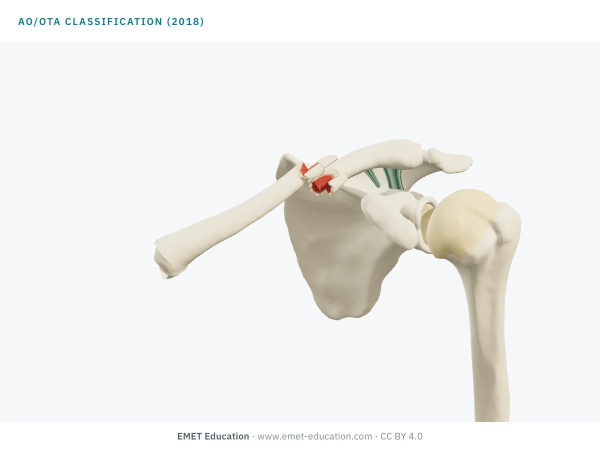
Which AO/OTA code is this?
Download and cite
Free infographics, for teaching and publication
Use them in a course, a presentation, a social media post or a scientific article, under the Creative Commons Attribution 4.0 license: free reuse, including commercial, provided EMET Education is credited with a link to this page.
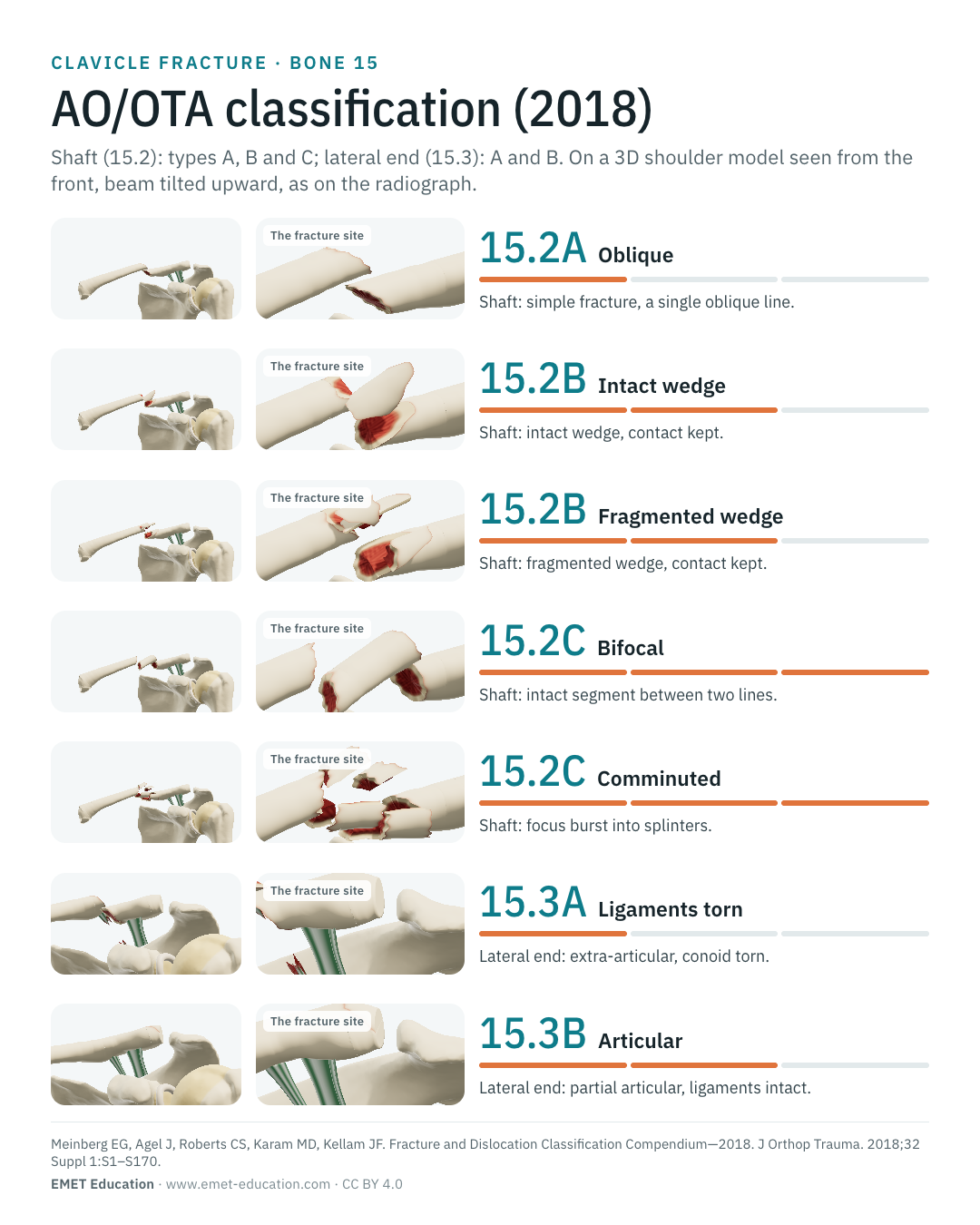
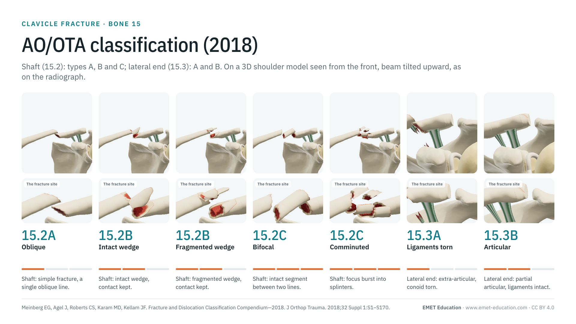
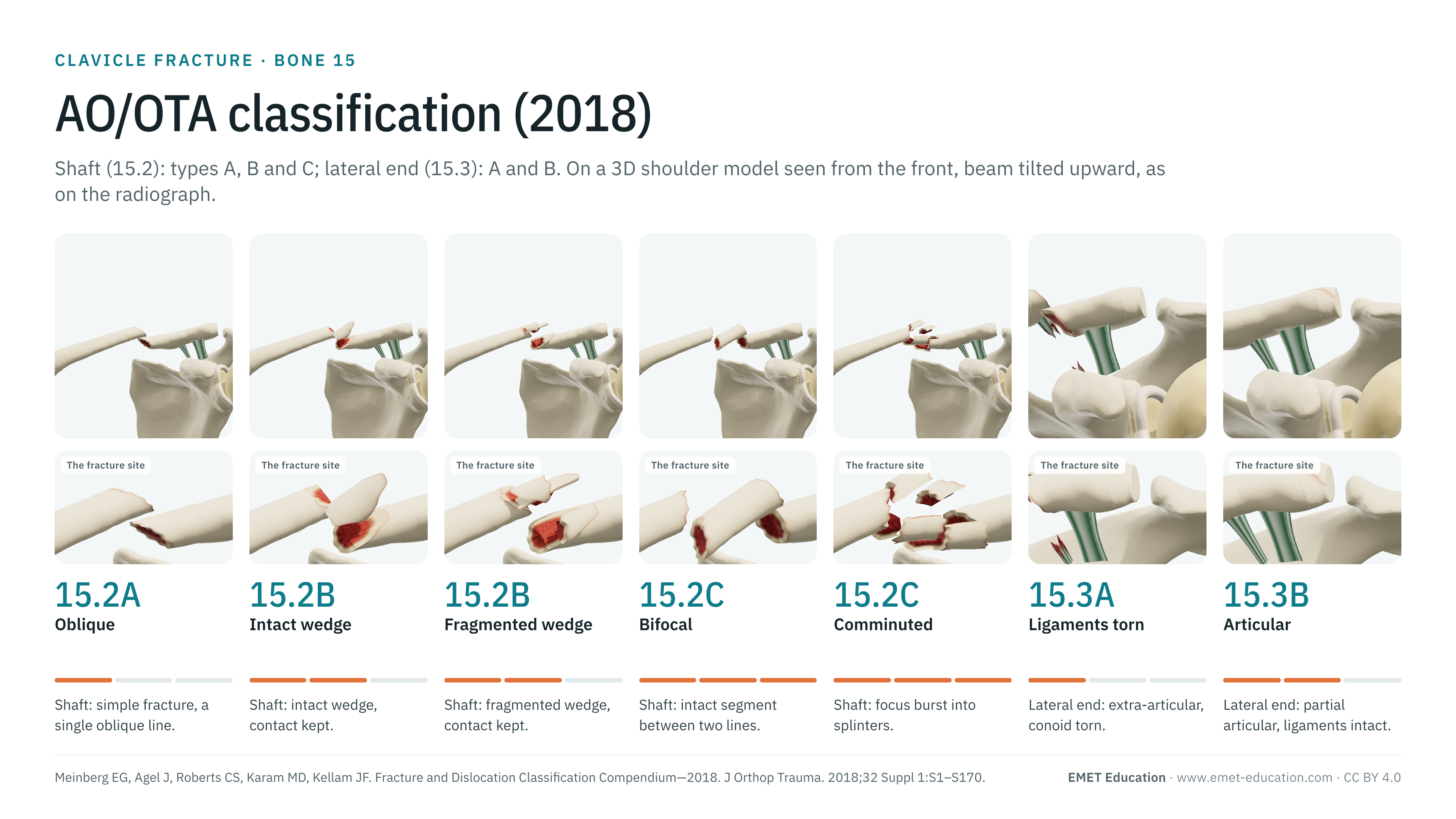
Cite this figure
Ready to paste. For a journal figure, add the compendium reference (Meinberg et al., 2018).
EMET Education. AO/OTA classification of clavicle fractures: 3D visualization [Internet]. 2026 [cited]. Available from: https://www.emet-education.com/en/classifications/ao-ota-clavicle/. CC BY 4.0.EMET Education. (2026). AO/OTA classification of clavicle fractures: 3D visualization [Infographic]. https://www.emet-education.com/en/classifications/ao-ota-clavicle/ (CC BY 4.0)<a href="https://www.emet-education.com/en/classifications/ao-ota-clavicle/"><img src="https://www.emet-education.com/en/classifications/ao-ota-clavicle/img/infographic.webp" alt="AO/OTA classification of clavicle fractures in 3D" width="960"></a><br>Figure: <a href="https://www.emet-education.com/en/classifications/ao-ota-clavicle/">EMET Education</a>, CC BY 4.0Questions
Frequently asked questions
Why does the shaft have no subgroups?
In the 2018 version, the AO Surgery Reference specifies that the classification does not subdivide shaft fractures into spiral, oblique or transverse (A), intact or fragmented wedge (B), or subtypes of multifragmentary (C) (AO Surgery Reference). The model's patterns are descriptions, filed under their type.
What is the difference between 15.2B and 15.2C?
In 15.2B, a wedge sits between two main fragments that keep a contact, whether the wedge is intact or fragmented. In 15.2C, the fracture is multifragmentary: an intact segment between two lines, a fragmented spiral line or a focus burst into splinters.
What is the difference between 15.3A and 15.3B?
15.3A is extra-articular, 15.3B partial articular: the line enters the acromioclavicular joint (AO Surgery Reference). The state of the coracoclavicular ligaments is added in brackets: (c) for torn ligaments.
How do I go from an AO/OTA code to Robinson or Neer?
Not through an official table: they were made for different questions. The model displays them together: for the shaft, Robinson 2A while contact is kept, 2B beyond; for the lateral end, one Neer type per code (EMET Education's reading). See the Neer page.
Is there a code for the medial third?
The medial segment is 15.1; the AO Surgery Reference details no type for it. The model does not cover it.
Is the AO/OTA code enough to decide treatment?
No: for the same code, management may vary, and the shaft code carries neither displacement nor shortening. It is used to describe and compare.
Can I reuse the infographics?
Yes, under CC BY 4.0, including in an article or commercially, provided EMET Education is credited with a link to this page (see "Cite this figure"). The images are renderings of the EMET model: they reproduce no figure from the AO/OTA compendium.
Sources
Author and references
- Meinberg EG, Agel J, Roberts CS, Karam MD, Kellam JF. Fracture and Dislocation Classification Compendium—2018. J Orthop Trauma. 2018;32 Suppl 1:S1–S170. doi:10.1097/BOT.0000000000001063 · PMID 29256945
- AO Foundation. AO Surgery Reference: clavicle fractures (adult trauma). surgeryreference.aofoundation.org. Consulté le 4 novembre 2026 / accessed November 4, 2026.
- Robinson CM. Fractures of the clavicle in the adult. Epidemiology and classification. J Bone Joint Surg Br. 1998;80(3):476–84. doi:10.1302/0301-620x.80b3.8079 · PMID 9619941
- Allman FL Jr. Fractures and ligamentous injuries of the clavicle and its articulation. J Bone Joint Surg Am. 1967;49(4):774–84. doi:10.2106/00004623-196749040-00024 · PMID 6026010
- Postacchini F, Gumina S, De Santis P, Albo F. Epidemiology of clavicle fractures. J Shoulder Elbow Surg. 2002;11(5):452–6. doi:10.1067/mse.2002.126613 · PMID 12378163
- Rauer T, Boos M, Neuhaus V, et al. Inter- and intraobserver agreement of three classification systems for lateral clavicle fractures – reliability comparison between two specialist groups. Patient Saf Surg. 2020;14:4. doi:10.1186/s13037-019-0228-y · PMID 31911821
- Lin J, Sun WJ, Chen JH, et al. Comparison of interobserver agreement of four classification systems for lateral clavicle fractures between two groups of surgeons: a multicenter study. Orthop Surg. 2023;15(8):2138–43. doi:10.1111/os.13659
- Stenson J, Baker W. Classifications in brief: the modified Neer classification for distal-third clavicle fractures. Clin Orthop Relat Res. 2021;479(1):205–9. doi:10.1097/CORR.0000000000001456
- Neer CS 2nd. Fractures of the distal third of the clavicle. Clin Orthop Relat Res. 1968;58:43–50. PMID 5666866
3D models
Show the fracture to your patient
The images on this page come from an interactive model. In clinic, choose the site, type and displacement from your patient's radiograph, then show the proposed treatment: nonoperative treatment, plate, intramedullary screw or coracoclavicular fixation.
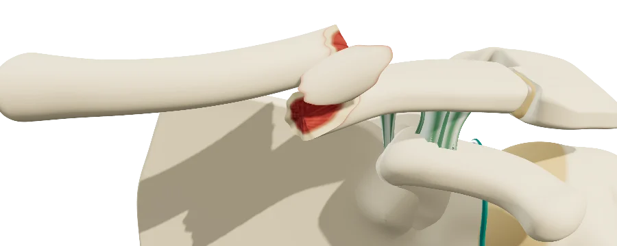 ShoulderClavicle fractureShaft and lateral end, AO/OTA, Robinson and Neer, displacement, healing, plating and coracoclavicular fixation.
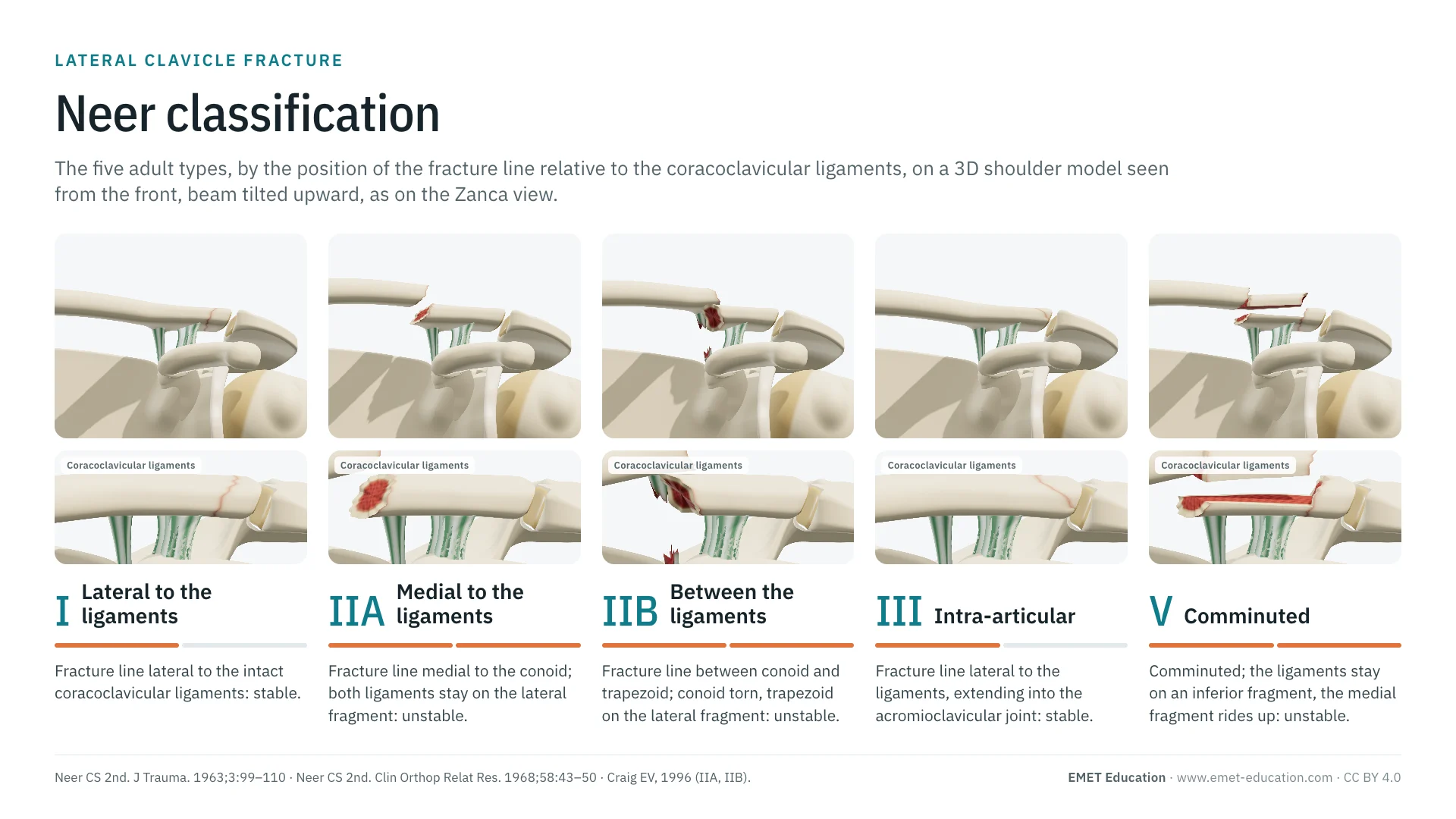 ClassificationsNeer classification (clavicle)The types I to V of the lateral end, by the coracoclavicular ligaments.
 ClassificationsAll 3D classificationsArranged by joint.
Explain the fracture in 3D
The clavicle fracture model opens in your browser, at the code you choose, with no sign-up.
Share this page with a colleague or a resident: