- EMET Education
- 3D classifications
- Eaton-Littler classification (thumb basal joint)
Classification · Thumb basal joint osteoarthritis
Eaton-Littler Classification of Thumb CMC Arthritis in 3D
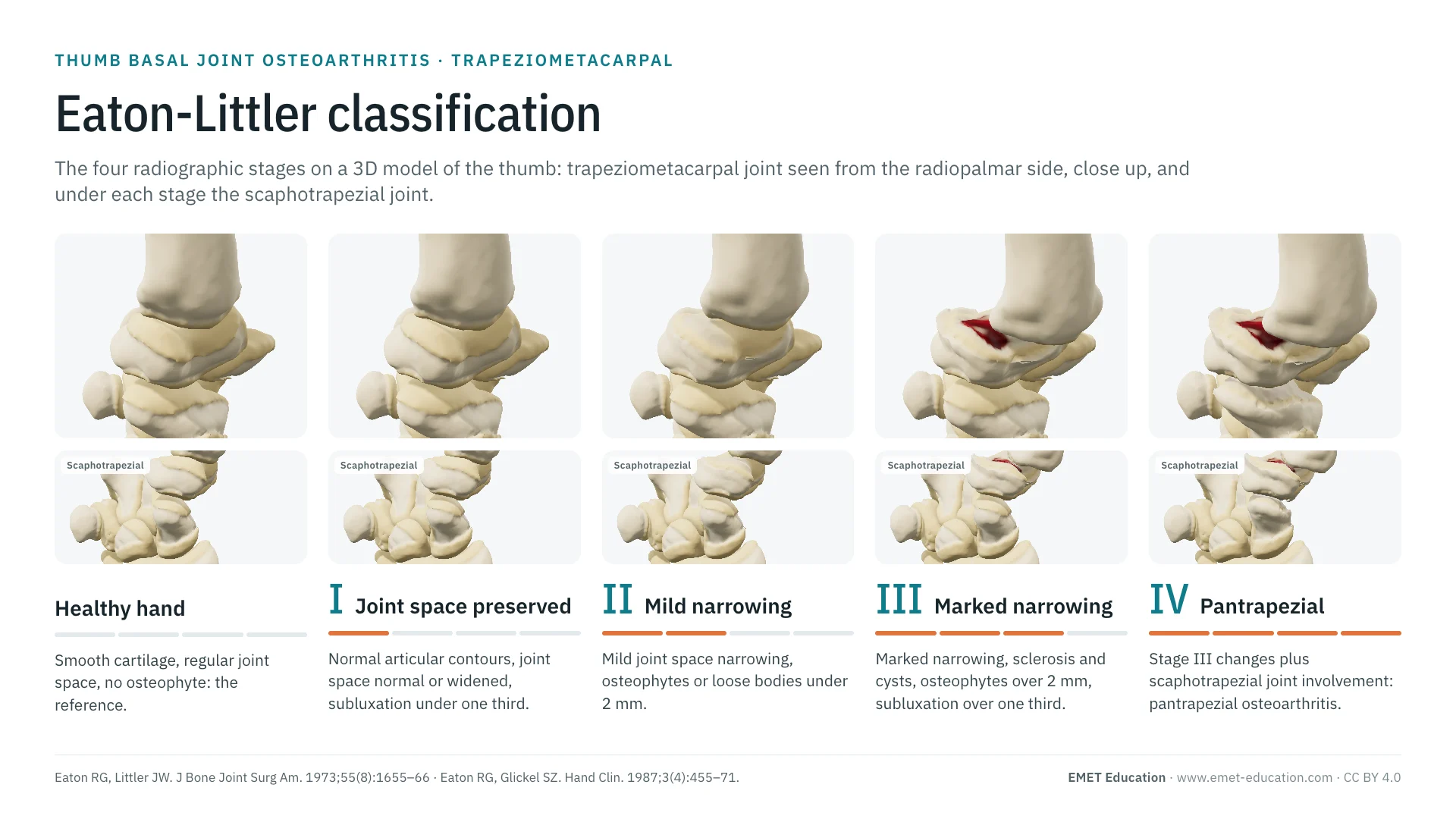
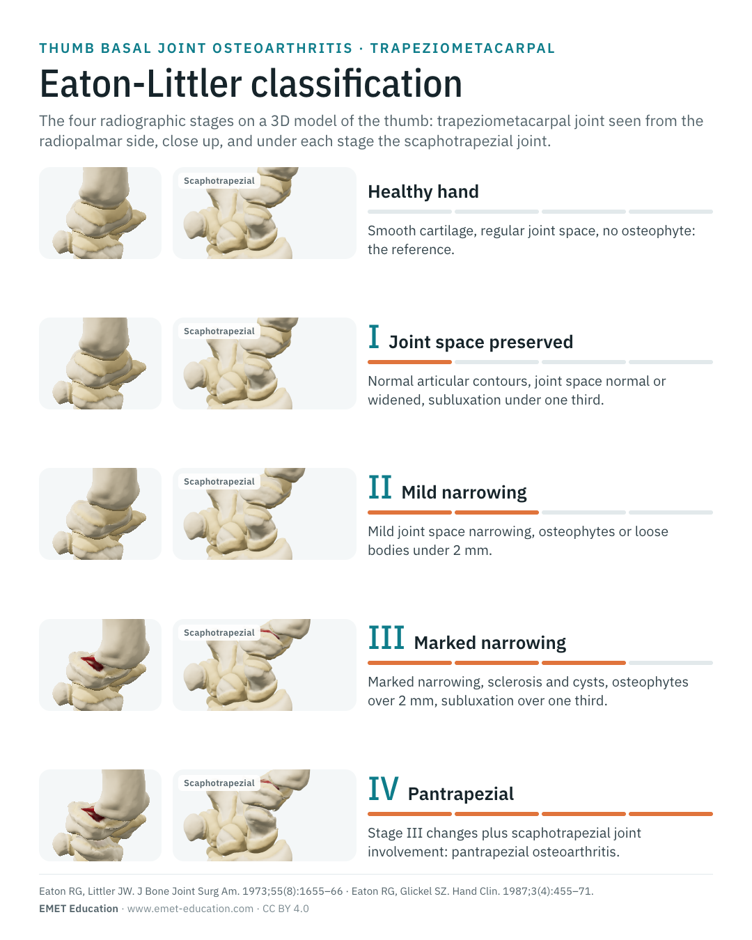
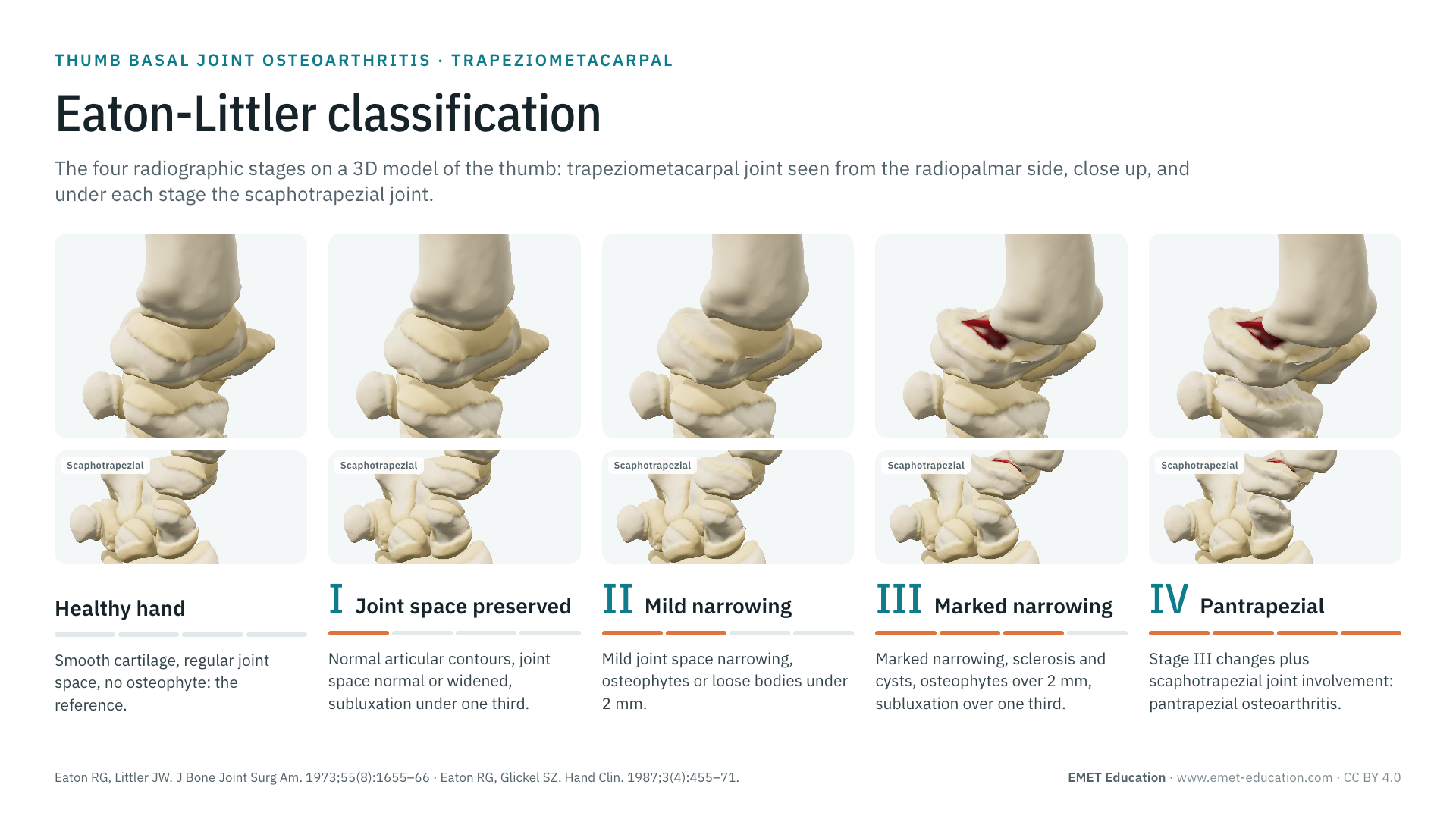
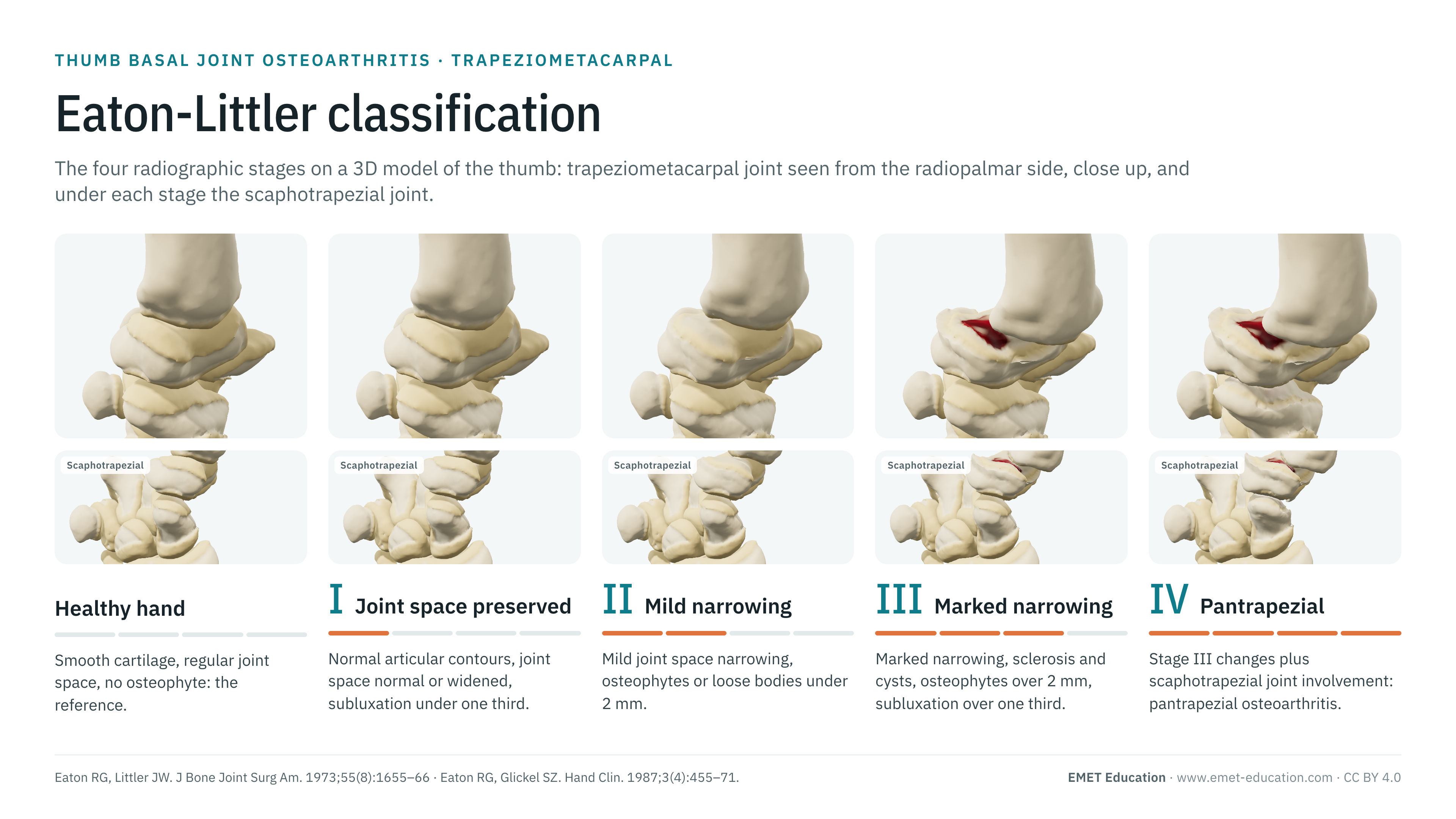
The four Eaton-Littler radiographic stages of thumb basal joint (trapeziometacarpal) osteoarthritis, on the rhizarthrosis 3D model, seen close up from the radiopalmar side, with the scaphotrapezial joint that defines stage IV. With the definition of each stage, the three published versions, kappas, the link with treatment, grading pitfalls and free infographics for teaching and publication.
Designed by an orthopedic surgeon
The 3D model opens in your browser, at the stage shown, with no sign-up. Infographics are free to reuse under CC BY 4.0.
- 1973Eaton and Littler describe the stages in the Journal of Bone and Joint Surgery (Eaton and Littler); Eaton and Glickel revise them in 1987 (Hand Clin)
- 4Radiographic stages; stage IV is defined by the scaphotrapezial joint in the 1987 version
- 926Citations of the 1973 article (OpenAlex, October 31, 2026); 490 for the 1987 one
- κ 0.11–0.56Interobserver agreement, poor to fair (systematic review of four studies)
The stages
From stage I to stage IV, in 3D
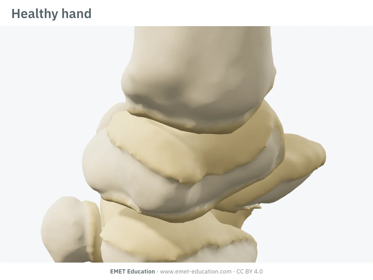
Each image is a rendering of the EMET Education rhizarthrosis 3D model at that stage: the trapeziometacarpal joint close up, seen from the radiopalmar side, long fingers hidden, the way the joint space, osteophytes and subluxation of the first metacarpal are read on the radiograph. The healthy hand is the reference: there is no stage 0 in the classification. Definitions follow the 1987 Eaton and Glickel version (Kennedy et al., Carità et al.).
-
Healthy hand (reference)
- In the 3D model
- Smooth cartilage, regular joint space, base of the first metacarpal (M1) centred on the trapezial saddle, no beak. This is the model without lesions, not a stage.
-
IJoint space preserved or widened
- Definition
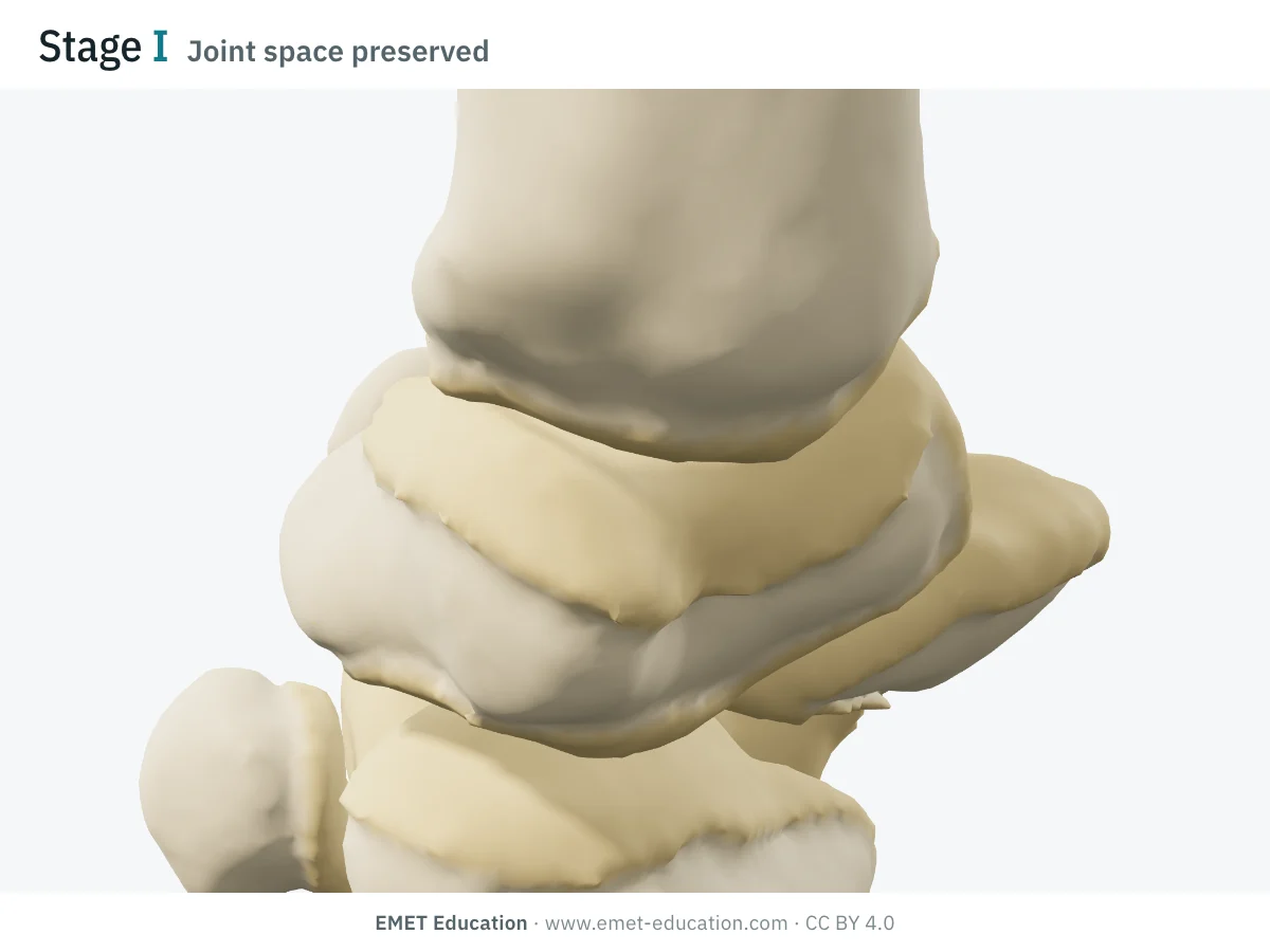
- Normal articular contours; joint space normal or slightly widened (effusion, laxity); subluxation of less than one third of the base of M1.
- In the 3D model
- Dull cartilage on the palmar part of the joint, joint space and contours preserved, base of M1 in place. The model does not draw the widened joint space or synovitis.
- Reported management
- Medical treatment and a thumb base splint first (Kroon et al.). For intractable pain, Eaton and Littler proposed ligament reconstruction with a strip of flexor carpi radialis at stages I and II: 95% good or excellent results in their series of 50 reconstructions, at 7 years on average (Eaton et al., 1984).
-
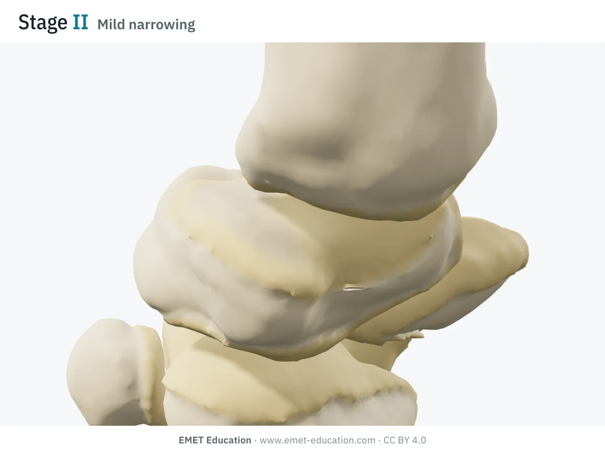
IIMild narrowing
- Definition
- Mild joint space narrowing; osteophytes or loose bodies under 2 mm; subluxation around one third ("up to" or "at least" one third depending on the table).
- In the 3D model
- Cartilage thinned by half in the palmar compartment, first osteophytes (about 1.5 mm); the base of M1 slides outward and backward by 22% of its width (gauge "up to one third").
- Reported management
- Same options as stage I (Eaton et al., 1984). This is the most misleading stage: in an arthroscopic series, 26 of the 31 stage II joints had Outerbridge grade 3 or 4 cartilage (Carità et al.).
-
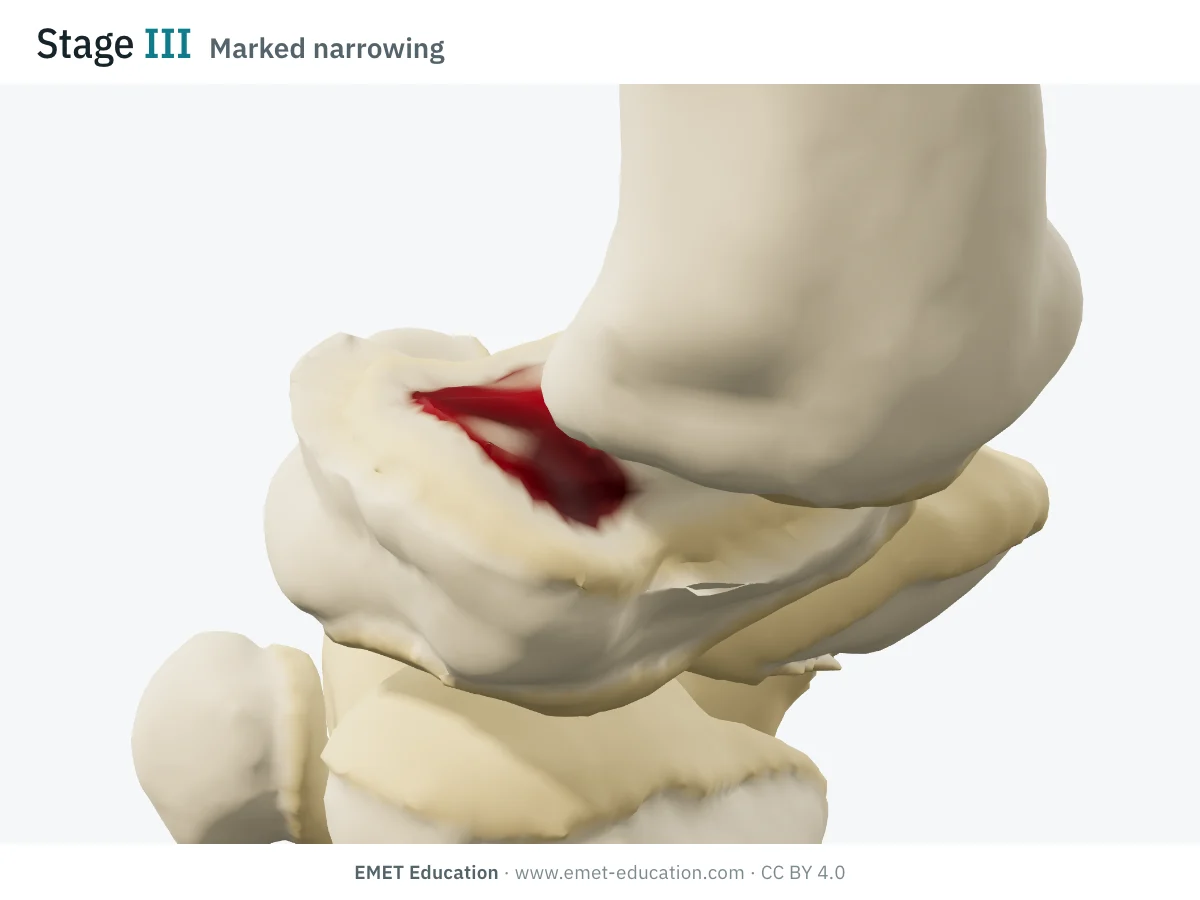
IIIMarked narrowing
- Definition
- Marked joint space narrowing, subchondral sclerosis and cysts; osteophytes or loose bodies over 2 mm; subluxation over one third; scaphotrapezial joint preserved.
- In the 3D model
- Bare bone in the palmar compartment (dark red, sclerotic bone), cartilage thinned everywhere, osteophytes of about 3 mm, base of M1 subluxated by 45% of its width (gauge "over one third"). Cysts are not drawn.
- Reported management
- Less consistent results with ligament reconstruction: 74% good or excellent for stages III and IV combined (Eaton et al., 1984). Options: trapeziectomy, with or without ligament reconstruction and tendon interposition (Burton and Pellegrini); trapeziometacarpal prosthesis, which Badia and Sambandam reserved for stage III and early stage IV in older, low-demand patients (26 thumbs). No surgical technique has proved superior to the others (Kroon et al.).
-
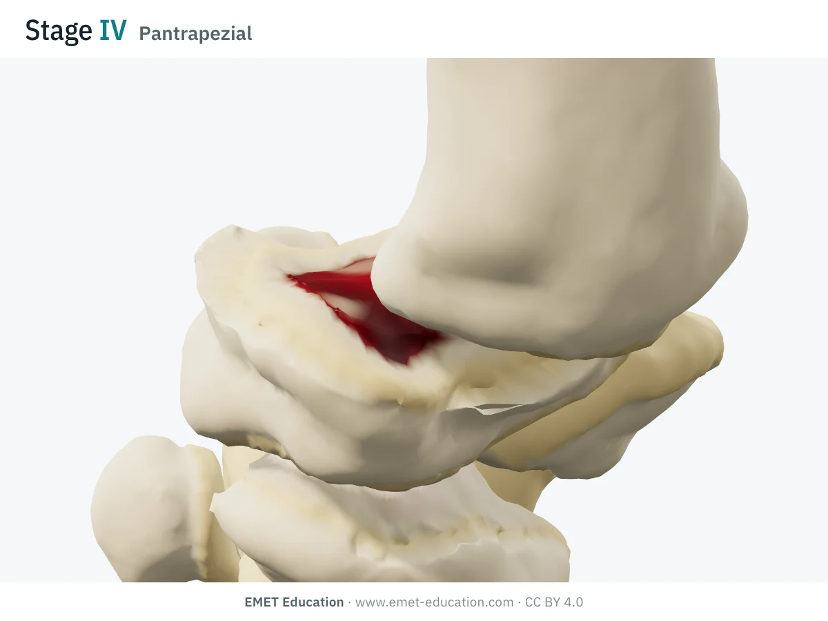
IVScaphotrapezial involvement (pantrapezial)
- Definition
- Stage III changes plus involvement of the scaphotrapezial joint (narrowing, cysts, sclerosis): pantrapezial osteoarthritis.
- In the 3D model
- Stage III plus scaphotrapezial osteoarthritis: narrowing, beaks of about 1.8 mm, slight tilt of the scaphoid, visible in the small view under each stage of the infographic. The model's gauge turns the scaphotrapezial joint on at step IV.
- Reported management
- Eaton and Littler recommended arthroplasty or arthrodesis rather than ligament reconstruction (Kennedy et al.). A moderately worn scaphotrapezial joint, left in place during trapeziometacarpal arthroplasty, did not progress (18 thumbs, 8 years of follow-up); but the radiograph matched the operative state of that joint in only 76% of cases (Glickel et al., 1992).
Schematic rendering, for teaching: the model illustrates each stage on a single hand, it does not reproduce a radiograph. Definitions reworded from Kennedy et al. (2016), Carità et al. (2024) and Holzbauer et al. (2026); the figures of the articles are not reproduced. Subluxation is a separate gauge in the model: here it is set at 0 for stage I, 22% of the base for stage II, 45% for stages III and IV (one-third threshold). Not drawn: the widened joint space of stage I, synovitis, cysts. The bar shows the stage: one segment for I, four for IV.
The original article
From Eaton and Littler (1973) to Eaton and Glickel (1987)
The staging was born in the operating room. In Ligament reconstruction for the painful thumb carpometacarpal joint (J Bone Joint Surg Am, 1973; doi:10.2106/00004623-197355080-00010), R. G. Eaton and J. W. Littler describe an extra-articular reconstruction that stabilises the trapeziometacarpal joint with a strip of flexor carpi radialis routed through the base of M1, performed since 1967 (Eaton et al., 1984). They grade the joint in four radiographic stages, which serve to indicate the operation.
Three texts fixed the classification:
- 1973: four stages. Stage IV there designates advanced degeneration of the trapeziometacarpal joint with major subluxation; the scaphotrapezial joint enters the definition only in 1987 (after Kennedy et al.; the original text was not re-read for this page);
- 1984: Eaton, Lane, Littler and Keyser grade the first 50 reconstructions from I to IV (mean follow-up 7 years): 95% good or excellent results at stages I and II, 74% at stages III and IV. Reconstruction is now recommended only for stages I and II (Eaton et al., 1984);
- 1987: Eaton and Glickel present staging as the rationale for treatment, read mainly on the lateral view, and to be adjusted if inspection at surgery shows more damage than the radiograph (Eaton and Glickel). Their version redefines stage IV: involvement of the scaphotrapezial joint, in addition to the trapeziometacarpal joint (Kennedy et al.);
- the versions differ in whether and how much subluxation is included, the stage at which sclerosis appears, the degree of narrowing and the definition of stage IV (Nelson et al., 2025). A 2026 study compares the three published versions on the original texts (Holzbauer et al.).
The 1973 article has 926 citations and the 1987 one 490 (OpenAlex, October 31, 2026). It is the most widely used radiographic severity scale for thumb basal joint osteoarthritis (Melville et al.); the 1987 article has no DOI, find it by PMID 3693416.
Reliability
How reproducible are the stages?
Moderately. A systematic review of four studies (163 patients) finds poor to fair interobserver agreement (kappa 0.11 to 0.56) and fair to moderate intraobserver agreement (0.54 to 0.66) (Berger et al.). Later studies do no better: disagreement lies in the intermediate stages II and III.
| Study | Measure | Value |
|---|---|---|
| Kubik and Lubahn, 2002 | Intra- then interobserver kappa, 3 hand surgeons and 3 residents, 40 radiographs | 0.66 · 0.53 |
| Dela Rosa et al., 2004 | Intra- then interobserver kappa, 6 hand surgeons, 40 sets, depending on the number of views (values from the table of Berger et al.) | 0.59–0.61 · 0.37–0.56 |
| Spaans et al., 2011 | Interobserver kappa, 5 radiologists and 8 hand surgeons, 40 cases (value from the table of Berger et al.); agreement on the treatment chosen was only fair | 0.50 |
| Hansen et al., 2013 | Mean intra- then interobserver kappa on radiographs, 5 readers, 50 hands; with CT: 0.76 then 0.22 | 0.54 · 0.17 |
| Becker et al., 2016 | Interobserver agreement (s* statistic, not kappa), 92 hand surgeons, 30 radiographs: cutting the scale to the first three stages (no scaphotrapezial joint) brings it close to substantial agreement | s* |
| Holzbauer et al., 2026 | Three versions (1973, 1984, 1987), 8 readers, 100 lateral views: interobserver kappa 0.39 (1973) then 0.45 (1984 and 1987); intraobserver kappa 0.51 to 0.98 | 0.39 · 0.45 |
| Nelson et al., 2025 | Eaton-Glickel, one surgeon and one trained student, 40 series: intraobserver agreement moderate (student) to substantial (surgeon), agreement between the two moderate (kappas not given in the abstract) | moderate |
In the study by Holzbauer et al., a reader agrees fairly well with himself, but readers agree little with one another, especially for stages II and III; the authors conclude that this classification cannot guide treatment on its own. CT improves a reader's agreement with himself, not agreement between readers, and the distinction between stages III and IV remains poor (Hansen et al.).
Variants and neighbours
The three Eaton versions, and other scales
"Eaton-Littler", "Eaton" and "Eaton-Glickel" designate three versions published in 1973, 1984 and 1987, whose definitions do not fully overlap (Holzbauer et al.). Most recent work cites the 1987 version (Nelson et al.), often under the name Eaton-Littler.
Other scales describe the same joint, on the radiograph, at arthroscopy or in the population:
| Scale | Principle | Note |
|---|---|---|
| Eaton and Littler, 1973 | Four radiographic stages described with ligament reconstruction; stage IV: advanced trapeziometacarpal degeneration | Original text not re-read for this page; interobserver agreement 0.39 (Holzbauer). |
| Eaton et al., 1984 | Grading I to IV of 50 ligament reconstructions, followed for 7 years on average | A version of its own for Holzbauer; interobserver agreement 0.45. |
| Eaton and Glickel, 1987 | Staging as the rationale for treatment; stage IV: scaphotrapezial joint involved; read on the lateral view | The most widely used version, and the one in the 3D model. |
| Dell, Brushart and Smith, 1978 | 91 thumbs classified by radiographic severity; mild disease treated with anti-inflammatory drugs and a C-splint, trapezium resection with shortening of abductor pollicis longus in 16 thumbs | Predates Eaton-Glickel; its classes are not described here (abstract only read). |
| Badia, 2006 | Arthroscopic classification of the trapeziometacarpal joint, with a treatment algorithm | Sees the cartilage, which the radiograph underestimates; its stages are not described here (abstract only read). |
| Allieu, 2021 | Three radiographic parameters scored 0 to 3: trapeziometacarpal narrowing, instability-subluxation, scaphotrapeziotrapezoid damage | Points out the shortcomings of Dell and Eaton-Littler; needs prospective follow-up. |
| Curtis, 2025 | Stages I, II, IIIA, IIIB and IV, with thresholds measured on digital radiographs (subluxation in %, osteophytes in mm, narrowing relative to the scaphotrapezial joint); embeds the Eaton-Glickel scale | Agreement between a surgeon and a student: almost perfect then substantial, against moderate for Eaton-Glickel (40 series); needs verification by other teams. |
| Kellgren-Lawrence applied to the trapeziometacarpal joint (knee scale) | Grades 2 to 4 in a Finnish cohort of 3,595 subjects aged 30 and over | Epidemiology: 7% of men and 15% of women (age-adjusted); radiographic signs did not predict work disability. |
Use
In research and in clinical practice
In research
The Eaton-Glickel scale describes the radiographic severity of symptomatic thumb basal joint osteoarthritis cohorts, but it does not predict the patient's experience: in 85 patients (106 sets of radiographs), no patient-reported score (Michigan Hand Questionnaire, pain, PROMIS) correlated with the stage (Nelson et al., 2025). Classification precision is first a matter of method: state the version, the views, the readers and how subluxation was measured.
In clinical practice
Clinical examination and radiographs are usually enough for the diagnosis (Melville et al.). The stage says where the joint stands on the film: at stage I or II, a joint still preserved, which the Eaton and Littler ligament reconstruction sought to protect; at stage III, a severely damaged joint; at stage IV, disease that goes beyond the trapeziometacarpal joint (Eaton et al., 1984, Kennedy et al.).
Treatment starts non-operatively, whatever the stage: prolonged thumb base splinting has a beneficial effect, intra-articular corticosteroid or hyaluronic acid injections at the thumb base showed no clear benefit, and no surgical technique proved superior to another in trials, the more complex ones causing more complications (Kroon et al., 2018).
The stage therefore guides little: when 8 hand surgeons had to choose a treatment for 40 cases, their agreement was only fair (Spaans et al., 2011). The decision rests on symptoms, impact and failure of non-operative treatment; the stage locates the lesion, and the scaphotrapezial joint is also judged at surgical inspection (Eaton and Glickel).
Grading pitfalls
Five pitfalls when grading thumb basal joint osteoarthritis
- State the version. The 1973, 1984 and 1987 versions do not overlap: stage of sclerosis, degree of narrowing, subluxation, definition of IV (Nelson et al.). Published tables write stage II subluxation as "up to" or "at least" one third: write down the version and the threshold.
- II and III get confused. These are the least reproducible stages (Holzbauer et al.). Measuring subluxation as a percentage of the width of the base of M1 and the osteophyte in millimetres, as the Curtis scale does, avoids eyeballing (Nelson et al.).
- Stage IV is the scaphotrapezial joint. Look at that joint separately; the radiograph matched surgical inspection in only 76% of cases (Glickel et al., 1992), CT does not make the distinction between III and IV more reproducible (Hansen et al.), and without the scaphotrapezial joint the scale becomes more reliable (Becker et al.).
- Views matter. Eaton and Glickel read mainly the lateral view (Eaton and Glickel); Becker et al. had readers grade on Robert, lateral and oblique views (Becker et al.), Dela Rosa et al. compared AP and lateral wrist views with the Bett (Gedda) view: agreement between the two readings was only fair, and tended to improve with all three views together (Dela Rosa et al.).
- The radiograph is neither the cartilage nor the pain. In 51 arthroscopies, 26 of the 31 stage II joints had Outerbridge grade 3 or 4 cartilage (Carità et al.); 5 of 20 joints were discordant in a smaller series (Sun et al.), and there was no correlation between stage and cartilage damage on 20 operated surfaces (März et al.). Do not dismiss a very symptomatic patient because the stage is low.
Self-test
Which stage?
The same hand seen from the radiopalmar three-quarter view, at the four stages, in random order. The clues: cartilage and joint space, beaks, subluxation of the base of M1, and the scaphotrapezial joint at stage IV. The answer comes with the definition.
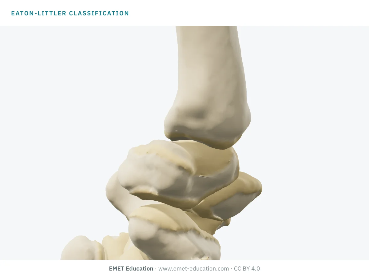
Which Eaton-Littler stage is this?
Download and cite
Free infographics, for teaching and publication
Use them in a course, a presentation, a social media post or a scientific article, under the Creative Commons Attribution 4.0 license: free reuse, including commercial, provided EMET Education is credited with a link to this page. The license covers EMET Education's images, not the classification itself.
Cite this figure
Ready to paste. For a journal figure, add the original reference (Eaton and Littler, 1973).
EMET Education. Eaton-Littler classification of thumb basal joint osteoarthritis: 3D visualization [Internet]. 2026 [cited]. Available from: https://www.emet-education.com/en/classifications/eaton-littler/. CC BY 4.0.EMET Education. (2026). Eaton-Littler classification of thumb basal joint osteoarthritis: 3D visualization [Infographic]. https://www.emet-education.com/en/classifications/eaton-littler/ (CC BY 4.0)<a href="https://www.emet-education.com/en/classifications/eaton-littler/"><img src="https://www.emet-education.com/en/classifications/eaton-littler/img/infographic.webp" alt="Eaton-Littler classification of thumb basal joint osteoarthritis in 3D" width="960"></a><br>Figure: <a href="https://www.emet-education.com/en/classifications/eaton-littler/">EMET Education</a>, CC BY 4.0Questions
Frequently asked questions
What is the Eaton-Littler classification?
It is a four-stage radiographic scale of thumb basal joint (trapeziometacarpal) osteoarthritis, described by Eaton and Littler in 1973 and revised by Eaton and Glickel in 1987. It grades joint space narrowing, osteophytes, subluxation of the base of the first metacarpal and, at stage IV, involvement of the scaphotrapezial joint.
What is the difference between Eaton-Littler and Eaton-Glickel?
They are two versions of the same scale, from 1973 and 1987, with an intermediate 1984 version. They differ in the role of subluxation, the stage at which sclerosis appears, the degree of narrowing and above all the definition of stage IV, which became involvement of the scaphotrapezial joint in 1987. In practice, "Eaton-Littler" most often means the 1987 version: state it in a paper.
What distinguishes stage III from stage IV?
At stage IV the scaphotrapezial joint is involved (narrowing, cysts, sclerosis) in addition to the severely damaged trapeziometacarpal joint of stage III: it is pantrapezial osteoarthritis. It is also the least reproducible distinction, and the radiograph matches the operative state of the scaphotrapezial joint in only 76% of cases.
On which radiographic view is it graded?
Eaton and Glickel stress the lateral view of the trapeziometacarpal joint, completed depending on the team by the Robert view, the Bett (Gedda) view and an oblique. Agreement between readers tends to improve when several views are combined; in research, state which.
Which treatment according to the Eaton-Littler stage?
Treatment starts with a splint and medical treatment at all stages. Eaton and Littler reserved ligament reconstruction for stages I and II; at stages III and IV, trapeziectomy, with or without ligament reconstruction and tendon interposition, or a prosthesis is offered. No technique has proved superior and surgeons agree little on the choice: the stage guides, it does not decide.
Does the radiographic stage match the symptoms?
Poorly. In a series of 85 patients, no patient-reported score correlated with the stage; at arthroscopy, 26 of 31 stage II joints had severely damaged cartilage (Outerbridge 3 or 4). A low stage does not exclude a painful joint or severe cartilage damage.
Can I reuse these images in a course or an article?
Yes. The images and infographics on this page are renderings of the EMET Education thumb basal joint osteoarthritis 3D model, under the Creative Commons Attribution 4.0 license: free reuse, including commercial, with credit to EMET Education and a link to this page. They reproduce no figure from the original articles. The "Cite this figure" box gives ready-to-paste citations.
Sources
Author and references
- Eaton RG, Littler JW. Ligament reconstruction for the painful thumb carpometacarpal joint. J Bone Joint Surg Am. 1973;55(8):1655–66. doi:10.2106/00004623-197355080-00010
- Eaton RG, Glickel SZ. Trapeziometacarpal osteoarthritis. Staging as a rationale for treatment. Hand Clin. 1987;3(4):455–71. PMID 3693416
- Eaton RG, Lane LB, Littler JW, Keyser JJ. Ligament reconstruction for the painful thumb carpometacarpal joint: a long-term assessment. J Hand Surg Am. 1984;9(5):692–9. doi:10.1016/s0363-5023(84)80015-5
- Kennedy CD, Manske MC, Huang JI. Classifications in Brief: The Eaton-Littler classification of thumb carpometacarpal joint arthrosis. Clin Orthop Relat Res. 2016;474(12):2729–33. doi:10.1007/s11999-016-4864-6
- Holzbauer M, Behawy M, Diepold J, Mihalic J. Intrarater and interrater reliability of three published versions of Eaton classification for trapeziometacarpal joint osteoarthritis. J Hand Surg Am. 2026;51(5):502.e1–502.e8. doi:10.1016/j.jhsa.2025.12.026
- Berger AJ, Momeni A, Ladd AL. Intra- and interobserver reliability of the Eaton classification for trapeziometacarpal arthritis: a systematic review. Clin Orthop Relat Res. 2014;472(4):1155–9. doi:10.1007/s11999-013-3208-z
- Kubik NJ 3rd, Lubahn JD. Intrarater and interrater reliability of the Eaton classification of basal joint arthritis. J Hand Surg Am. 2002;27(5):882–5. doi:10.1053/jhsu.2002.35310
- Dela Rosa TL, Vance MC, Stern PJ. Radiographic optimization of the Eaton classification. J Hand Surg Br. 2004;29(2):173–7. doi:10.1016/j.jhsb.2003.09.003
- Spaans AJ, van Laarhoven CM, Schuurman AH, van Minnen LP. Interobserver agreement of the Eaton-Littler classification system and treatment strategy of thumb carpometacarpal joint osteoarthritis. J Hand Surg Am. 2011;36(9):1467–70. doi:10.1016/j.jhsa.2011.06.017
- Hansen TB, Sørensen OG, Kirkeby L, Homilius M. Computed tomography improves intra-observer reliability, but not the inter-observer reliability of the Eaton-Glickel classification. J Hand Surg Eur Vol. 2013;38(2):187–91. doi:10.1177/1753193412443502
- Becker SJ, Bruinsma WE, Guitton TG, van der Horst CM. Interobserver agreement of the Eaton-Glickel classification for trapeziometacarpal and scaphotrapezial arthrosis. J Hand Surg Am. 2016;41(4):532–40.e1. doi:10.1016/j.jhsa.2015.12.028
- Nelson B, Zama R, Snee I, Sanghavi KK. Development and assessment of the Curtis thumb carpometacarpal osteoarthritis radiographic classification. J Hand Surg Glob Online. 2025;7(4):100733. doi:10.1016/j.jhsg.2025.03.007
- Nelson B, Zama R, Sanghavi KK, Giladi AM. Assessing correlations between radiographic classification and patient-reported outcome measures for symptomatic thumb carpometacarpal osteoarthritis. J Hand Surg Glob Online. 2025;7(3):100702. doi:10.1016/j.jhsg.2025.01.011
- Carità E, Donadelli A, Laterza M, Rossettini G, Villafañe JH, Perazzini PG. Assessing the predictive accuracy of the Eaton–Littler classification in thumb carpometacarpal osteoarthritis: a comparative analysis with the Outerbridge classification in a cohort of 51 cases. Diagnostics (Basel). 2024;14(16):1703. doi:10.3390/diagnostics14161703
- März V, Könneker S, Tamulevicius M, Vogt PM. Intraoperative cartilage analysis of the first carpometacarpal joint - comparison with conventional staging according to Eaton and Littler. Arch Orthop Trauma Surg. 2024;144(11):4947–54. doi:10.1007/s00402-024-05587-w
- Glickel SZ, Kornstein AN, Eaton RG. Long-term follow-up of trapeziometacarpal arthroplasty with coexisting scaphotrapezial disease. J Hand Surg Am. 1992;17(4):612–20. doi:10.1016/0363-5023(92)90303-7
- Haara MM, Heliövaara M, Kröger H, Arokoski JPA, Manninen P, Kärkkäinen A, et al. Osteoarthritis in the carpometacarpal joint of the thumb. Prevalence and associations with disability and mortality. J Bone Joint Surg Am. 2004;86(7):1452–7. doi:10.2106/00004623-200407000-00013
- Dell PC, Brushart TM, Smith RJ. Treatment of trapeziometacarpal arthritis: results of resection arthroplasty. J Hand Surg Am. 1978;3(3):243–9. doi:10.1016/s0363-5023(78)80088-4
- Badia A. Trapeziometacarpal arthroscopy: a classification and treatment algorithm. Hand Clin. 2006;22(2):153–63. doi:10.1016/j.hcl.2006.02.006
- Allieu Y. Anatomically based radiological classification of thumb basal joint arthritis. Hand Surg Rehabil. 2021;40S:S15–S20. doi:10.1016/j.hansur.2020.04.013
- Badia A, Sambandam SN. Total joint arthroplasty in the treatment of advanced stages of thumb carpometacarpal joint osteoarthritis. J Hand Surg Am. 2006;31(10):1605–14. doi:10.1016/j.jhsa.2006.08.008
- Kroon FPB, Carmona L, Schoones JW, Kloppenburg M. Efficacy and safety of non-pharmacological, pharmacological and surgical treatment for hand osteoarthritis: a systematic literature review informing the 2018 update of the EULAR recommendations for the management of hand osteoarthritis. RMD Open. 2018;4(2):e000734. doi:10.1136/rmdopen-2018-000734
- Melville DM, Taljanovic MS, Scalcione LR, Eble JM. Imaging and management of thumb carpometacarpal joint osteoarthritis. Skeletal Radiol. 2015;44(2):165–77. doi:10.1007/s00256-014-1997-0
- Sun M, Yoshida R, Tsai E, Kulber D. Discrepancy between radiographic and arthroscopic findings of thumb basilar joint arthritis: a preliminary clinical report. Plast Reconstr Surg Glob Open. 2023;11(3):e4877. doi:10.1097/GOX.0000000000004877
- Burton RI, Pellegrini VD Jr. Surgical management of basal joint arthritis of the thumb. Part II. Ligament reconstruction with tendon interposition arthroplasty. J Hand Surg Am. 1986;11(3):324–32. doi:10.1016/s0363-5023(86)80137-x
3D models
Show the arthritic thumb to your patient
The images on this page come from an interactive model, the thumb basal joint osteoarthritis 3D model. In clinic, set the Eaton-Littler stage, the subluxation of the base of the first metacarpal, the Z deformity and scaphotrapezial involvement, then turn the hand to show the worn joint.
HandThumb basal joint osteoarthritisCartilage wear, osteophytes, subluxation of M1, Z deformity and scaphotrapezial osteoarthritis.
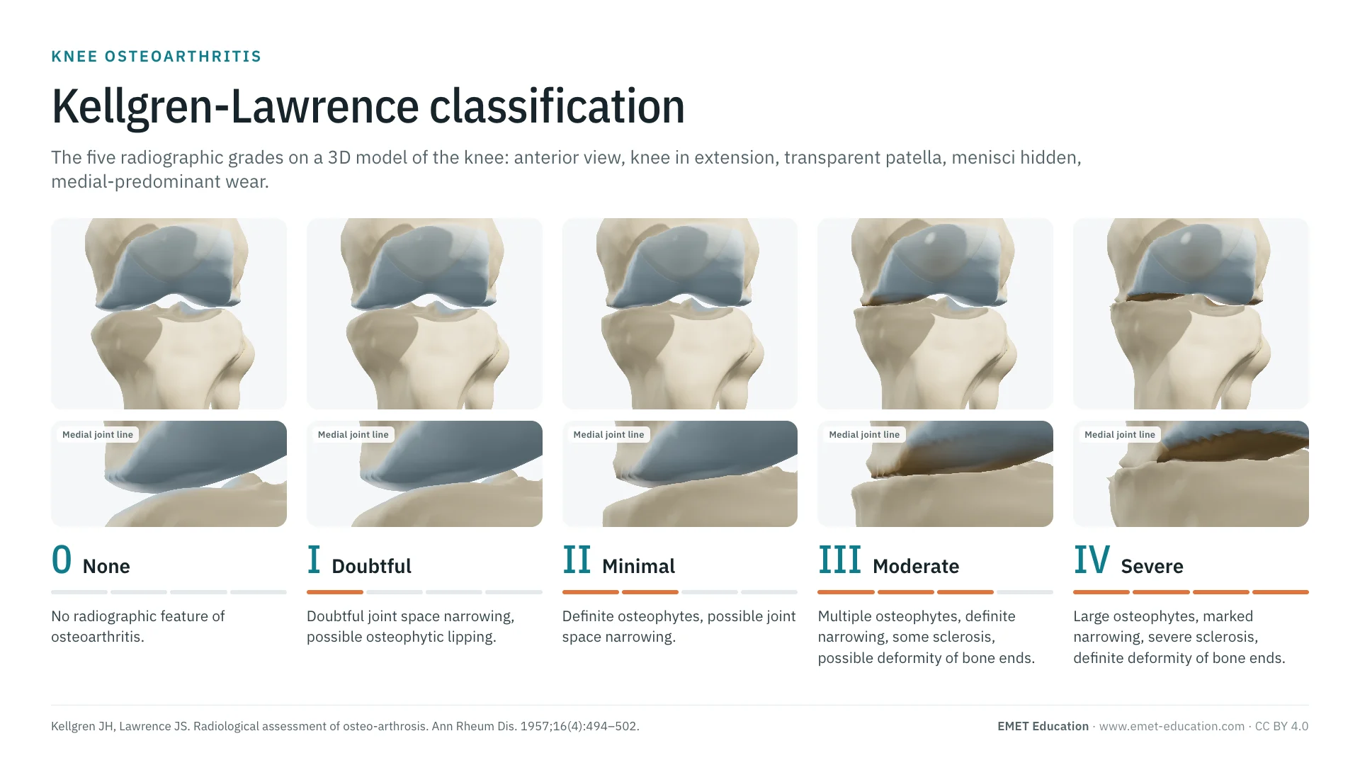 ClassificationsKellgren-Lawrence classification of the kneeThe other radiographic osteoarthritis scale, from 0 to 4, in 3D.
 ClassificationsAll 3D classificationsArranged by joint.
Explain the arthritic thumb in 3D
The thumb basal joint osteoarthritis model opens in your browser, at the stage you choose, with no sign-up.
Share this page with a colleague or a resident: