- EMET Education
- 3D classifications
- Herbert classification (scaphoid)
Classification · Acute scaphoid fracture
Herbert Classification of Scaphoid Fractures in 3D
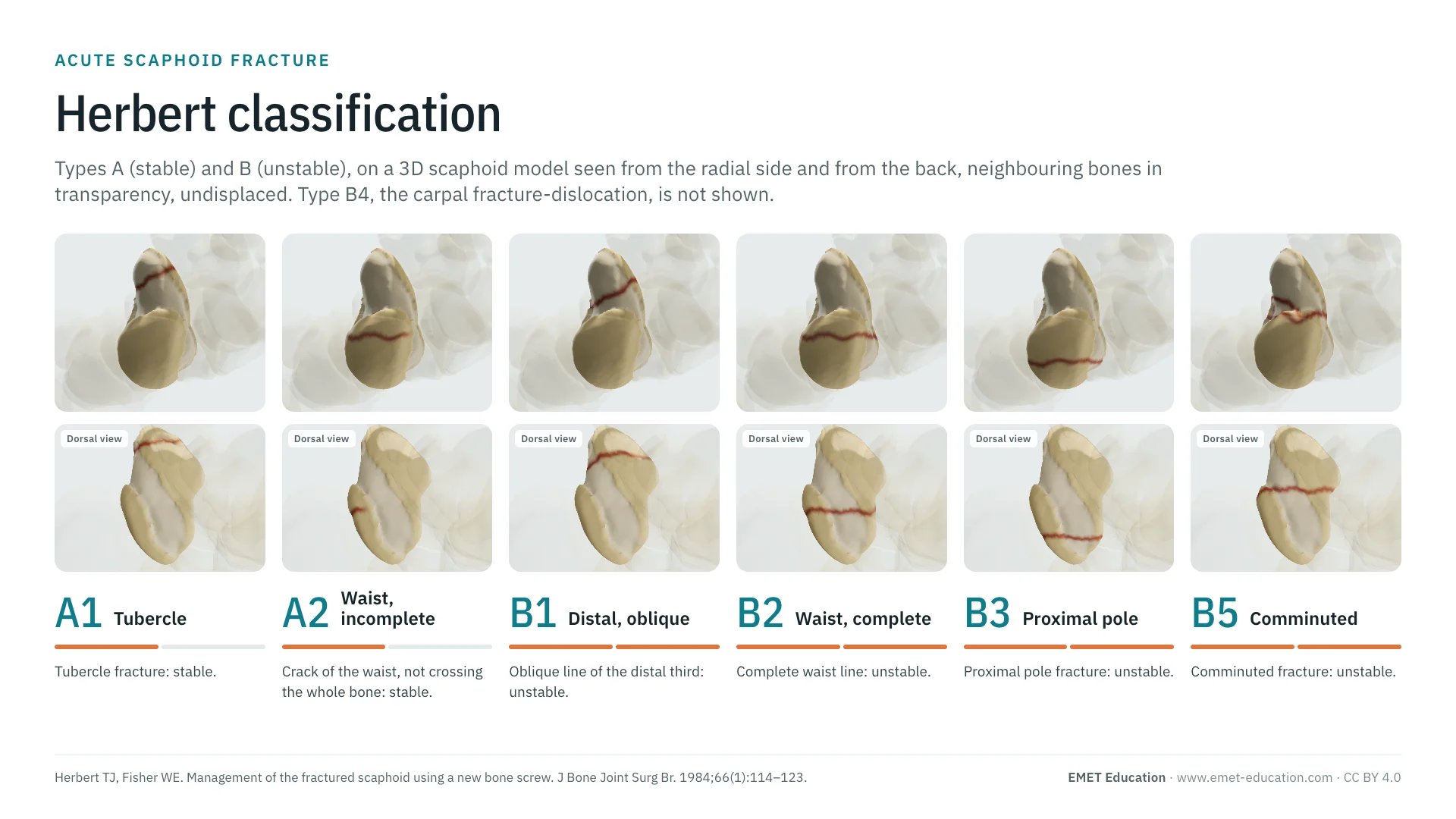
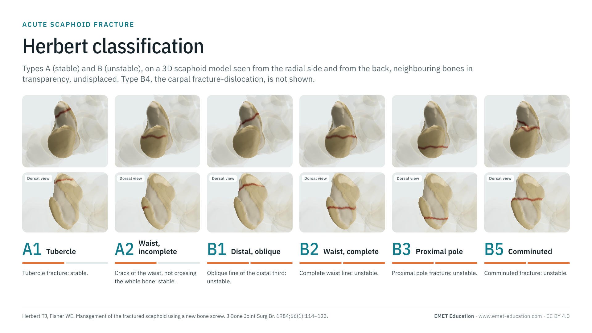
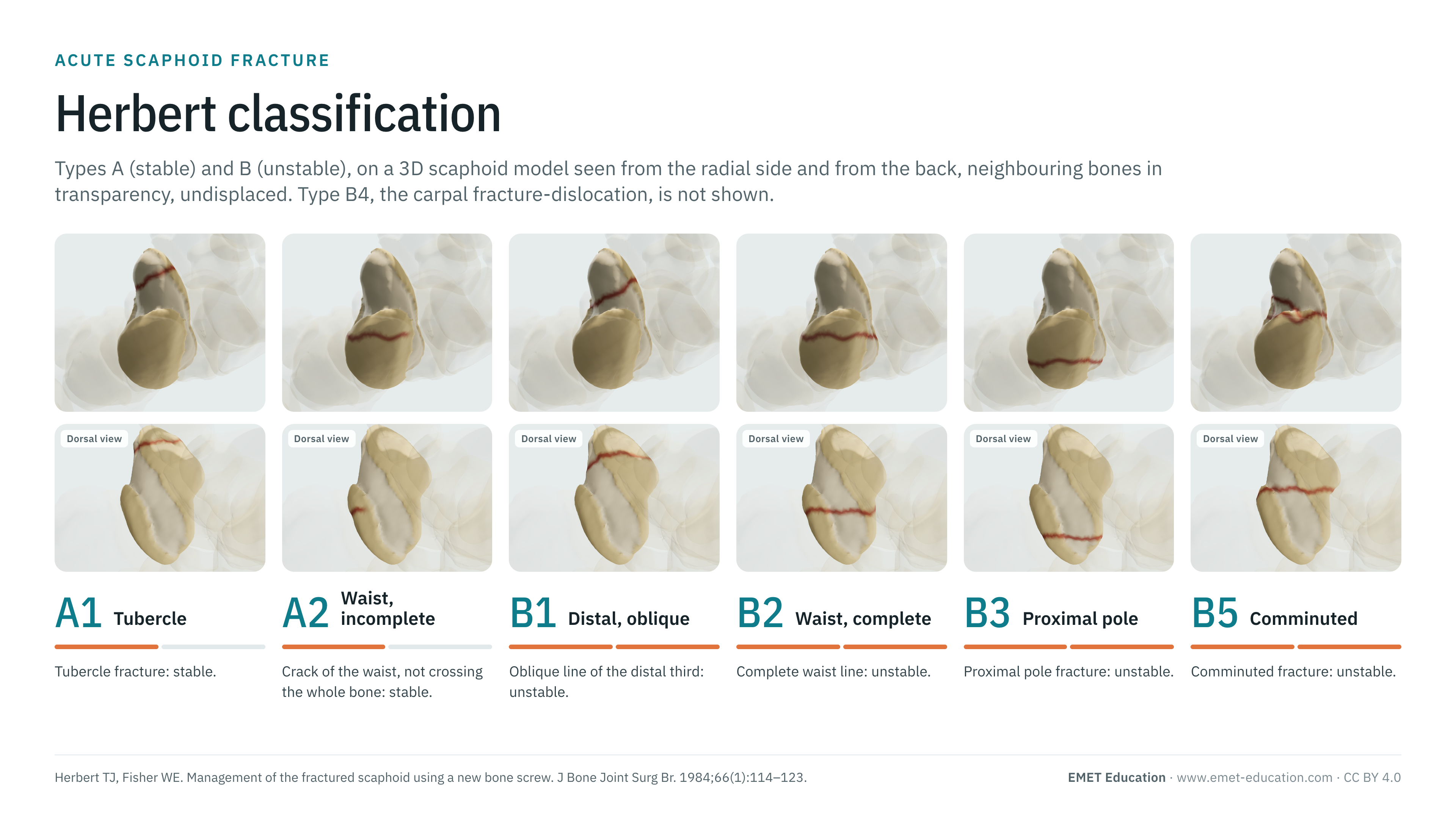
The types of the Herbert classification, stable (A1, A2) and unstable (B1 to B5) acute scaphoid fractures, on the scaphoid fracture 3D model, seen from the radial side and from the back, neighbouring bones in transparency. With the 1984 paper and its screw, how to read each type, reliability, grading pitfalls and free infographics for teaching and publication.
Designed by an orthopedic surgeon
The 3D model opens in your browser, at the type shown, with no sign-up. Infographics are free to reuse under CC BY 4.0.
- 1984Described by Herbert and Fisher in the Journal of Bone and Joint Surgery (Br) (Herbert and Fisher)
- A–DTypes: A1 and A2 stable; B1 to B5 unstable; C and D for delayed union and nonunion
- 933Citations of the 1984 paper (OpenAlex, October 31, 2026)
- κ 0.31–0.37Interobserver agreement of Herbert on radiographs, in two recent studies
The types
Types A and B, in 3D
Each image is a rendering of the EMET Education scaphoid fracture model at the site and pattern of that type: the scaphoid seen from the radial side (slightly palmar) on the left, from the back on the right, neighbouring bones in transparency; the red line is the fracture. Both views are needed: a crack (A2) runs only on the palmar and radial faces, so it stops short of the dorsal face, and the wedge of a comminuted fracture (B5) is palmar, so invisible from the back. The model draws six of the seven types; B4, the carpal fracture-dislocation, is described without an image. All types are shown undisplaced, to show only the fracture line; displacement can be set in the model.
-
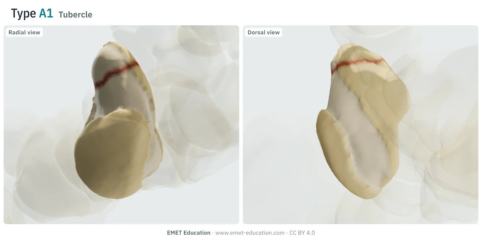
A1Tubercle
- Definition
- Type A (stable acute fracture), A1: fracture of the scaphoid tubercle, at the distal palmar end of the bone.
- Stability
- Stable: immobilization is usually enough (type A).
- In the 3D model
- A small tubercle fragment, detached 4.5 mm from its tip; undisplaced.
-
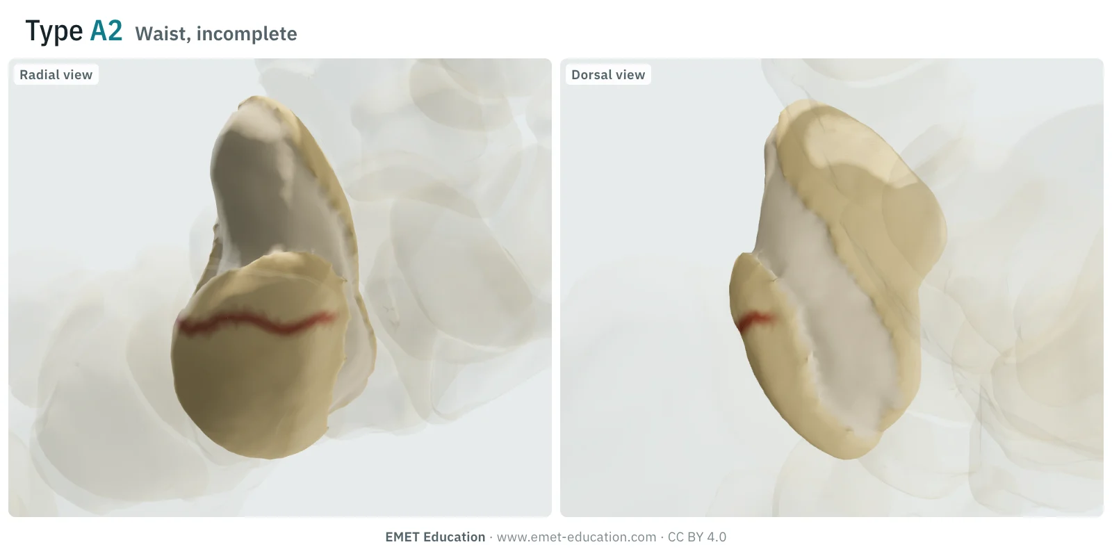
A2Waist, incomplete
- Definition
- Type A, A2: incomplete fracture of the waist, a crack that does not cross the whole bone, undisplaced.
- Stability
- Stable: immobilization is usually enough (type A).
- In the 3D model
- Waist fracture line limited to the palmar and radial faces; the model does not displace it. From the back, it stops at the radial edge.
-
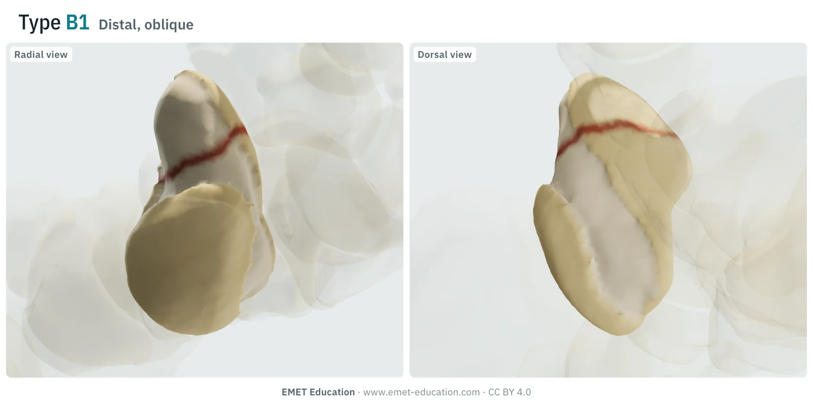
B1Distal, oblique
- Definition
- Type B (unstable acute fracture), B1: oblique fracture of the distal third.
- Stability
- Unstable (type B): risk of displacement and nonunion.
- In the 3D model
- Oblique line, dipping toward the palm, at 70% of the height of the bone from the proximal pole; undisplaced.
-
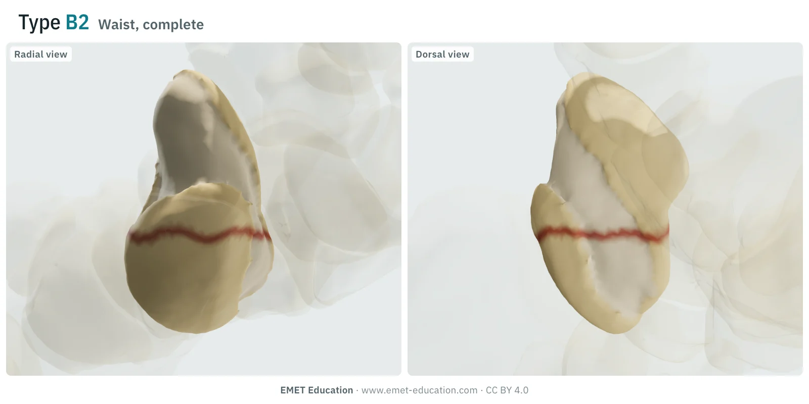
B2Waist, complete
- Definition
- Type B, B2: complete fracture of the waist. Some reviews write "displaced or mobile" (Steinmann and Adams): the instability is the one expected of a line that crosses the whole bone.
- Stability
- Unstable (type B): risk of displacement and nonunion.
- In the 3D model
- Complete transverse waist line, at 38% (proximal half) or 52% (distal half) of the height of the bone; the model codes any complete waist line as B2, displaced or not. Displacement can be set: 1.3 mm shift, then 15° extension tilt of the proximal fragment with the lunate, as on CT (Schwarcz et al.).
-
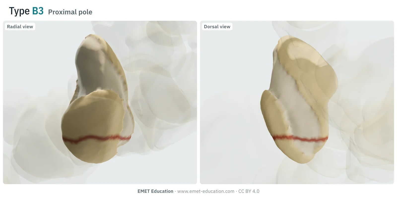
B3Proximal pole
- Definition
- Type B, B3: proximal pole fracture. The pole has no common definition: the proximal fifth of the bone has been proposed.
- Stability
- Unstable (type B): risk of displacement and nonunion.
- In the 3D model
- Transverse line at 20% of the height of the bone from the proximal pole; undisplaced. With the blood supply shown, every vessel reaching the proximal fragment is cut.
-
B4Carpal fracture-dislocation
- Definition
- Type B, B4: carpal fracture-dislocation, most often a trans-scaphoid fracture with perilunate dislocation. In the series by Herzberg et al. (166 perilunate dislocations and fracture-dislocations), dorsal trans-scaphoid fracture-dislocations made up 61% of the whole, and the diagnosis had first been missed in a quarter of cases.
- Stability
- Unstable (type B): risk of displacement and nonunion.
- In the 3D model
- Not drawn: the model does not show the carpus dislocated around the lunate (planned for later).
-
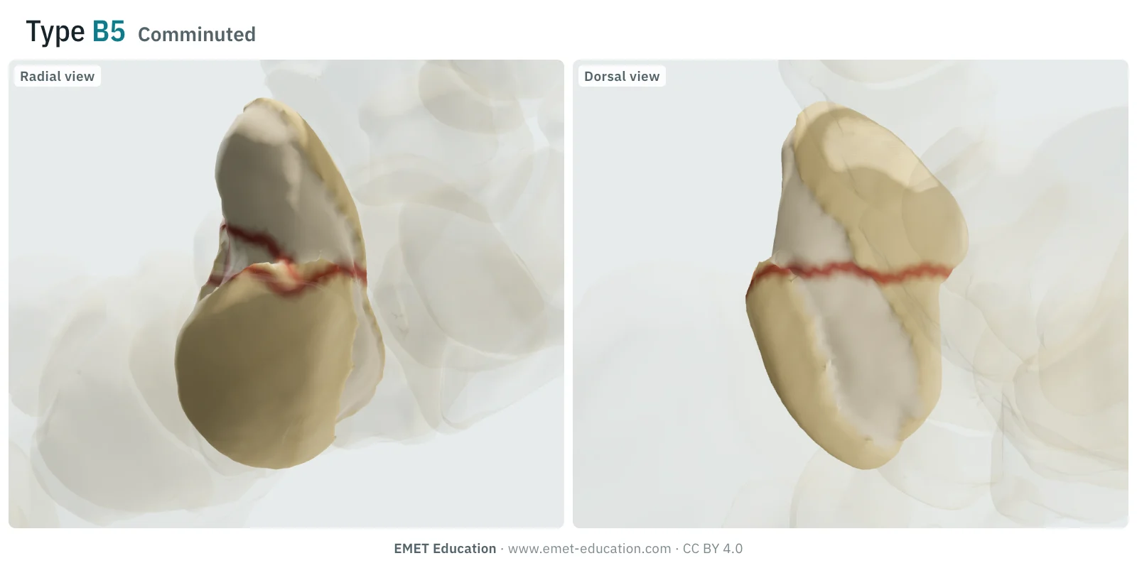
B5Comminuted
- Definition
- Type B, B5: comminuted fracture of the scaphoid, in several fragments.
- Stability
- Unstable (type B): risk of displacement and nonunion.
- In the 3D model
- Waist line (at 52% of the height of the bone) with a palmar wedge against the line, in two splinters; it is seen from the radial side, but not from the back.
Schematic rendering, for teaching: the model illustrates each type on a single scaphoid, it does not reproduce a radiograph. Definitions reworded from Steinmann and Adams, Ten Berg et al. and Clementson et al.; neither the figures nor the sentences of the Herbert and Fisher paper are reproduced. The bar shows stability: one segment for type A, two for type B.
The original paper
From the Herbert screw to the classification
The classification comes from a surgical paper, Herbert and Fisher (1984): it describes a double-threaded screw that fixes all types of scaphoid fracture rigidly, so much so that a cast is rarely needed afterwards, and it proposes a classification. The indications include acute unstable fractures, delayed unions and nonunions (with grafting). In the prospective series of 158 operations done from 1977 to 1981, all the acute fractures had united, and 83% of the whole. A series of 431 patients operated on by the same surgeon over 13 years followed (Filan and Herbert, 1996). In the review by Ten Berg et al., the Herbert classification is the most cited of the 13 systems found for acute fractures, ahead of Russe and Mayo; all rely on radiography.
The principle is stability: the risk that the fracture moves and fails to unite in the cast, more than the mere site of the line. The letters read as follows (Steinmann and Adams):
- A, stable acute fracture: A1, tubercle fracture; A2, incomplete fracture of the waist;
- B, unstable acute fracture: B1, oblique of the distal third; B2, complete of the waist; B3, of the proximal pole; B4, carpal fracture-dislocation; B5, comminuted;
- C, delayed union, after more than 6 weeks of plaster immobilization;
- D, established nonunion, fibrous (D1) or sclerotic (D2). This page covers only acute fractures (A and B), the only ones the model shows.
The types are not equally common. In Edinburgh, among 151 adult scaphoid fractures in one year (incidence of 29 per 100,000 per year), the most common was B2 (55 fractures, 36.4%) (Duckworth et al., 2012). In a series of 415 fractures, 64% were at the waist and 18.1% at the tubercle (Garala et al., 2016). The proximal pole is rare: 5 to 10% of fractures according to Clementson et al., 5.8% of the 1,147 acute fractures treated nonoperatively in the meta-analysis by Eastley et al..
Reliability
How reproducible is the Herbert type?
Modestly. As early as 1999, Desai et al. found the reproducibility of the Compson, Herbert and Russe classifications only fair, and none predicted union. Recent studies give interobserver kappas of about 0.3 to 0.4 for Herbert on radiographs, that is, fair agreement; in the study by Dean et al., CT does not improve it, but a reader agrees better with himself.
| Study | Measure | Value |
|---|---|---|
| Desai et al., 1999 | Inter- and intraobserver reproducibility of Compson, Herbert and Russe, radiographs (the abstract gives no coefficients) | “fair” |
| Dean et al., 2018 | Interobserver kappa, 6 readers in 2 centres, 60 acute fractures without dislocation: radiographs, then CT (Mayo: 0.57 · 0.54; Russe: 0.13 · 0.07) | 0.37 · 0.35 |
| Dean et al., 2018 | Intraobserver kappa, 2 readers: radiographs, then CT (Mayo: 0.82 · 0.86; Russe: 0.58 · 0.55) | 0.69 · 0.84 |
| Drijkoningen et al., 2019 | Retrospective study of 871 patients; interobserver kappa on posteroanterior radiographs: Herbert, then a simplified three-site classification (0.37). Readers' stated doubt was 3.6 out of 10 for Herbert, 2.1 for the simplified one | 0.31 |
| Daniels et al., 2020 | Intraclass correlation coefficient of the Herbert classification, high-resolution peripheral CT, 4 readers, 7 patients all diagnosed with a fracture (95% CI 0.42–0.94) | ICC 0.73 |
In the study by Dean et al., Mayo, which grades the site, was the most reproducible of the established classifications (interobserver 0.57), ahead of Herbert (0.37) and Russe (0.13), and a simple measure of the position of the line along the long axis did better (ICC 0.76). The same authors found only moderate agreement on comminution (ICC 0.47 on radiographs, 0.53 on CT). In practice: the letter (A or B) to communicate, the type only with imaging that allows it, and for research a written definition of each boundary.
Related classifications
What the other classifications add
Herbert answers one question, "is this fracture stable?". The other scaphoid classifications describe the site or the orientation of the line, and are not mutually exclusive: Ten Berg et al. count thirteen, based on site, orientation of the fracture plane or stability. The 2018 AO/OTA code also has a chapter for the carpal bones; EMET Education has not reviewed it and does not reproduce its codes.
The main ones:
| Classification | What it grades | Typical use |
|---|---|---|
| Russe (1960) | The orientation of the line: transverse, horizontal oblique, vertical oblique. | Vertical obliques (under 5% of fractures) are highly unstable (Clementson et al.). The lowest reproducibility of the three in Dean et al.. |
| Mayo (Cooney et al., 1980) | The site: proximal pole, middle third, distal third, distal tubercle, distal intra-articular; plus instability factors as an addition. | More reproducible than Herbert and Russe in Dean et al.; used in some departments as a morphological description (Clementson et al.). The same 1980 paper sets the threshold of 1 mm of offset, or tilting of the lunate, for calling a fracture displaced. |
| Prosser et al. (1988) | Fractures of the distal articular surface of the scaphoid: avulsions of the palmar tip of the tuberosity, impaction of the radial half. | Concerns the distal end, an area close to Herbert types A1 and B1; recently modified by Clementson et al. |
| Schernberg | Numbered zones along the scaphoid; the model shows it live, next to Herbert. | French-language use, for example "zones 2 to 4" for nonunions (Taleb et al.). Original definition not reproduced on this page. |
| Simplified classification (Drijkoningen et al., 2019) | Three sites on the posteroanterior radiograph: proximal pole (line proximal to the distal scapholunate interval), waist, distal tubercle. | Research; agreement slightly better than Herbert (κ 0.37 versus 0.31) and less doubt among readers. |
| Long-axis measurement (Dean et al., 2018) | The distance of the line, measured along the long axis of the scaphoid, rather than a type. | Research; interobserver ICC 0.76, better than Mayo, Herbert and Russe in the same study. |
Use
In research and in clinical practice
In research
Herbert is the language of most scaphoid series and trials. It lacks precision about the proximal pole: its definition varies, and the nonunion risk with it. In the meta-analysis by Eastley et al., 34% of acute proximal pole fractures treated nonoperatively progressed to nonunion, with a relative risk of 7.5 compared with more distal fractures, and they propose defining the pole as the proximal fifth of the bone. For Chong et al., the relative risk of nonunion went from 2.3 ("proximal third") to 3.4 ("proximal pole") depending on the term used, and, all terms pooled, the nonunion rate was 6% after surgery against 18% without: state in any publication how the pole is defined.
In clinical practice
The letter guides treatment. Type A fractures, stable, can be treated by immobilization; distal fractures most often heal in 4 to 6 weeks depending on their type (Clementson et al.). Type B fractures were, in the spirit of the classification, candidates for fixation; but Clementson et al. judge that treating every bicortical fracture, apart from the tubercle, as unstable can lead to overtreatment. The SWIFFT trial (439 adults, bicortical waist fracture, 61% undisplaced) found no clinically relevant difference at 52 weeks between fixation and cast (PRWE score 11.9 versus 14.0; adjusted difference −2.1, 95% CI −5.8 to 1.6), with few nonunions (1 after surgery, 4 after cast): for undisplaced or minimally displaced waist fractures, the cast remains an option.
Displacement is not read in the letter, but it decides: according to Clementson et al., about 90% of undisplaced waist fractures, or those displaced by 0.5 mm or less, unite at 6 weeks after proper nonoperative care, and fixation is recommended from 1.5 mm of displacement. The proximal pole should generally be fixed (Clementson et al.): its blood supply comes mostly from radial artery branches entering through the dorsal ridge, and a proximal line exposes the fragment to ischaemia (Gelberman and Menon). A comminuted fracture is more often displaced and unstable at arthroscopy (Buijze et al.; Bulstra et al.), and may need a graft (Schernberg). A fracture-dislocation (B4) calls for surgical reduction and fixation, and delay worsens the result: in the series by Herzberg et al., the best radiographic results came from open reduction and internal fixation, but post-traumatic arthritis reached 56% of cases treated early.
Grading pitfalls
Five pitfalls when grading a scaphoid fracture
- "Stable" is not "undisplaced". Herbert stability is the one expected of the fracture, not what the film shows: at arthroscopy, 11 of 31 undisplaced fractures were nonetheless unstable (mobile), and all displaced fractures were (Buijze et al.). Conversely, a complete waist line, B2, can be treated without surgery when slightly displaced (SWIFFT).
- A2 or B2: is the line complete? An incomplete crack runs around only part of the circumference; on a single view it can be taken for a complete line, just as a real line can be missed. Classification is best done on CT, with coronal and sagittal reconstructions along the long axis of the bone (Clementson et al.).
- Displacement is not part of the type. It is graded separately, with a written threshold: 1 mm of offset or tilting of the lunate for Cooney et al., 0.5 mm or less for "undisplaced" according to Clementson et al.. CT improves the reliability of this diagnosis (interobserver kappa 0.43 versus 0.27 on radiographs) but not much its accuracy, which stays under 80% (Lozano-Calderón et al.). The angle of the line is a minor factor in displacement of waist fractures (Aruse et al.).
- B3 or B2: where does the waist end? The proximal pole has no common definition; depending on whether the proximal fifth or third is used, the nonunion risk attributed to it changes (Eastley et al.; Chong et al.). A line between the two is graded with a written rule (for example the position of the line along the long axis), not by eye.
- B4 and B5: look for the carpus and for splinters. A scaphoid fracture on a doubtful film requires a search for perilunate dislocation, first missed in 25% of cases (Herzberg et al.). Comminution is poorly graded on radiographs (moderate agreement): with a suspected fracture and normal initial films, Clementson et al. recommend MRI or CT within three to five days.
Self-test
Which type?
The same scaphoid seen from the radial side and from the back, at the model's six types, in random order. Find the type of each one; the answer comes with the definition.
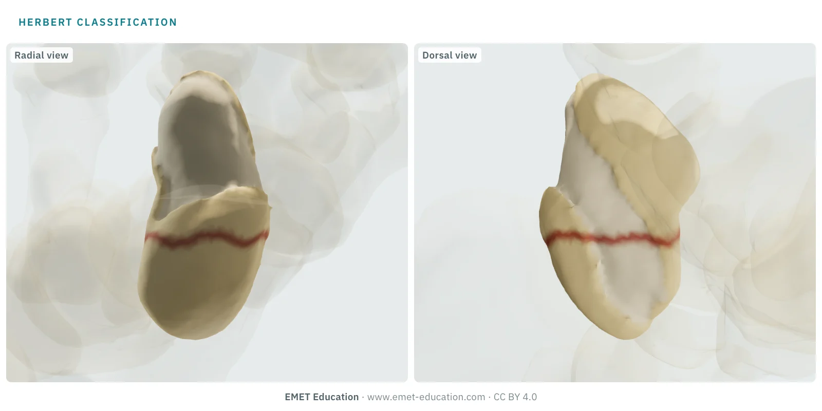
Which Herbert type is this?
Download and cite
Free infographics, for teaching and publication
Use them in a course, a presentation, a social media post or a scientific article, under the Creative Commons Attribution 4.0 license: free reuse, including commercial, provided EMET Education is credited with a link to this page. The license covers EMET Education's images, not the classification itself.
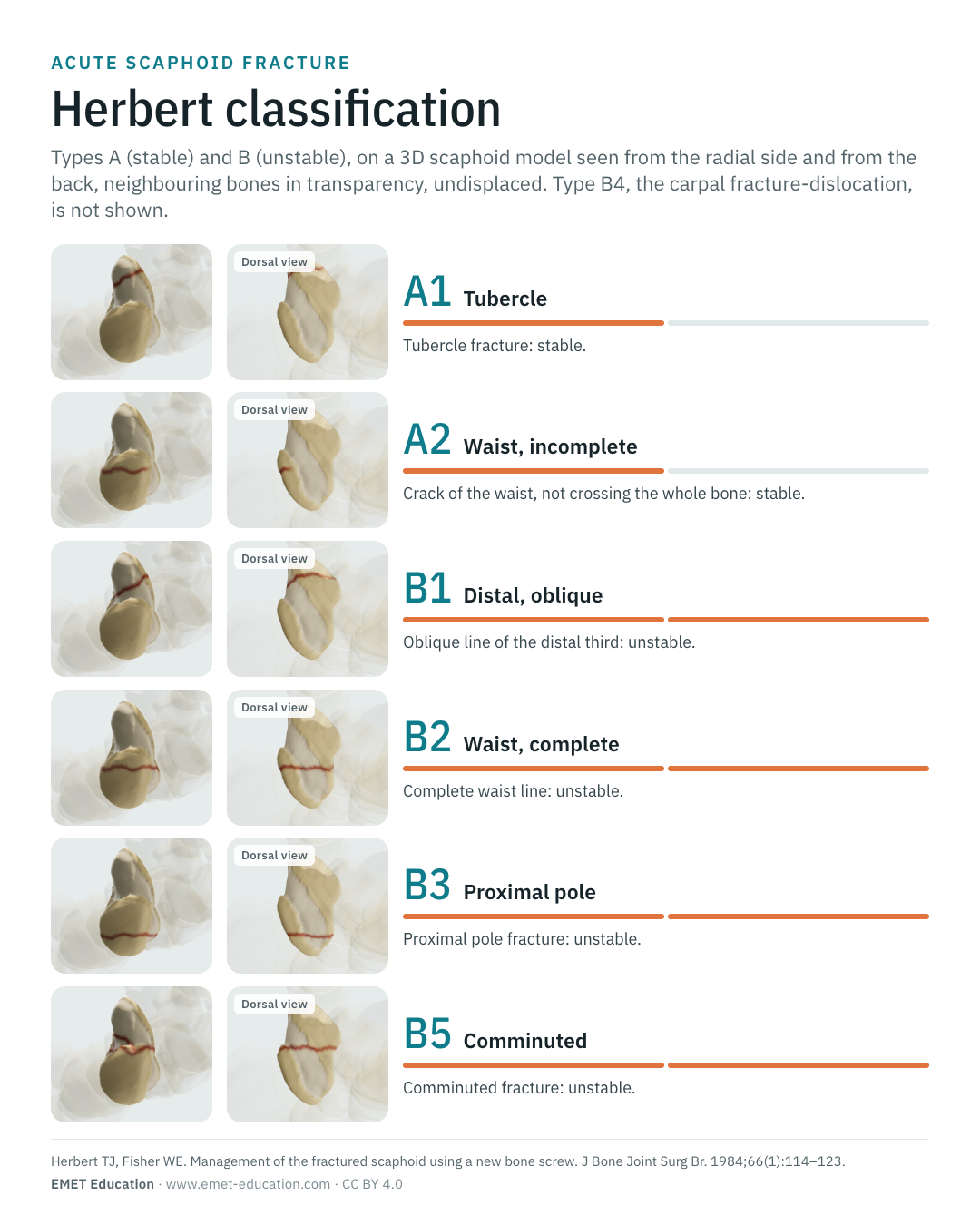
Cite this figure
Ready to paste. For a journal figure, add the original reference (Herbert and Fisher, 1984).
EMET Education. Herbert classification of scaphoid fractures: 3D visualization [Internet]. 2026 [cited]. Available from: https://www.emet-education.com/en/classifications/herbert-scaphoid/. CC BY 4.0.EMET Education. (2026). Herbert classification of scaphoid fractures: 3D visualization [Infographic]. https://www.emet-education.com/en/classifications/herbert-scaphoid/ (CC BY 4.0)<a href="https://www.emet-education.com/en/classifications/herbert-scaphoid/"><img src="https://www.emet-education.com/en/classifications/herbert-scaphoid/img/infographic.webp" alt="Herbert classification of scaphoid fractures in 3D" width="960"></a><br>Figure: <a href="https://www.emet-education.com/en/classifications/herbert-scaphoid/">EMET Education</a>, CC BY 4.0Questions
Frequently asked questions
What do A1, A2, B1, B2, B3, B4 and B5 mean?
The letter gives stability: A for a stable acute fracture, B for an unstable acute fracture. The number gives the site or type: A1 tubercle, A2 incomplete fracture of the waist; B1 oblique of the distal third, B2 complete of the waist, B3 proximal pole, B4 carpal fracture-dislocation, B5 comminuted. The letters C (delayed union) and D (nonunion) complete the classification.
What is the difference between an A2 and a B2 fracture?
The line is incomplete for A2, a crack that does not cross the whole bone, and complete for B2. A complete waist line is classed as unstable, whatever its displacement; but on a single view the difference is hard to read, hence the value of CT with reconstructions along the long axis of the scaphoid.
Does a B2 waist fracture always need surgery?
No. The Herbert classification puts bicortical fractures among the unstable ones, but the SWIFFT trial (439 adults) found no clinically relevant difference at 52 weeks between fixation and cast for undisplaced or minimally displaced waist fractures. The guidelines by Clementson et al. keep fixation for displacement of 1.5 mm or more. The decision rests with the surgeon and the patient.
Why do proximal pole fractures (B3) lead to nonunion and necrosis?
Because the supply of the scaphoid comes mostly from radial artery branches entering through the dorsal ridge: in the anatomical study by Gelberman and Menon (15 specimens), they provided 70 to 80% of the intraosseous vascularity, including the whole proximal pole. A proximal line therefore exposes the fragment to ischaemia. In a meta-analysis, 34% of proximal pole fractures treated nonoperatively progressed to nonunion, with a relative risk of 7.5 compared with more distal fractures.
Herbert, Mayo or Russe: which one to use?
They do not answer the same question: Herbert grades stability, Mayo the site, Russe the orientation of the line. Herbert is the most cited, Mayo is somewhat more reproducible, Russe is the least in the study by Dean et al. None is reproducible enough to replace a written description of the site, line, displacement and comminution.
Can I reuse these images in a course or an article?
Yes. The images and infographics on this page are under the Creative Commons Attribution 4.0 license: free reuse, including commercial, with credit to EMET Education and a link to this page. They reproduce no figure from the Herbert and Fisher paper. The "Cite this figure" box gives ready-to-paste citations.
How were the 3D images made, and what do they not show?
They are renderings of the EMET Education scaphoid fracture model: a scaphoid taken from CT, cut into fragments according to the site and pattern of each type, neighbouring bones in transparency. They are schematic views, not radiographs. The model does not draw type B4 (carpal fracture-dislocation), nor types C and D; it shows all types undisplaced; it codes any complete waist line as B2; and its live Herbert reading depends on the site and pattern alone, with no other criterion.
Sources
Author and references
- Herbert TJ, Fisher WE. Management of the fractured scaphoid using a new bone screw. J Bone Joint Surg Br. 1984;66(1):114–123. doi:10.1302/0301-620X.66B1.6693468
- Ten Berg PW, Drijkoningen T, Strackee SD, Buijze GA. Classifications of acute scaphoid fractures: a systematic literature review. J Wrist Surg. 2016;5(2):152–159. doi:10.1055/s-0036-1571280
- Steinmann SP, Adams JE. Scaphoid fractures and nonunions: diagnosis and treatment. J Orthop Sci. 2006;11(4):424–431. doi:10.1007/s00776-006-1025-x
- Clementson M, Björkman A, Thomsen NOB. Acute scaphoid fractures: guidelines for diagnosis and treatment. EFORT Open Rev. 2020;5(2):96–103. doi:10.1302/2058-5241.5.190025
- Duckworth AD, Jenkins PJ, Aitken SA, Clement ND, Court-Brown CM, McQueen MM. Scaphoid fracture epidemiology. J Trauma Acute Care Surg. 2012;72(2):E41–E45. doi:10.1097/TA.0b013e31822458e8
- Garala K, Taub NA, Dias JJ. The epidemiology of fractures of the scaphoid: impact of age, gender, deprivation and seasonality. Bone Joint J. 2016;98-B(5):654–659. doi:10.1302/0301-620X.98B5.36938
- Dean BJF, Riley ND, McCulloch ER, Lane JCE, Touzell AB, Graham AJ. A new acute scaphoid fracture assessment method: a reliability study of the ‘long axis’ measurement. BMC Musculoskelet Disord. 2018;19:310. doi:10.1186/s12891-018-2236-y
- Desai VV, Davis TR, Barton NJ. The prognostic value and reproducibility of the radiological features of the fractured scaphoid. J Hand Surg Br. 1999;24(5):586–590. doi:10.1054/jhsb.1999.0197
- Drijkoningen T, Mohamadi A, van Leeuwen WF, Schwarcz Y, Ring D, Buijze GA. Scaphoid fracture patterns—part two: reproducibility and demographics of a simplified scaphoid fracture classification. J Wrist Surg. 2019;8(6):446–451. doi:10.1055/s-0039-1692470
- Daniels AM, Wyers CE, Janzing HMJ, et al. The interobserver reliability of the diagnosis and classification of scaphoid fractures using high-resolution peripheral quantitative CT. Bone Joint J. 2020;102-B(4):478–484. doi:10.1302/0301-620X.102B4.BJJ-2019-0632.R3
- Russe O. Fracture of the carpal navicular: diagnosis, non-operative treatment, and operative treatment. J Bone Joint Surg Am. 1960;42-A:759–768. doi:10.2106/00004623-196042050-00002
- Cooney WP, Dobyns JH, Linscheid RL. Fractures of the scaphoid: a rational approach to management. Clin Orthop Relat Res. 1980;(149):90–97. PMID 6996886
- Prosser AJ, Brenkel IJ, Irvine GB. Articular fractures of the distal scaphoid. J Hand Surg Br. 1988;13(1):87–91. doi:10.1016/0266-7681(88)90061-7
- Schernberg F. Fractures récentes du scaphoïde (moins de trois semaines). Chir Main. 2005;24(3-4):117–131. doi:10.1016/j.main.2005.05.001
- Taleb C, Bodin F, Collon S, Gay A, Facca S, Liverneaux P. Retrograde percutaneous screw fixation for scaphoid type II non-union in Schernberg zones 2 to 4: a series of 38 cases. Chir Main. 2015;34(1):32–38. doi:10.1016/j.main.2014.10.150
- Eastley N, Singh H, Dias JJ, Taub N. Union rates after proximal scaphoid fractures; meta-analyses and review of available evidence. J Hand Surg Eur Vol. 2013;38(8):888–897. doi:10.1177/1753193412451424
- Chong HH, Kulkarni K, Shah R, Hau MYT, Athanatos L, Singh HP. A meta-analysis of union rate after proximal scaphoid fractures: terminology matters. J Plast Surg Hand Surg. 2022;56(5):298–309. doi:10.1080/2000656X.2021.1979016
- Dias J, Brealey S, Cook L, et al. Surgical fixation compared with cast immobilisation for adults with a bicortical fracture of the scaphoid waist: the SWIFFT RCT. Health Technol Assess. 2020;24(52):1–234. doi:10.3310/hta24520
- Herzberg G, Comtet JJ, Linscheid RL, Amadio PC, Cooney WP, Stalder J. Perilunate dislocations and fracture-dislocations: a multicenter study. J Hand Surg Am. 1993;18(5):768–779. doi:10.1016/0363-5023(93)90041-Z
- Buijze GA, Jørgsholm P, Thomsen NO, Björkman A, Besjakov J, Ring D. Factors associated with arthroscopically determined scaphoid fracture displacement and instability. J Hand Surg Am. 2012;37(7):1405–1410. doi:10.1016/j.jhsa.2012.04.005
- Bulstra AEJ, Al-Dirini RMA, Turow A, et al. The influence of fracture location and comminution on acute scaphoid fracture displacement: three-dimensional CT analysis. J Hand Surg Eur Vol. 2021;46(10):1072–1080. doi:10.1177/17531934211040962
- Aruse O, Immerman I, Badir O, Haj ME, Volk I, Luria S. Scaphoid fracture displacement is not correlated with the fracture angle. J Hand Surg Eur Vol. 2021;46(6):607–615. doi:10.1177/17531934211004434
- Lozano-Calderón S, Blazar P, Zurakowski D, Lee SG, Ring D. Diagnosis of scaphoid fracture displacement with radiography and computed tomography. J Bone Joint Surg Am. 2006;88(12):2695–2703. doi:10.2106/JBJS.E.01211
- Schwarcz Y, Peleg E, Joskowicz L, Wollstein R, Luria S, et al. Three-dimensional analysis of acute scaphoid fracture displacement: proximal extension deformity of the scaphoid. J Bone Joint Surg Am. 2017;99(2):141–149. doi:10.2106/JBJS.16.00021
- Gelberman RH, Menon J. The vascularity of the scaphoid bone. J Hand Surg Am. 1980;5(5):508–513. doi:10.1016/s0363-5023(80)80087-6
- Filan SL, Herbert TJ. Herbert screw fixation of scaphoid fractures. J Bone Joint Surg Br. 1996;78(4):519–529. doi:10.1302/0301-620X.78B4.0780519
3D models
Show the fracture to your patient
The images on this page come from an interactive model, the scaphoid fracture 3D model. In clinic, choose the site and the pattern, set the displacement from your patient's imaging, show the blood supply of the proximal pole, then show the proposed treatment: resin cast, percutaneous screw or open reduction with screw.
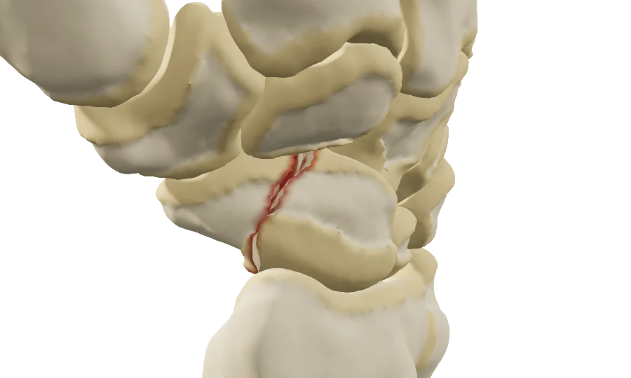
Explain the fracture in 3D
The scaphoid fracture model opens in your browser, at the type you choose, with no sign-up.
Share this page with a colleague or a resident: